2. Bath et al. / Journal of Applied Pharmaceutical Science 5 (07); 2015: 130-135 131
This study was undertaken to develop an accurate, simple
and sensitive HPLC method to measure coumarin hydroxylase
activity as a marker of CYP2A6 activity then apply the method to
investigate the potential effect of RES, SF, and TQ on hepatic
CYP2A6 activity in Spontaneously Hypertensive Rat (SHR)
model.
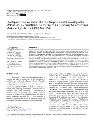
Fig. 1: CYP2A6-mediated COUM hydroxylation to 7-OH COUM.
This specific animal model of hypertension is currently
used in our lab to investigate the antihypertensive effect of certain
phytochemicals. The importance of this study is emphasized by
the findings that certain phytochemicals in vegetables and fruits
have the capacity to modulate drug metabolizing enzymes and
significantly affect pharmacokinetics, and hence
pharmacodynamics, of concomitantly administered medications.
EXPERIMENTAL
Materials
Chemicals and reagents
Coumarin (COUM), 7-hydroxycoumarin (7-OH COUM),
6-hydroxychlorzoxazone (6-OH CZX), thymoquinone (TQ) and
all chemicals used for microsomal preparation, determination of
microsomal protein content, and enzyme assays were purchased
from Sigma-Aldrich (ST. Louis, MO).
Purified sulforaphane (SF) was obtained from LKT
laboratories (St. Paul, MN). Resveratrol (RES) was purchased
from TCI (Portland, OR). Acetonitrile and methanol were HPLC
grade and obtained from Fisher Scientific (Pittsburg, PA). A Milli
Q Synthesis (Millipore, Bedford, MA) water purification system
provided purified deionized water. All other chemicals used were
analytical grade.
Animals
Male 14-week-old SHR were obtained from Charles
River Laboratories (Wilmington, MA). All animals were
maintained under controlled housing conditions of light (6 AM-6
PM) with temperature (22 °C) and received standard laboratory
chow and water ad libitum. All rats were allowed at least 1 week
to become acclimated to the housing conditions and ensure steady
and reliable blood pressure readings before use in experiments.
All procedures were approved by the Institutional Animal Care
and Use Committee of Pacific University and Oregon Health &
Science University. Animals were randomly divided into 7 groups
of 5 animals each. One group served as the control group, whereas
the remaining groups were provided with either low dose (20
mg/kg) or high dose (40 mg/kg) of SF, RES, or TQ in the drinking
water for 8 weeks. Preliminary stability studies using LC-MS
indicated that RES, SF, and TQ solutions are stable in water at
room temperature for at least 5 days (data not shown). Fresh
solutions were made from stock twice a week. The selection of
RES, SF, or TQ doses was based on preliminary studies in our
laboratory and previous studies (Elbarbry et al., 2014);
Kulkarni, 2014).
Methods
Preparation of Hepatic Microsomes
Hepatic microsomes were prepared as described
previously (Elbarbry et al., 2012). Briefly, 0.5 g of liver was
homogenized in 2 mL homogenization buffer (50mM Tris buffer,
150mM KCl, 0.1mM dithiothreitol, 1mM EDTA, 20% glycerol,
and 0.1mM phenylmethyl sulfonylfluoride).
The homogenate was centrifuged at 9000×g for 30 min in
a Beckman L8-55 Ultracentrifuge (Palo Alto, CA, USA). The
supernatant was carefully transferred to clean ultracentrifuge tubes
and centrifuged at 100,000×g for 30 min. The pellet was washed in
2 mL of 150mM KCl and centrifuged again at 100,000×g for 30
min. The pellet was resuspended in 2 mL of 0.25M sucrose
solution and 400 μL aliquots were transferred to cryogenic
microcentrifuge tubes (1.5 mL). Microsomal suspensions were
stored at −80 °C until use.
Determination of Microsomal Protein Content
Microsomal protein concentrations were determined by
the method of Lowry et al.(Lowry et al., 1951) using bovine serum
albumin as the standard. Absorbance was measured at 750 nm on a
Synergy2®
micro-plate reader using Gen5 Software (BioTek,
Winooski, VT).
Chromatographic Conditions
A Shimadzu LC 20A HPLC system (Maryland, USA)
consisting of an auto-sampler, a degasser, a binary pump, a model
SPD-20A photodiode array detector. Chromatographic separation
was carried out on Zorbax Eclipse XDB C18 column (150 4.6
mm I.D., 5 m particle size).
The column was kept at 40o
C. The isocratic mobile phase
consisted of methanol and 1% glacial acetic acid mixture (35:65,
v/v) was run at a flow rate of 0.6 ml/min. Absorbance was
monitored at 320 nm. This wavelength was found adequate to
monitor COUM, 7-OHCOUM and 6-OHCZX as indicated by
using the PDA detector.
Preparation of Standard Solutions and Calibration Standards
COUM and 7-OH COUM were dissolved in dimethyl
sulfoxide (DMSO) to prepare stock solutions (10 mM) each. The
stock solutions were stored at - 20°C where analytes were found to
be stable for at least 8 weeks. Using these stock solutions,
calibration standards were prepared in drug-free and heat-
inactivated rat hepatic microsomes at concentrations of 0, 0.025,
0.05, 0.1, 0.25, 0.50, 1.0, 2.5, and 5.0 µM of each analyte. Three
3. 132 Bath et al. / Journal of Applied Pharmaceutical Science 5 (07); 2015: 130-135
quality control (QC) samples were prepared by spiking drug-free
rat hepatic microsomes with COUM and 7-OH COUM for method
validation studies.
These QC samples were prepared to contain 0.25 µM
(QC1); 1.0 µM (QC2); and 2.5 µM (QC3) of each analyte. A
working internal standard (IS) solution was prepared by diluting 6-
OH CZX stock solution (1 mM) in methanol to obtain 100 µM.
Microsomal Incubation
7-Hydroxylase activity was determined by quantification
of 7-OH COUM formation rate in rat hepatic microsomes.
Preliminary experiments were conducted to determine linear
metabolite formation kinetics with respect to COUM
concentration, incubation time and microsomal protein
concentration. Microsomal incubation mixtures consisted of
COUM (20 μM), 0.4 mg/ml liver microsomal protein, 2mM
MgCl2, 1mM NADPH, and 50mM phosphate buffer, pH 7.4, in a
final volume of 0.5 ml. After a pre-incubation period of 3 min at
37°C, the reaction was started by addition of NADPH and
incubated at 37°C for 15 min in a shaking water bath. The reaction
was terminated by addition of 50μL ice-cold 50% tri-chloroacetic
acid as described before(Yuan et al., 2002). Metabolite formation
rate was calculated by dividing the amount of the metabolites
formed by the incubation time and microsomal protein content
(nmol/min/mg).
Sample Preparation
To 200μL calibration standards, QC samples, or
microsomal incubation mixtures, 50 μL of the working internal
standard solution was added. The mixtures were vortex mixed for
20 seconds. After centrifugation at 10,000×g in an Eppendorf
microcentrifuge (Model 5417 C, Brinkmann instruments,
Westbury, NY, USA) for 15 min, 20 μL of the supernatant was
injected directly onto the analytical column for immediate HPLC
analysis.
Method Validation
Method validation procedures were performed according
to FDA guidelines
(http://www.fda.gov/downloads/drugs/guidancecomplianceregulat
oryinformation/guidances/ucm070107.pdf, accessed Dec. 1, 2014)
and as explained previously (Elbarbry et al., 2009;Elbarbry and
Shoker, 2007) to evaluate the suitability of the method for the
quantitative determination of COUM and its 7-OH metabolite, in
rat hepatic microsomes. Specificity was tested by analysis of five
independent drug-free rat hepatic microsomes supplemented only
with IS to ensure the absence of endogenous compounds with the
same retention times at the analytes of interest. The linearity of the
method was evaluated by processing eight-point calibration curves
range 0.025-5.0 µM on six different days. The peak height ratios
of each analyte to the internal standard were plotted against the
analyte’s nominal concentration. A linear least-squares regression
analysis was conducted to determine slope, intercept and
coefficient of determination (R2
) to demonstrate linearity of the
method. The lower limit of detection (LLOD) was defined as the
lowest detectable concentration, taking into consideration a signal-
to-noise (S/N) ratio of 3. Lower limit of quantification (LLOQ)
was determined as the lowest concentration at which the precision,
expressed as % coefficient of variation (CV), is less than 20%,
accuracy is 80-120% and S/N ratio is at least 10. The accuracy
and precision of the proposed method were determined by analysis
of the QC samples. The intra-day accuracy and precision were
assessed from the results of five replicate analyses of QC samples
on a single assay day. The inter-day accuracy and precision were
determined from the same QC samples analyzed on 6 consecutive
days. Accuracy (%) is expressed as [(calculated amount/predicted
amount) x100]. The mean value should be within 15% of the
predicted value except at LLOQ, where it should not deviate by
more than 20%. Precision is expressed as % CV, and it should not
exceed 15% except for the LLOQ, where it should not exceed
20%.
Stability of QC samples was evaluated after long-term
(frozen at -20°C) and short-term (bench top, room temperature)
storage, and after going through three freeze and thaw cycles. The
stability of stock solutions of COUM, 7-OH COUM and IS was
also evaluated as described below:
i. Freeze and Thaw Stability: Three aliquots of each QC sample
were stored at -20°C for 24 hours and thawed unassisted at
room temperature. The freeze–thaw cycle was repeated two
more times, and then analyzed on the third cycle.
ii. Short-Term Temperature Stability: Three aliquots of each QC
sample were thawed at room temperature and analyzed at 4
and 12 hours (based on the expected duration that samples
will be maintained at the HPLC auto-sampler during analysis)
iii. Long-Term Temperature Stability: Three aliquots of each QC
sample were kept at -20°C for two weeks (the estimated time
between the date of first sample collection and the date of last
sample analysis).
iv. Stock Solution Stability: The stability of stock solutions of
COUM, 7-OH COUM, and the internal standard were
evaluated at room temperature for 6 hours and at -20°C for 2
weeks.
Data Analysis
Data are reported as mean ± SD. Difference in CYP2A6
activity among groups was assessed by one-way Analysis of
Variance (ANOVA) with Turkey's post-hoc test for pair-wise
multiple comparisons. Statistical analysis was conducted using
GraphPad Prism 5.0 (GraphPad Software Inc., San Diego, CA).
RESULTS AND DISCUSSION
As studying the effect of herbal remedies on the activity
of drug metabolizing enzymes is a routine experiment in our
laboratory, development of simple, cost-effective, accurate, fast
and sensitive HPLC methods is a key. Due to its clinical and
toxicological relevance, CYP2A6 is one of the most important
P450 enzymes. Hydroxylation of COUM to 7-OH COUM has
4. Bath et al. / Journal of Applied Pharmaceutical Science 5 (07); 2015: 130-135 133
been used as a marker for CYP2A6 in biological samples (Soucek,
1999). In our preliminary experiments using published
fluorescence-based HPLC methods, selectivity was an issue as
some of the studied herbal remedies or their metabolites co-elute
with COUM or 7-OH COUM. Additionally, accuracy and
precision were frequently below the accepted standards and a use
of IS in the HPLC analysis was needed to improve them.
Therefore, we described in this article a new UV-based HPLC
assay for COUM and its 7-OH metabolite as a marker of CYP2A6.
After trying several mobile phase compositions and different
columns, methanol and 1% glacial acetic acid (35/65) was chosen
as a mobile phase and Zorbax Eclipse XDB C18 was selected as a
stationary phase on the basis of shorter retention time, symmetric
peaks, good resolution, and excellent selectivity. 6-OH CZX is
structurally-similar to COUM and its metabolite, and was found to
be a good internal standard in this method. Under the
chromatographic conditions explained in the methods section, 6-
OH CZX, 7-OH COUM, and COUM were eluted and well
separated at retention times 6.2, 7.5, and 10.9 min respectively
(Figure 2a).
Fig. 2: Typical HPLC chromatograms of COUM and its metabolite (7-OH
COUM) in rat hepatic microsomes samples. (a) Blank rat hepatic microsomes
sample spiked with internal standard (6-OH CZX), 7-OH COUM (0.025 µM),
and COUM (0.025 µM). (b) Blank rat hepatic microsomes sample spiked only
with the internal standard (20µM) shows no interfering peaks at retention times
of COUM or 7-OH COUM. (c) Blank rat hepatic microsomes sample without
any spiking shows no interfering peaks at retention times of the analytes or the
internal standard.
Method Validation
Selectivity
Selectivity of the proposed analytical method was
indicated by the absence of any endogenous interference at
retention times of peaks of interest as evaluated by chromatograms
of blank rat hepatic microsomes (Figure 2c) and blank rat hepatic
microsomes spiked only with the internal standard (Figure 2b).
Linearity
Calibration curves were constructed by plotting peak
height ratios of the analyte to IS versus concentration of the
analyte. The calibration curves of COUM and its metabolite in rat
hepatic microsomes were linear over concentration ranges of
0.025-5 µM (Table 1). The coefficient of determination (R2
) was
greater than 0.999 in all calibration curves (n = 6). The mean
regression equations were y = 1.212x - 0.0221 for 7-OH COUM
and y = 2.71x - 0.0197 for COUM. Lower limit of
detection (LLOD) and quantitation (LLOQ) were determined
based upon Signal-to-Noise (S/N) ratio of 3 and 10, respectively.
Spiked rat hepatic microsomes (for COUM and its metabolite)
were assayed for all analytes in decreasing concentration.
Chromatograms from the assay of blank hepatic microsomal
samples were used for the establishment of the background noise.
LLOD of COUM and 7-OH COUM was 0.001 and 0.005 µM,
respectively. However, LLOQ for both analytes, where maximal
intra- and inter-day variation in accuracy and precision is 20%,
was 0.025 µM. The range and sensitivity level of calibration
curves in our method were sufficient to measure coumarin
hydroxylase activity in rat hepatic microsomes.
5. 134 Bath et al. / Journal of Applied Pharmaceutical Science 5 (07); 2015: 130-135
Accuracy and Precision
To determine intra-day and inter-day accuracy and
precision of the assay, replicate set (n = 5 and 6, respectively) of
three concentrations (low, medium and high) of each analyte in rat
hepatic microsomes were analyzed. Accuracy and precision were
calculated as described in the materials and methods section and
results are summarized in Table 2. The intra-assay and inter-assay
precision for COUM and its metabolite were less than 10% in all
cases. Blindly assayed QC samples of both COUM and 7-OH
COUM show an average intra-day and inter-day accuracy in the
range of 98-112% (Table 2). These data showed that the proposed
method was both accurate and precise for the determination of
COUM and its metabolite in rat hepatic microsomes.
Stability
To evaluate the stability of COUM, 7-OH COUM, and IS
under storage and handling conditions, separate QC standards,
covering the low (0.05µM), medium (1 µM), and high ranges
(2.5µM), were prepared for different temperatures and times.
The accuracy of COUM and 7-OH COUM obtained after
three freeze-thaw cycles, short-term storage (4-12 hr.) at room
temperature, and long-term storage (2 weeks) at -20°C
was 95-105%, as shown Table 3. Additionally, the standard stock
solution of both analytes and IS were found to be stable for at
least 6 hr. at room temperature and for 2 weeks at -20°C
(Table 3). The stability of both analytes and IS was
consistent with other reports (Kim et al., 2005;Soucek, 1999).
Application of the Proposed Method
COUM is extensively metabolized by CYP2A6 isozyme
to 7-OH COUM (Yuan et al., 2002). Accordingly, COUM
hydroxylase is a commonly used marker for CY2A6 activity. Our
validated HPLC-UV method is adequately sensitive, accurate, and
precise to evaluate COUM hydroxylase activity in rat hepatic
microsomes.
Fig. 3: Representative HPLC chromatograms of rat hepatic microsomes after
incubation with COUM (20 μM) in the absence (a) and presence (b) of
NADPH. Incubation conditions are explained in the methods section
Figures 3a and 3b indicated that COUM hydroxylation
was exclusively mediated by microsomal CYP enzymes and
required NADPH for electron transfer. Recent research in our lab
has indicated that certain phytochemicals such as sulforaphane
(SF), resveratrol (RES), and thymoquinone (TQ) modulate the
CYP-mediated metabolism of arachidonic acid in SHR model, a
mechanism through which these phytochemicals protect SHR rats
against the progressive rise in blood pressure (Elbarbry et al.,
2014).
In the current study, we have utilized the developed
HPLC method to investigate the effect of these phytochemical on
Table. 1: Linearity data for the quantification of COUM and its metabolite, 7-OH COUM in in rat hepatic microsomes by the proposed HPLC method
Intercept Slope R2
LLOQ LLOD Linearity Range
Average* SD Average* SD Average* SD (µM) (µM) (µM)
7-OH COUM -0.0221 0.018 1.22 0.287 0.99960 0.000274 0.025 0.005 0.025-5
COUM -0.0197 0.023 2.71 0.391 0.99964 0.000232 0.025 0.001 0.025-5
*n=6
Table. 2: Intra-day (n = 5) and inter-day (6 consecutive days) accuracy and precision data for the quantitation of COUM and its metabolite, 7-OH COUM in in
rat hepatic microsomes by the proposed HPLC method.
Nominal Conc. Intra-day (n =5) Inter-day (n =6)
(µM) Average SD Accuracy % CV% Average SD Accuracy % CV%
7-OH COUM 0.25 0.281 0.022 112 7.74 0.268 0.022 107 8.18
1 0.986 0.074 98.5 7.55 1.047 0.052 104 5.02
2.5 2.652 0.144 106 5.43 2.823 0.148 112 5.23
COUM 0.25 0.254 0.015 102 5.87 0.259 0.025 103 9.67
1 1.04 0.05 104 7.12 1.085 0.073 108 6.77
2.5 2.54 0.107 101 7.17 2.505 0.107 100 4.28
Table. 3: Stability of COUM, 7-OH COUM, and 6-OH CZX in solution under different storage and handling conditions.
Freeze-Thaw Stability Short-Term Stability
Long-Term
Stability
Stock Solution Stability
After 4 hr After 12 hr Room Temp. for 6 hr. -20°C for 2 weeks
COUM
7-OH
COUM
COUM
7-OH
COUM
COUM
7-OH
COUM
COUM
7-OH
COUM
COUM
7-OH
COUM
6-OH
CZX
COUM
7-OH
COUM
6-OH
CZX
QC1 95.5 ±2.5 101 ± 2.5 99.5 ± 2 100.5 ± 3 97.5 ± 3 101.5±1.5 98 ±3.5 102 ± 3 97 ± 4.5 104 ± 1.5 105 ±4.5 104 ± 2.5 100 ± 2.5 100 ± 2.5
QC2 99 ± 2 102 ± 1.5 97.5 ± 1.5 102.5 ± 2 99 ±2.5 97 ± 3 102 ±3 97 ± 1.5
QC3 98 ± 3.5 98 ± 3 101.5 ± 2 99.5 ±2.5 103 ± 3 95 ± 2.5 95 ± 1.5 100 ± 5
6. Bath et al. / Journal of Applied Pharmaceutical Science 5 (07); 2015: 130-135 135
CYP2A6 activity through measuring the rate of 7-OH COUM
formation in hepatic microsomes of SHR following administration
of different doses of these phytochemicals in their drinking water.
As shown in Figure 4, no significant effect of either SF or TQ was
observed on CYP2A6 activity.
Fig. 4: Effect of administration of dietary doses of sulforaphane (SF),
Resveratrol (RES), and Thymoquinone (TQ) on hepatic microsomal CYP2A6
activity in SHR rats. Male SHR rats (n = 5) were administered 20 mg/kg or 40
mg/kg of SF or RES or TQ in their drinking water for 8 weeks and livers were
harvested at the end of the study. Activities of CYP2A6 enzyme was
determined using the proposed HPLC method and as explained in the method
section. All groups were compared using one-way ANOVA followed
by multiple comparisons. *Significant difference from control with P<0.05.
On the other hand, 20 mg/kg and 40 mg/kg RES treatment resulted
in 28% and 33% inhibition of CYP2A6 activity, respectively. This
CYP enzyme is the principal enzyme responsible for the oxidation
of nicotine. Additionally, it is also involved in the clearance of
several pharmaceuticals and carcinogens (Le et al., 2003).
Inhibition of CYP2A6 activity by RES could pose a risk of drug-
herb interactions and a caution should be exercised in prescribing
compounds metabolized by CYP2A6 enzyme in patients who are
taking RES or RES-congaing natural products. On the other hand,
RES may offer protection against carcinogens such as aflatoxin B1
and nitrosamines that are bio-activated by CYP2A6 (Higashi et al.,
2007;Oscarson, 2001).
CONCLUSION
In this study, we described an HPLC-UV method for the
simultaneous determination of COUM and its 7-OH metabolite as
a surrogate marker for CYP2A6 enzyme activity. This new
method offers the advantages of simplicity, specificity, precision,
adequate sensitivity, and low sample volume. We successfully
applied the method to determine CYP2A6 activity in rat hepatic
microsomes and to investigate the effect of three commonly used
phytochemicals SF, RES, and TQ on CYP2A6 activity in a rat
model of hypertension.
ACKNOWLEDGEMNTS
We thank Anik Amin and Margarita Can for microsomal
preparation. This work was supported in part by the Medical
Research Foundation of Oregon (MRF), Collins Medical Trust,
Faculty Development Grant from Pacific University, Oregon and
Dean’s Incentive Grant from Pacific University School of
Pharmacy, Oregon (F.E.).
REFERENCES
Elbarbry F, Attia A, Shoker A. Validation of a new HPLC
method for determination of midazolam and its metabolites: application to
determine its pharmacokinetics in human and measure hepatic CYP3A
activity in rabbits. J Pharm Biomed Anal 2009; 50: 987-993.
Elbarbry F, Ragheb A, Marfleet T, Shoker A. Modulation of
hepatic drug metabolizing enzymes by dietary doses of thymoquinone in
female New Zealand White rabbits. Phytother Res 2012; 26: 1726-1730.
Elbarbry F, Vermehren-Schmaedick A, Balkowiec A.
Modulation of arachidonic Acid metabolism in the rat kidney by
sulforaphane: implications for regulation of blood pressure. ISRN
Pharmacol 2014; 2014: 683508.
Elbarbry FA, Shoker AS. Liquid chromatographic
determination of mycophenolic acid and its metabolites in human kidney
transplant plasma: pharmacokinetic application. J Chromatogr B Analyt
Technol Biomed Life Sci 2007; 859: 276-281.
Higashi E, Nakajima M, Katoh M, Tokudome S, Yokoi T.
Inhibitory effects of neurotransmitters and steroids on human CYP2A6.
Drug Metab Dispos 2007; 35: 508-514.
Killard AJ, O'Kennedy R, Bogan DP. Analysis of the
glucuronidation of 7-hydroxycoumarin by HPLC. J Pharm Biomed Anal
1996; 14: 1585-1590.
Kim D, Wu ZL, Guengerich FP. Analysis of coumarin 7-
hydroxylation activity of cytochrome P450 2A6 using random
mutagenesis. J Biol Chem 2005; 280: 40319-40327.
Kulkarni SS, Canto C. The molecular targets of Resveratrol.
Biochim Biophys Acta 2014.
Le GA, Dreano Y, Lucas D, Berthou F. Diversity of selective
environmental substrates for human cytochrome P450 2A6: alkoxyethers,
nicotine, coumarin, N-nitrosodiethylamine, and N-
nitrosobenzylmethylamine. Toxicol Lett 2003; 144: 77-91.
Lowry OH, ROSEBROUGH NJ, FARR AL, RANDALL RJ.
Protein measurement with the Folin phenol reagent. J Biol Chem 1951;
193: 265-275.
Lutz ES, Markling ME, Masimirembwa CM. Monolithic silica
rod liquid chromatography with ultraviolet or fluorescence detection for
metabolite analysis of cytochrome P450 marker reactions. J Chromatogr B
Analyt Technol Biomed Life Sci 2002; 780: 205-215.
Oscarson M. Genetic polymorphisms in the cytochrome P450
2A6 (CYP2A6) gene: implications for interindividual differences in
nicotine metabolism. Drug Metab Dispos 2001; 29: 91-95.
Ragheb A, Attia A, Eldin WS, Elbarbry F, Gazarin S, Shoker A.
The protective effect of thymoquinone, an anti-oxidant and anti-
inflammatory agent, against renal injury: a review. Saudi J Kidney Dis
Transpl 2009; 20: 741-752.
Ragheb A, Elbarbry F, Prasad K, Mohamed A, Ahmed MS,
Shoker A. Attenuation of the development of hypercholesterolemic
atherosclerosis by thymoquinone. Int J Angiol 2008; 17: 186-192.
Schneider E, Clark DS. Cytochrome P450 (CYP) enzymes and
the development of CYP biosensors. Biosens Bioelectron 2013; 39: 1-13.
Soucek P. Novel sensitive high-performance liquid
chromatographic method for assay of coumarin 7-hydroxylation. J
Chromatogr B Biomed Sci Appl 1999; 734: 23-29.
Waxman DJ, Chang TK. Spectrofluorometric analysis of
CYP2A6-catalyzed coumarin 7-hydroxylation. Methods Mol Biol 2006;
320: 91-96.
Yuan R, Madani S, Wei XX, Reynolds K, Huang SM.
Evaluation of cytochrome P450 probe substrates commonly used by the
pharmaceutical industry to study in vitro drug interactions. Drug Metab
Dispos 2002; 30: 1311-1319.
How to cite this article:
Sundeep Bath, Wael Ebied, Hytham Ahmed, Fawzy Elbarbry.
Development and Validation of a New Simple Liquid
Chromatographic Method for Determination of Coumarin and its 7-
Hydroxy Metabolite as a Marker of Cytochrome P450 2A6 in Rats.
J App Pharm Sci, 2015; 5 (07): 130-135.