1. pubs.acs.org/jmc Published on Web 07/20/2009 r 2009 American Chemical Society
4694 J. Med. Chem. 2009, 52, 4694–4715
DOI: 10.1021/jm900259h
Discovery of Leukotriene A4 Hydrolase Inhibitors Using Metabolomics Biased Fragment
Crystallography†
Douglas R. Davies,‡
Bjorn Mamat,§
Olafur T. Magnusson,
)
Jeff Christensen,‡
Magnus H. Haraldsson,
)
Rama Mishra,§
Brian Pease,§
Erik Hansen,‡
Jasbir Singh,§
David Zembower,§
Hidong Kim,‡
Alex S. Kiselyov,§
Alex B. Burgin,‡
Mark E. Gurney,
)
and Lance J. Stewart*,‡
‡
deCODE biostructures, Inc., 7869 NE Day Road West, Bainbridge Island, Washington 98110, §
deCODE chemistry, Inc., 2501 Davey Road,
Woodridge, Illinois 60517, and
)
deCODE genetics, Inc., Sturlugata 8, IS-101 Reykjavik, Iceland
Received March 2, 2009
We describe a novel fragment library termed fragments of life (FOL) for structure-based drug discovery.
The FOL library includes natural small molecules of life, derivatives thereof, and biaryl protein
architecture mimetics. The choice of fragments facilitates the interrogation of protein active sites,
allosteric binding sites, and protein-protein interaction surfaces for fragment binding. We screened the
FOL library against leukotriene A4 hydrolase (LTA4H) by X-ray crystallography. A diverse set of
fragments including derivatives of resveratrol, nicotinamide, and indole were identified as efficient
ligands for LTA4H. These fragments were elaborated in a small number of synthetic cycles into potent
inhibitors of LTA4H representing multiple novel chemotypes for modulating leukotriene biosynthesis.
Analysis of the fragment-bound structures also showed that the fragments comprehensively recapitu-
lated key chemical features and binding modes of several reported LTA4H inhibitors.
Introduction
The wealth of data deposited by structural gnenomics
initiatives indicates that ∼20% of protein crystal structures
contain stably bound small molecules. These endogenous
ligands include substrates, cofactors, products, bound metal
ions,1
andevencomponentsofthecrystallant.Forexample, of
∼3500 structures deposited by the Protein Structure Initiative
(as of January 2009), approximately 342 contain natural
ligands and 280 contain cofactors (http://smb.slac.stanford.
edu/jcsg/Ligand_Search/). Not infrequently, electron density
deriving from an unknown ligand is present in deposited
structures. Moreover, there are numerous reports of natural
molecules that mediate powerful biological effects by stabiliz-
ing protein-protein interactions.2,3
Evolution produced con-
served protein motifs that optimally bind small molecules
representing preferred or privileged chemical architectures.
Kuntz et al.4
showed empirically that for ligands that bind
strongly to proteins, each non-hydrogen atom contributes
about -1.5 kcal/mol to the free energy of ligand binding to
about 15 non-hydrogen atoms (∼180 MW), the maximum
size of most enzyme substrates or metabolites. Furthermore,
the dominant interactions were van der Waals interactions
that will depend upon shape complementarity and hydropho-
bic effects. Thus, the coevolution of protein structures with
chemical architectures represented in the metabolome will
select for optimization of van der Waals interactions based on
shape complementarity. These preferred chemical architec-
tures will bind proteins with high ligand efficiency. This
suggests that the natural molecules of life represented by the
metabolome5
can be a preferred starting point for fragment
based drug discovery (FBDDa
).6-8
We therefore constructed a screening set for FBDD that we
have called “fragments of life” (FOL). Fragment-based drug
discovery has gained acceptance within the pharmaceutical
industry (as reviewed by Congreve et al.9
), as it provides an
alternative to expensive and sometimes inefficient high-
throughput screening (HTS) methods for chemical hit identi-
fication.10
The general concept ofFBDD involves screeningof
low molecular weight “rule of three” compounds (MW <
300, e3 rotatable bonds, e3 hydrogen bond donor/acceptor-
(s), TPSA < 60, ClogP < 3)11
against target macromolecules
using a variety of detection methods. These include X-ray
crystallography, nuclear magnetic resonance (NMR), surface
plasmon resonance, differential thermal denaturation, fluor-
escence polarization, and other techniques.9,12-15
†
This manuscript describes structural information from 20 unique
LTA4H structures with bound ligands, all of which have been entered
into the Protein Data Bank and listed here in the following format
(compound number bound to LTA4H,PDBID code): 2, 3FTS; 3, 3FTU;
3 þ 1, 3FTX; 4, 3FTV; 4 þ acetate, 3FTW; 5, 3FTY; 6, 3FU0; 7, 3FU3;
8, 3FU5; 9, 3FU6; 10, 3FUD; 11, 3FUH; 12, 3FUE; 13, 3FUF; 14,
3FUI; 15, 3FUJ; 16, 3FUK; 17, 3FUM; 18, 3FUN; 19, 3FH7.
*To whom correspondence should be addressed. Phone: 206-780-
8911. Fax: 206-780-8547. E-mail: lstewart@decode.com.
a
Abbreviations: LTA4H, leukotriene A4 hydrolase; LCMS, liquid
chromatography mass spectrometry; FOL, fragments of life; FBDD,
fragment based drug discovery; TPSA, total polar surface area; NMR,
nuclear magnetic resonance; PDB ID, Protein Data Bank identification
number; ClogP, calculated log of octanol/water partition coefficient;
LTB4, leukotriene B4; FOL-Nat, members of the fragments of life
library that are natural molecules of life; FOL-NatD, members of the
fragments of life library that are synthetic derivatives or isosteres of
natural molecules of life; FOL-Biaryl, members of the fragments of life
library that are synthetic biaryl molecules, many of whose energy
minimized structures mimic R-, β-, and γ-turns; HWB, human whole
blood; rt, room temperature.
2. Article Journal of Medicinal Chemistry, 2009, Vol. 52, No. 15 4695
The design of reasonably sized fragment libraries is essential
for successful FBDD. Considering both the concepts of ligand
efficiency16
and the size of feasible chemical space for frag-
ments,17
we strived to establish a compact fragment set (∼1300
molecules) that (i) encompasses diverse biological and chemical
thought, (ii) facilitates chemical optimization, and (iii) has
solubility properties that make the library compatible with
rapid screening methods (Figure 1). We selected this library
size reasoning that at least half of the marketed drug mole-
cules can be constructed from a limited set of privileged
structures.18-21
In assembling the fragment set, we also took
note of independent research findings on the origin of con-
served metabolism,22
ligand-protein binding data from inter-
national structural genomics efforts,23
and scaffold framework
diversity calculations for organic molecules.24
To determine the utility of the FOL library, it was screened
against leukotriene A4 hydrolase (LTA4H) by X-ray crystal-
lography. LTA4H is a 69 kDa bifunctional enzyme with
aminopeptidase and epoxide hydrolase activities that utilize
a single zinc active site.25-27
The leukotrienes are a group of
proinflammatory lipid mediators that have been implicated in
the pathogenesis and progression of atherosclerosis.28
For
example, it hasbeen shownthata haplotype(HapK) spanning
the LTA4H gene confers an increased risk of myocardial
infarction,29
a finding that is consistent with a growing body
ofbiochemical andcell biologicalresearch.30
Singlenucleotide
polymorphisms spanning the LTA4H gene have also been
correlated with increased risk of asthma and allergy suscept-
ibility.31
The importance of LTA4H as a therapeutic target is
exemplified by the development of multiple inhibitors repre-
senting different chemotypes.32-43
The active site pocket of LTA4H is a 17 A˚ long L-shaped
cleft capable of binding the leukotriene A4 substrate. The
crystal structure of LTA4H26
was originally solved bound to
bestatin (1), a low molecular weight secondary metabolite of
Streptomyces olivoreticuli44
and a component of the FOL
library. The catalytic domain of LTA4H shares a high degree
of structural similarity with the metalloprotease thermolysin
including a conserved active site zinc binding motif (HEXXH-
X18-E) (Figure 2A). Very little is known about the biological
relevance of the LTA4H aminopeptidase activity. It is specu-
lated that the enzyme may process peptides related to inflam-
mation and host defense.45,46
As a hydrolase, the enzyme
converts the oxirane ring of LTA4 to the respective diol,
leukotriene B4 (LTB4), which acts as a chemoattractant and
an activator of inflammatory responses mediated by binding
to G-protein-coupled receptors BLT1 and BLT2.47-51
In this report we show that the FOL library yielded frag-
ment hits including natural molecules such as resveratrol and
derivatives of nicotinamide. We also show how FOL hits
could be elaborated into potent LTA4H inhibitors with drug-
like properties representing novel chemotypes. These frag-
ment-based inhibitors show good ligand efficiency for
inhibition of peptidase and hydrolase activities in vitro.
Results
FOL Library Design. In order to facilitate use of the FOL
library in a diverse set of applications, each of its members
was determined to have solubility in methanol at greater than
50 mM (see Materials and Methods). Individual members of
the FOL library were maintained as 50 mM stocks in
methanol which allowed them to be dispensed and dried into
powders for subsequent dissolution into new formulations
with no residual solvent (e.g., crystal soaking conditions).
Within the FOL library, we defined natural molecules of life
“FOL-Nat” to be any small molecule (MW j 350) which is
known to be produced by any living organism and therefore
represented within the metabolome.5
Without bias for any
particular metabolic pathway, the current FOL-Nat set
includes 218 molecules of life. It is noteworthy that the first
LTA4H crystal structure was obtained with bestatin 1, a
natural metabolite bound in the active site, and there are
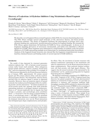
Figure 1. Conceptual organization of key components of the fragments of life library, with example fragments. The current FOL library (1329
molecules) contains chemically tractable natural small molecule metabolites such as bestatin 1 (FOL-Nat), metabolite-like compounds and
their bioisosteres (FOL-NatD), and protein architecture mimetics (FOL-Biaryl). Any natural molecule with MW < 350 Da that exists as a
substrate, natural product, or allosteric regulator of any metabolic pathway in any cell type is a potential candidate FOL-Nat fragment.
Example fragments 11 and 13 that contain the privileged scaffold indole98
from the serotonin and auxin biosynthetic pathways are shown.
FOL-NatD fragments are defined as heteroatom containing derivatives, analogues, or fragments of any FOL-Nat molecule. FOL-Biaryl
fragments were selected from a variety of biaryl compounds that are potential mimics of protein R, β, or γ turns.59-61
At the right-hand side of
the figure, compound 10 can be seen superimposed on a β-turn of a protein structure (residues Ala20-Ala21-Asp22-Ser23 of PDB ID 1RTP).
An additional FOL-Biaryl component is also shown, superimposed on an R-turn (residues Ser65-Ile66-Leu67-Lys68 of PDB ID 1RTP).
3. 4696 Journal of Medicinal Chemistry, 2009, Vol. 52, No. 15 Davies et al.
numerous similar examples of natural molecules leading
to the design of drug candidates that modulate protein
activity.52-54
Evolutionary concepts of substrate ambiguity
and enzyme promiscuity suggest that many enzyme active
sites have evolved flexibility to transiently bind substrates
and products.55-58
We therefore decided to add heteroatom
derivatives and isosteres of natural molecules of life to our
fragment set. Termed “FOL-NatD”, these synthetic deriva-
tives or isosteres of natural molecules of life are anticipated
to be fortified with small modifications that could stabilize
binding to enzyme active sites. The current FOL-NatD set
includes 666 fragments. In order to emulate 3D protein
architecture and to further diversify the FOL library, we
have included a set of 445 synthetic biaryl molecules, many of
Figure 2. Continued
4. Article Journal of Medicinal Chemistry, 2009, Vol. 52, No. 15 4697
whose energy minimized structures mimic R-, β-, and
γ-turns.59-61
This subset of the FOL library was named
“FOL-Biaryl.” All of the listed FOL concepts are summar-
ized in Figure 1.
Our final 1329 member FOL library generally conforms to
the “rule of three” guidelines for fragments11
(see Supporting
Information Figure 1 for physical and chemical properties of
the FOL library) and contains approximately 400 distinct
chemotypes (see Supporting Information Figure 2 for calcu-
lated diversity properties of the FOL library). Members of
the complete FOL library have an average molecular weight
of 182.5, ClogP of 0.96, 1.49 hydrogen bond donors, 2.56
Figure 2. Continued
5. 4698 Journal of Medicinal Chemistry, 2009, Vol. 52, No. 15 Davies et al.
hydrogen bond acceptors, 1.7 rings, and 2.7 rotatable bonds.
The only parameter that lies outside the rule of three is polar
surface area with an average value of 118.9 A˚ 3
. This is reflec-
tive of the water-soluble properties of many small molecules
of life. We reasoned that members of the FOL library repre-
sent “molecular rulers” providing information on the archi-
tecture and electronics of protein binding sites. Thus, our
FOL library is designed to interrogate both enzyme active
sites along with the more intriguing opportunities to probe
allosteric pockets and protein-protein interaction surfaces.
LTA4H Target FOL Screening. To validate the FOL
library as a screening set in a proof of concept study,
Figure 2. Continued
6. Article Journal of Medicinal Chemistry, 2009, Vol. 52, No. 15 4699
approximately 15% of the fragments were screened against
LTA4H by soaking respective crystals with randomly se-
lected pools each containing eight different structurally
diverse FOL fragments (see details of Computational and
Physical Pooling in Materials and Methods). The resulting
crystals were frozen, complete X-ray diffraction data sets
were collected, and the models were partially refined using an
LTA4H protein-only model. When unaccounted electron
density was observed, candidate fragments from the pool
that were consistent with the shape of the density were
soaked individually and additional complete data sets were
collected. When definitive electron density from a single
Figure 2. Continued
7. 4700 Journal of Medicinal Chemistry, 2009, Vol. 52, No. 15 Davies et al.
compound soak was observed, the fragment was built into
the electron density and the complete protein-fragment
model was refined. Thirteen unique hits were identified from
these experiments, 12 of which are summarized in
Figure 2B-M, representing an overall hit rate of 6%. The
13th fragment, an FOL-Biaryl member, will be described in a
separate publication.43
Crystallographic data collection
and refinement statistics for all structures are listed in
Supporting Information Table 1. LTA4H inhibition assay
data for all compounds in this manuscript are listed in
Supporting Information Table 2 and are also summarized
in Figure 2.
Figure 2. Continued
8. Article Journal of Medicinal Chemistry, 2009, Vol. 52, No. 15 4701
Representative fragments from all categories of FOL were
observed bound to LTA4H (FOL-Nat, FOL-NatD, and
FOL-Biaryl). Nine of the 13 FOL hit compounds were
discovered to bind independently within the L-shaped sub-
strate binding cleft. The FOL-Biaryl compound 10 was
found to bind on the surface of LTA4H, far from the active
site. Compound 7 was simultaneously bound to both the
surface and within the L-shaped cleft of LTA4H (see Sup-
porting Information Figure 3, an image of LTA4H sur-
face binding of 7). Three additional FOL-NatD molecules
(11-13) were accommodated in the substrate binding cleft
only upon concomitant binding of an additional inhibitor,
Figure 2. Continued
9. 4702 Journal of Medicinal Chemistry, 2009, Vol. 52, No. 15 Davies et al.
bestatin (1). The binding modes of individual fragments are
briefly described below.
The natural product resveratrol (compound 2),62,63
a
plant-derived polyphenol, was identified as a hit in our
screen (Figure 2B). This natural molecule has been impli-
cated in cardioprotection and antiaging.64-70
The polyphe-
nol nature of 2 suggests that it could make multiple hydrogen
bonds with the target including conserved water molecules at
the bottom of the cleft and the backbone carbonyl of Gln136.
In our hands, 2 was a relatively weak binder of LTA4H with
IC50 values of 366 and 212 μM in the peptidase and hydrolase
assays, respectively. The observed electron density for the
molecule was of intermediate quality. The model of the
bound fragment in the crystal structure refined with rela-
tively high temperature factors. Analysis of the structure of
the resveratrol complex further suggested that the bound
compound exists in a strained conformation due to the rigid
C7-C8 double bond. From this observation, we postulated
that dihydroresveratrol (compound 3), a major metabolite of
2 observed in mammals,64,71,72
would have more rotational
freedom and may bind more effectively to LTA4H. We syn-
thesized 3 and soaked LTA4H protein-only crystals. The
resulting crystal structure showed very clear electron density
for 3, and the fully refined model exhibited lower relative
temperature factors compared to 2 (Figure 2C). Specifically,
bound 3 displayed a conformation inaccessible to 2, with
a torsion angle of -161.2° around the C7-C8 bond
(Figure 3A). The observed better fit of 3 for binding LTA4H
did not translate to an increase in binding affinity, possibly
because of an energetic trade-off between an improved fit
and an entropic penalty to be paid by constraining a more
flexible ligand. The IC50 of 3 was comparable to that of the
parent resveratrol 2: 145 μM in the peptidase assay and 247
μM in the hydrolase assay.
N-(Pyridin-3-ylmethyl)aniline 4 is classified as an FOL-
NatD fragment. It shares structural similarities with nicoti-
namide, namely, a pyridin-3-carboxamide moiety. It was
bound at the bottom of the substrate binding cleft of LTA4H
near the resveratrol binding site (Figure 2D). Most interac-
tions of 4 with the protein were hydrophobic except for a
potential hydrogen bond between the aniline NH and the
backbone carbonyl of Pro374. Compound 4 displayed IC50
values of 1.32 and 1.67 mM in the peptidase and hydrolase
assays, respectively. Notably, 4 displayed structural similar-
ity compared to the phenoxymethylbenzene pharmacophore
of the previously reported inhibitors of LTA4H40
(PDB IDs
3CHO, 3CHQ). Superposition of 3CHQ or 3CHO structures
with our LTA4H-3 and LTA4H-4 complexes demonstrated
that the corresponding atoms of the phenoxymethylbenzene
were all within 0.7 A˚ of their counterparts in 3 or 4 (see
Supporting Information Figure 4). The structurally related
fragment 3-(phenylmethoxy)pyridin-2-amine (5) identified
in our screen featured a similar binding mode (Figure 2E).
The 2-amino function of the pyridine ring is likely to medi-
ate a potential 2.8 A˚ hydrogen bond with the backbone
carbonyl of Pro374. This bond is shorter and has more
favorable geometry compared to that between 4 and
Pro374. Notably, the interaction between the amine and
Pro374 moiety slightly perturbed the binding pose of the
biaryl scaffold. Better binding interactions of 5 with the
target translated into its better potency (IC50 values of 308
and 619 μM in peptidase and hydrolase assays). It is note-
worthy that the hydroxamic acid inhibitors of LTA4H,
which also contain phenoxymethylbenzene, do not appear
to occupy similar space in LTA4H, most likely because of the
dominant interactions that the hydroxamic acid component
has with the catalytic zinc ion73
(see Supporting Information
Figure 4).
We categorized the pyridine analogue 6 as an FOL-NatD
molecule. It features a pyridine ring present in several
molecules of life including niacin, vitamin B3 (pyridine-3-
carboxylic acid), and 4-pyridoxic acid, the catabolic break-
down product of vitamin B6. In our studies, fragment 6 was
bound to the hydrophobic center of the LTA4H cleft, in the
same location as 4. The carbonyl oxygen of 6 was in a
suboptimal position, namely, within 3.6 A˚ of the carbonyl
oxygen of Trp311 (Figure 2F). This unfavorable interaction
likely contributed to poor activity of the fragment in the
biological assays (IC50 =3.7 and 5.3 mM in peptidase and
hydrolase assays, respectively).
Figure 2. Panels showing ligand binding to LTA4H for compounds described in the manuscript. Enzyme assay IC50 values are in μM, human
whole blood cell assay IC50 values are in nM when reported, and ligand efficiency (LE) values are in kcal/(mol 3 heavy atom). Compound
structures are displayed as yellow stick structures, and LTA4H is displayed in gray. Green mesh corresponds to the Fo - Fc (difference) electron
density at the 3.0σ level of the crystal structure with the compound omitted from the model. Polar contacts with LTA4H and/or bound water
molecules are shown as red dashed lines. PDB IDs for each structure are indicated.
10. Article Journal of Medicinal Chemistry, 2009, Vol. 52, No. 15 4703
Two additional FOL-NatD fragment hits (compounds 7
and 9, Figure 2G and Figure 2I) and two FOL-Biaryls
(compounds 8 and 10, Figure 2H and Figure 2J) featured
thiazole (a component of thiamine, vitamin B1) or thiophene
rings. Although several thiazole derivatives have been iden-
tified as promiscuous protein binders,74
this chemotype
provided valuable structural insight into LTA4H. Specifi-
cally, molecules 7-9 displayed distinctly different binding
modes to the enzyme. They mapped out space occupancy of
the entire L-shaped substrate cavity of LTA4H (see Support-
ing Information Figure 5). For example, 7 was bound deep
within the LTA4H cavity (Figure 2G). The aminothiazole
group displaced two of the three structurally conserved water
molecules at the bottom of the substrate binding cleft. The
amino substituent of the thiazole group occupied a position
roughly between binding sites of the two displaced water
molecules and was within hydrogen bonding distance of the
carbonyl oxygen of Phe362. Thiophene derivative 8 was
bound at the other end of the LTA4H substrate binding
cleft near the zinc binding site (Figure 2H). The primary
amino group of the fragment was less than 4 A˚ from the zinc
atom, 2.9 A˚ from Oε1 of Gln136, and 2.7 A˚ from a carboxyl
oxygen of Glu271. Finally, the binding site of 9 was inter-
mediate between 7 and 8, with the thiophene ring making
hydrophobic contact with Phe314 and Leu369 (Figure 2I).
The amino group of 9 did not make any hydrogen bonds with
LTA4H. The thiophene ring did not displace the structurally
conserved water molecules, as was observed for 8. Both 8 and
9 were relatively potent ligands of LTA4H with measured
IC50 values equal to or below 100 μM in both peptidase and
hydrolase assays. Compound 7 afforded poor inhibitory
activity with IC50 values greater than 1 mM. This could be
a result of its binding mode being deep within the substrate
binding cleft, perhaps making it unable to interfere with
substrate binding in either of our LTA4H enzyme assays.
Interestingly, a second molecule of 7 appeared to be bound in
the LTA4H-7 cocrystal complex on the surface of the
protein. Its phenol moiety was in position to make polar
contact with the carboxyl side chain of Glu501 and the
backbone carbonyl of Leu343 (see Supporting Information
Figure 3). This molecule was also well situated to make polar
contact with imidazole bound between Glu501 and Glu348.
Imidazole is present in the crystallization buffer and is
included in most LTA4H structures observed at medium or
higher resolution.
Of all the fragments identified by crystallographic screen-
ing, only compound 10 (FOL-Biaryl) did not bind in the
substrate binding cavity (Figure 2J). It interacted with the
surface of the protein, in a pocket formed by Arg24, Ile188,
and Asp182. We have further confirmed the LTA4H binding
of 10 and 7 (which binds a different pocket on the surface of
LTA4H) by both surface plasmon resonance and saturation
transfer difference NMR studies75
(data not shown). Mole-
cule 10 was not competitive with bestatin (1) an LTA4H
competitive inhibitor (data not shown) and displayed little
or no inhibitory activity against LTA4H (IC50 > 1 mM).
Therefore, the two shallow surface pockets mapped out by
compounds 10 and 7 are unlikely to be useful targets for
inhibition of LTA4H. However, our findings suggest that the
application of FOL methodology may yield additional in-
sight into the topology of surface domains or allosteric sites.
It could be further used to develop modulators of protein-
protein interactions.76
Simultaneous LTA4H Binding of Two Fragments. The two
most common approaches in FBDD are fragment evolution
and fragment linking.9
The fragment evolution technique
involves chemical optimization of the initial fragment hit
to produce higher affinity ligands.77
Fragment linking is
focused on assembling adjacent independently bound frag-
ments into a single molecule to produce high affinity
ligands.78-81
In order to expedite the transition from frag-
ment hits to leadlike molecules, we explored the possibility of
identifying dual simultaneous FOL hits in LTA4H. This
“fragment plus fragment” approach was inspired by a grow-
ing number of cases where two small molecules bind within
protein targets and contact each other. For example, this can
be seen in the interaction of the plant hormone auxin (indole-
3-acetic acid)82
and an inositol hexakisphosphate cofactor in
the crystal structure of the TIR1-ASK1 complex.2
In one “fragment plus fragment” approach, we soaked
LTA4H crystals with FOL pools that were spiked with 1 mM
bestatin (IC50 = 178 nM in peptidase assay). Subsequent
Figure 3. Resveratrol structures. (A) The structure of resveratrol (2) (yellow) is superimposed upon dihydroresveratrol (3) (cyan). (B)
Superposition of the structure of dihydroresveratrol (3) is shown in the absence (cyan) and presence (magenta) of bestatin. Structurally
conserved water molecules and the side chain of residue Phe362 are also color-coded to show the differences in the structures with and without
bestatin. For crystallographic data, see Supporting Information Table 1.
11. 4704 Journal of Medicinal Chemistry, 2009, Vol. 52, No. 15 Davies et al.
screening of FOL yielded three different indole fragments
(compounds 11-13) bound in the substrate binding cleft
(Figure 2K-M). Notably, these fragments interacted with
LTA4H only in the presence of bestatin. A representative
example, 5-hydroxyindole (compound 11) was a component
of the FOL-NatD subset. This core is exhibited in the
5-hydroxyindole-3-acetic acid structure, which is a serotonin
metabolite83
(Figure 1). It was bound near residues Trp311
and Tyr378, with the hydroxyl oriented closest to the besta-
tin, 3.5 A˚ away (Figure 2K). Two halogenated analogues of
11, namely, 12 and 13 lined up with the target in a similar
orientation. Both chlorine and fluorine atoms of 12 and 13,
respectively, were ∼5.7 A˚ away from the nearest atom of the
phenyl ring of bestatin. The shift in the binding mode of these
indoles likely results from their decreased polar surface area
compared to 11. All obtained X-ray structures revealed well-
defined electron density maps for bestatin with a binding
mode essentially identical to that previously observed.26
In another approach, we sought to determine if the pre-
sence of bestatin could alter the binding mode of another
fragment hit whose binding pose would be expected to be
overlapping or incompatible with bestatin. Our rationale for
this approach was to determine if the binding mode(s) of any
fragments could be adjusted by induced-fit mechanisms
resulting from the bestatin interaction with Zn2þ
. With this
approach, we found that bestatin could induce dihydrores-
veratrol (3) to adopt a different pose compared to the
fragment 3 alone within the LTA4H substrate binding cavity
(Figure 3A). Specifically, a brief LTA4H crystal soak of 3
(25 mM) followed by a 4 h soak with a mixture of 3 (25 mM)
and bestatin (1 mM) resulted in a structure where 3 was
displaced by ∼2.5 A˚ from its original binding site. With
bestatin chelating the Zn2þ
in its typical binding mode,
molecule 3 was forced to bind at the bottom of the LTA4H
substrate binding cleft. It displaced two of the three structu-
rally conserved water molecules with its phenolic hydroxyl
groups (Figure 3B) and was within a distance of ∼3.3 A˚ to
bestatin. The torsional angle about the C7-C8 bond fea-
tured by the molecule in a LTA4H-3-bestatin structure
was approximately 169°. Resveratrol (compound 2) could
not be visualized in a similar crystal cosoak with bestatin. We
attributed this outcome to the lack of flexibility in 2 that is
critical to the binding at the bottom of the binding cleft in
the presence of bestatin. The discovery of a binding mode
unattainable without a second fragment was an interesting
outcome from the “fragment plus fragment” approach. In
this case, bestatin altered the topology of LTA4H binding
cavity and pushed dihydroresveratrol deeper into the cavity.
As a result, the molecule adopted a novel binding mode
requiring the side chain movement of Phe362. The new
observed binding mode is intriguing, particularly in the
displacement of structurally conserved waters. It is concei-
vable that fragment elaboration principles could be em-
ployed to produce potent inhibitor(s) that incorporates
design concepts of both bestatin and the novel dihydrores-
veratrol interaction.
Acetate is one of the smallest of fragments, with just four
non-hydrogen atoms and a molecular weight of 56. In
LTA4H, acetate ions in the crystallization solution were
often visualized binding to Yb3þ
ions that are located at a
key crystal lattice interface.26,27,73
Similarly, we have ob-
served acetate binding to the catalytic Zn2þ
in the high
resolution crystal structures of LTA4H with FOL com-
pound 9 (Figure 2I). We sought to extend the “fragment
plus fragment” strategy by employing acetate bound at the
catalytic zinc in combination with other fragments. Notably,
when 200 mM acetate was included as an additive in an
LTA4H crystal soak with compound 4, the final solved
structure revealed both acetate and compound 4 binding
simultaneously as anticipated from their previously observed
individual binding modes. It is interesting to note that the
acetate-Zn2þ
binding events together with the binding
modes of compounds 4, 5, and 9 were essentially super-
imposable (see Supporting Information Figure 6). On the
basis of this observation, we postulated that a proper linking
of carboxylic acid functionality to a hydrophobic biaryl
would yield a potent inhibitor. Of particular interest were
templates that could provide for the proper dihedral angle
and distances in positioning both biaryl and acetate phar-
macophores within the active site of LTA4H. Physicochemi-
cal properties, good ligand efficiency, and chemical feasibil-
ity were additional criteria we considered. In the course
of chemical studies, (R)-prolinol was selected as a tether
between a hydrophobic biaryl and a metal-binding carboxy-
late, yielding DG-051 (19), a drug molecule that has com-
pleted phase IIa clinical studies.43
Thus, the DG-051 mole-
cule (Figure 2S) displays two key pharmacophore features
suggested by our fragment-based studies, namely, a car-
boxylic acid moiety binding to the active site zinc and a
hydrophobic anchor at the bottom of the substrate binding
cavity.
Elaboration of FOL Fragment Hits. The abundance of
fragment screening data summarized above prompted us to
improve the potency of several FOL hits through a limited
medicinal chemistry effort. The rationale was to concep-
tually prove that the identified weak binders could be rapidly
evolved into a potent LTA4H inhibitors following structural
guidance obtained thus far. On the basis of the synthetic
feasibility, we selected three fragments (4, 6, and 11) as
templates for this chemical elaboration (see Schemes 1-3).
In selecting proper pharmacophores for our chemistry effort,
we considered both the trajectory for fragment linkage as
suggested by the structural studies as well as druglike char-
acter of the designed molecules. Midsized cyclic amines were
of particular interest, as they offered well-defined spatial
orientation for the pharmacophores. Furthermore, these
moieties were expected to mediate additional interaction
within the active site and to enhance solubility and perme-
ability of a fragment derivative across biological barriers. A
brief synthetic effort followed by crystallographic screening
converged on (R)-prolinol as a suitable tertiary amine tether.
For example, considering the LTA4H binding mode of 4, we
reasoned that amendment of 20 with the optimized (R)-
prolinol linker of DG-051 would yield a more potent mole-
cule, 14 (Scheme 1). Further, on the basis of the structural
data for 7, wherein its aminothiazole was found to displace
structurally conserved water molecules deep within the
LTA4H active site cavity, we amended 23 to introduce both
thiophene and (R)-prolinol groups to afford 18 (Scheme 1).
Synthesis of the prolinol derived analogue 14 was accom-
plished by the nucleophilic displacement of tosyl group in 24
with the phenolate derived from 4-benzylaminophenol 20
(Scheme 1). This step was followed by the removal of Boc-
protecting group from 25. The tosylate 24 was prepared
following the literature procedure from (R)-Boc-prolinol.
The Friedel-Crafts acylation of anisole with 4-iodobenzoyl
chloride followed by reaction of the methyl ether 22 with
boron tribromide in CH2Cl2 provided the phenol 23. The
12. Article Journal of Medicinal Chemistry, 2009, Vol. 52, No. 15 4705
sodium phenolate of 23, generated in situ with NaH in DMF,
was subsequently reacted with the tosylate 24 to provide the
BOC protected 4-iodobenzophenone derivative 26. Stille
coupling of 26 with thiophene-3-boronic acid followed by
deprotection of the N-BOC under acidic conditions provided
the compound 18 (Scheme 1).
Subsequent structural studies of 14 revealed that the
pyrrolidine moiety was bound at the bend in the LTA4H
substrate binding cavity (Figure 2N). It provided the neces-
sary kink for the molecule pivoting toward catalytic Zn2þ
. Its
binding mode resembled the pyrrolidine group orientation in
the DG-051-LTA4H complex (Figure 4). It was also similar
to the positioning of the phenyl ring of bestatin bound to
LTA4H. This allowed the lone electron pair of the nitrogen
to form a potential 3 A˚ hydrogen bond with Oε1 of Gln134.
This simple structure-guided modification immediately furn-
ished a 4000-fold improvement in affinity of 14 against the
enzyme with IC50 values of 207 and 157 nM in the peptidase
and hydrolase assays, respectively. Superimposing the struc-
ture of 14 with the coordinates of the “fragment plus frag-
ment” structure of 4 with acetate showed that there was a
1.9 A˚ gap between the methyl carbon of acetate and the
nearest carbon atom of the pyrrolidine (Figure 4), suggesting
that a single carbon atom linkage between the two compo-
nents would yield a molecule with LTA4H binding similar
to that of DG-051 (Figure 2S). Structural studies of 18
(Figure 2R) confirmed that, similar to 7, the thiophene moiety
displaced two water molecules from the bottom of the sub-
strate binding cavity (Figure 5). The biaryl core of 18 was
shifted compared to the parent molecule 6, moving carbonyl
oxygens 5.2 A˚ apart. This motion likely decreased the un-
favorable interaction with the backbone carbonyl of Trp311,
resulting in a potent inhibitor with IC50 values of 80 nM in the
peptidase assay and 189 nM in the hydrolase assay.
To further explore elaboration of fragment 6, we displaced
the fluorine atom in 6 with N-pyrrolidin-2-ethanol to furnish
Scheme 1. Synthetic Routes for New Molecules Inspired by the LTA4H Binding Modes of Compounds 4, 6, and 7a
a
Reagents and conditions: (a) AlCl3, 10 °C, nitrobenzene, 90%; (b), BBr3, CH2Cl2, -78 °C f 0 °C, 85%; (c) NaH/DMF, 0 °C f rt f 90 °C; (d) 1 M
HCl/Et2O, 80-97%; (e) thiophene-3-boronic acid (2 equiv), Pd(OAc)2 (25 mol %), K2CO3/EtOH/DME, 90 °C, 60%.
Scheme 2. Elaboration of Fragment 6a
a
Reagents and conditions used are as follows: (a) 1-(2-hydroxyethyl)pyrrolidine, K-Ot
Bu, DMSO, 90 °C, 16 h, 54%; (b) NaBH4, MeOH, 40 °C, 4 h.
Scheme 3. Elaboration of Fragment 11a
a
Reagents and conditions: (a) 1-(2-chloroethyl)pyrrolidine, K2CO3, acetone, 19%; (b) 1-bromo-2-chloroethane, K2CO3, 2-butanone, reflux 60 h,
17%; (c) piperidine-4-carboxylic acid ethyl ester, HCl (2 equiv), KI (0.3 equiv), K2CO3, DMF, rt f 90 °C, 57%; (d) aq NaOH, EtOH, rt.
13. 4706 Journal of Medicinal Chemistry, 2009, Vol. 52, No. 15 Davies et al.
the biaryl ketone 27 (Scheme 2). This intermediate was
reduced with sodium borohydride to afford the targeted
biaryl carbinol derivative 17 (Scheme 2).
The LTA4H cocrystal structure of 17 revealed a potential
hydrogen bond between the OH group in the molecule and
the carbonyl of Trp311 (Figure 2Q). Molecule 17 featured
an IC50 of 202 μM (peptidase assay) and 170 μM (hydrolase
assay), which was a 25- to 30-fold improvement over the
initial hit 6 but was an order of magnitude less potent than
molecule 18, the more fully elaborated derivative of fragment
6. This comparison suggests that the improved potency of
compound 18 likely results from a combination of water
molecule displacement deep within the LTA4H active site
pocket by the thiophene, plus the effects of hydrogen bond-
ing between Gln134 and the pyrrolidine of 18.
Encouraged by these data, we routinely used pyrrolidine-
containing moieties in our optimization effort. For example,
the 5-hydroxyindole fragment identified earlier as hit 11 was
modified with 1-(2-methoxyethyl)pyrrolidine in the presence
of K2CO3 in acetone (Scheme 3) to afford compound 15
(IC50 of about 321 and 234 μM in the peptidase and hydro-
lase assays, respectively). Structural data suggested that the
pyrrolidine moiety of 15 occupied the same space as observed
for 14, with the ring being in proximity to the side chain of
Gln134 (Figure 2O).
Considering both activity data and synthetic feasibility,
we attempted to replace the pyrrolidine ring of 15 with a
piperidine carboxylic acid moiety to furnish 16 (Scheme 3).
For the preparation of compound 16, 5-hydroxyindole 11
was first reacted with 1-bromo-2-chloroethane to provide
the 2-chloroethyl ether 28. Subsequent displacement of the
primary chloro group of 28 with piperidine-4-carboxylic
acid ethyl ester in refluxing 2-butanone furnished the
ester derivative 29. This compound was further hydrolyzed
with NaOH in EtOH to afford the targeted molecule 16.
Structural elucidation of 16 bound to LTA4H revealed that
its carboxylic acid moiety was bound to the active site Zn2þ
(Figure 2P). However, its potency of IC50 of about 1491 and
966 μM in the peptidase and hydrolase assays, respectively,
was 4-fold less than that of 15. Notably, the binding modes
for molecules 15 and 16 significantly differed. Positioning of
indole group (Figure 6) was driven by the orientation of
pyrrolidine (for 15) and piperidine-4-carboxylic acid (for 16)
pharmacophores in the active site of LTA4H.
Discussion
In this work we have combined the concepts of metabolo-
mics with fragment based drug discovery to assemble a
∼1300-member “fragments of life” (FOL) small molecule
library for screening by X-ray crystallography. This set in-
cludes highly soluble metabolites, natural products, their
derivatives, and biaryl molecules mimicking architectural
motifs found in proteins. Small molecules of life have coe-
volved with their respective macromolecular targets, and
therefore, such interactions are likely to be highly ligand
efficient. Inaddition,signalingpathwaysare knowntodisplay
allostericfeedbackmechanismsinvolving small molecules (for
a recent review, see Goodey and Benkovic84
). Consequently,
application of molecules of life in the FBDD approach may
lead to the identification of novel allosteric binding sites in
protein targets. In addition, many molecules of life are likely
to feature druglike properties, since these compounds display
properties necessary for intracellular activity (e.g., reasonable
target affinity, reversible binding, controlled on/off rates,
solubility, permeability, etc.).
The FOL library has been designed without target bias. In
the initial validation, we screened approximately 200 mole-
cules (15%) of the entire selection (1329 compounds) against
LTA4H using X-ray crystallography. Thirteen unique
Figure 4. Elaboration of 4. Refined crystal structures of LTA4H in complex with molecules in the compound 4 elaboration series are shown.
Compound 4 and acetate from the “fragment plus fragment” structure are displayed as yellow stick structures, while compound 14 is magenta
and compound 19 (DG-051) is green. The side chain conformations of LTA4H bound to each of these compounds are essentially
superimposable (gray side chains) with the exception of Gln134, which demonstrates some flexibility in its conformation as represented by
DG-051 (green), compound 4 and acetate (yellow), and compound 14 (magenta). Polar contacts are displayed as color-coded dashed lines.
14. Article Journal of Medicinal Chemistry, 2009, Vol. 52, No. 15 4707
fragments were discovered, representing a hit rate of 6%.
Most of the identified fragments (12 out of 13) were bound to
the active site pocket, while fragment 10 bound within a
shallow surface pocket. Fragment 7 interacted with both the
active site pocket and the surface of LTA4H. It is noteworthy
that the two fragments found binding to the surface of
LTA4H were biaryl structures with either a thiophene or
thiazole moiety plus at least one amino or hydroxyl group.
A similar observation has been reported recently for thiazole
derivatives binding at sites of protein-protein interaction,76
suggesting the possibility that these themes may be exploited
to develop specialized fragment libraries to screen for mod-
ulators of protein-protein interactions. Importantly, FOL
hits varied both structurally and in their source within the
library. Specifically, biological molecules FOL-Nat (e.g.,
resveratrol, dihydroresveratrol, 5-hydroxindole), their deriva-
tives FOL-NatD (e.g., 5-hydroxyindole, nicotinamide, and
isonicotinic acid derivatives), and FOL-Biaryls which mimic a
protein structure (e.g., fragment 10) were found to bind to
LTA4H in a diverse array of binding modes.
The structures of LTA4H with bound resveratrol (com-
pound 2) and its metabolite dihydroresveratrol64,71,72
3 were
of particular interest because of the potential therapeutic
implications. As LTA4H is involved in both increased risk
of myocardial infarction29
and inflammation,30
one may
consider attributing the apparent cardioprotective properties
of resveratrol and other sirtuin activators85-87
to LTA4H
modulation. Our finding suggests thatthe FOLapproach may
Figure 6. Elaboration of 5-hydroxyindole 11, showing superposition of the indole-containing fragment structures: 5-hydroxyindole 11
(dark green), 5-chlororindole 12 (magenta), 5-fluoroindole 13 (cyan), and derivative compounds 15 (yellow) and 16 (pink).
Figure 5. Elaboration of 6. (A) Superposition of compounds 6 (yellow), 17 (green), and 18 (cyan). Invariant portions of the LTA4H structure
are colored gray, and compound-specific polar contacts, side chain conformations, and bound water molecules are color-coded by compound.
(B) Superpostion of 18 (cyan) with the thiazole-containing fragment 7 (rose) and the pyrrolidine containing compound 14 (magenta). Both 7
and 18 displace the central structurally conserved water molecule, and both 14 and 18 have pyrrolidine moieties that occupy nearly the same
space in the LTA4H structures.
15. 4708 Journal of Medicinal Chemistry, 2009, Vol. 52, No. 15 Davies et al.
facilitate rational identification of small molecule agents that
simultaneously modulate several therapeutically relevant
pathways.8
The binding of dihydroresveratrol 3 also illustrates the
potential utility of “fragment plus fragment” mode of screen-
ing. For example, when 3 bound to LTA4H together with
bestatin, several structural water molecules were displaced
within the LTA4H active site pocket and Phe362 was found to
adopt an alternative side chain rotamer conformation. This
finding shows that when fragment cocktails are repooled with
other fragments, new induced-fit fragment binding modes can
be observed.
Using structural guidance from FOL screening, we showed
that fragment hits could be expeditiously modified to generate
potent LTA4H inhibitors in a short synthetic sequence. The
diverse chemical nature of the initial hits suggests that this
strategy is applicable to the identification of novel and
structurally distinct chemotypes. In a representative example,
structural information from diverse active fragments was
combined to evolve a fragment hit 6 of very poor activity
(IC50=5.3 mM) in the peptidase assay into a potent LTA4H
inhibitor 18 (IC50=80 nM) in two synthetic steps. Fragment
hit 4 was derivatized in a single step to afford 14, a novel and
potent inhibitor of LTA4H (IC50=150 nM in the hydrolase
assay) with comparable potency (IC50=131 nM) for inhibi-
tion of LTB4 production in human whole blood. Compound
14 featured high ligand efficiency of 0.44 kcal/(mol3 heavy
atom) vs 0.36 kcal/(mol 3 heavy atom) for the clinical candi-
date DG-051 19. Thus, a combination of properly selected
fragments with structural insight into their mode of binding
allowed for maintenance of ligand efficiency and druglike
potential of the optimized hits.
The FOL library was of value in expanding the known
chemical space of LTA4H inhibitors. Our structural studies
have consistently identified multiple fragments that exemplify
reported LTA4H binders. For example, the binding modes of
our compounds 4 and 5 were all superimposed and mimicked
the benzoxyphenyl components of LTA4H inhibitors re-
ported by Kirkland et al. (see Supporting Information Figure
4).40
Furthermore, our ligand docking studies suggest that the
predicted binding modes of the LTA4H inhibitors reported by
Grice et al. and Rao et al.39,88
could be emulated with FOL
compounds identified in this study. Namely, the indole core of
fragments 12 and 13 overlaps well with the benzoxazole or
benzothiazole templates described by these authors (see Sup-
porting Information Figure 7). The fluorophenyl ring of 6, the
phenol substituent of 2 and 3, the phenyl ring of 4, and 5 all
superimpose to emulate the binding mode of the phenoxy
pharmacophore featured in the inhibitors reported by Grice
etal.andKirklandetal.(seeSupportingInformationFigure8).
Given the importance of LTA4H activity in myocardial
infarction28,29
and inflammatory respiratory disease,88
we
previously reported the development of DG-051 (19,
Figure 2S). This molecule is a potent inhibitor of LTB4
production in human whole blood (IC50=37 nM). DG-051
was designed via structure-guided medicinal chemistry. It has
recently completed phase IIa clinical trials.43
The agent
comprises three functional components: a butanoic acid, a
pyrrolidine linker, and a lipophilic biphenyl ether moiety. As
suggested by our structural studies, these pharmacophores
were effectively captured with FOL hits. Reversible binding of
the butanoic acid moiety of DG-051 to Zn2þ
in the active site
of LTA4H was mimicked by the acetate ion in our “fragment
plus fragment” approach (Figure 4). The pyrrolidine linker
thatprovidesflexibilityfor DG-051 tonavigatethe bend inthe
substrate binding cavity was largely typified by the prolinol
derivatives 14 and 18 as well as the pyrrolidine derivatives 15
and 17. The biphenyl ether functionality of our clinical
candidate binds within the most hydrophobic part of the cleft
lined with aromatic amino acid residues including Tyr267,
Trp311, and Phe314. Its chlorine substituent resides within
∼3.2 A˚ from one of the three structurally conserved water
molecules (in the proximity of Lys364 and Leu365) at the
bottom of the L-shaped active site cavity.43
These binding
features of DG-051 were represented by fragments 2-9.
In summary, we introduced a concept of fragments of life
(FOL) library for structure-based drug discovery. In proof-
of-concept studies, we screened the FOL set against LTA4H
by X-ray crystallography. The resulting structural informa-
tion yielded insight into a novel series of LTA4H binders. It
also captured binding trajectories for multiple reported in-
hibitors of the enzyme. We further demonstrated that the
identified fragments could be optimized in a short reaction
sequence to yield molecules with high ligand efficiency and
potent biological activity. The fragments and their derivatives
feature binding efficiency for LTA4H and surface efficiency
indices that match those of marketed drugs16
(see Supporting
Information Figure 9). Further studies are required to deter-
mine the versatility of the FOL library screening against
diverse target classes. It is likely that further chemical amend-
ments to this selection will be needed to enhance hit rates. In
addition, the versatility of the FOL set is being tested in
different readout platforms including NMR and surface
plasmon resonance.
Materials and Methods
Purification of LTA4H. Recombinant human LTA4H
(residues 1-611) was expressed in E. coli BL21-AI/pRARE.
An amount of 7.5 g of cell paste was lysed by nitrogen cavitation
at 2200 psi for 1 h in a lysis buffer consisting of 50 mM Tris-HCl,
pH 8.0, 1 mM PMSF, 0.2 mg/mL lysozyme, and 1.5 U/mL
benzonase. The lysate was clarified by centrifugation at 42 000
rpm for 45 min at 4 °C. Clarified lysate was filtered using a
0.2 μM syringe-top filter. Lysate was then applied to 2 Â 5 mM
HisTrap HP columns (GE Healthcare) equilibrated with 20 mM
Tris-HCl, pH 8.0, and 200 mM NaCl. The column was washed
with six column volumes of 30 mM imidazole and eluted with a
linear gradient of 30-300 mM imidazole in 20 mM Tris-HCl,
pH 8.0, containing 200 mM NaCl. The pooled peak fractions
were >95% pure by SDS-PAGE at a concentration of 2.7 mg/
mL and used directly for crystallization without further con-
centration or dialysis.
Crystallization and Fragment Screening. LTA4H was crystal-
lized by sitting drop vapor diffusion against a crystallant
containing 100 mM imidazole, pH 6.5, 12.67% w/v PEG-
8000, 100 mM sodium acetate, and 5 mM YbCl3. Crystal growth
was accelerated through the use of a microseeding protocol.
Seed stocks were prepared by suspending several crystals in
20 μL of crystallant and crushing them by vortexing in a Seed
Bead tube (Hampton Research). Crushed crystals were diluted
1:100 into fresh crystallant and vortexed again to form the final
seed stock. Crystallization drops were set up in a Compact Jr.
96-well crystallization plate (Emerald BioSystems) by combin-
ing 0.7 μL of LTA4H (2.7 mg/mL) with 0.7 μL of crystallant and
0.2 μL of seed stock over a reservoir volume of 100 μL.
X-ray Crystallography. Twenty-five pools of eight com-
pounds from the FOL library were screened against LTA4H
by X-ray crystallography. Pool stocks consisted of cocktails of
eight fragments, each at 6.25 mM in methanol. Individual FOL
compounds are stored in barcode labeled sealed septum vials as
16. Article Journal of Medicinal Chemistry, 2009, Vol. 52, No. 15 4709
50 mM stocks in pure methanol and stored at -20 °C. Approxi-
mately 30% of the fragment stocks have been analyzed over
a period of 12 months by mass spectrometry to demonstrate
general stability of the compounds in methanol (data not
shown). Fragment cocktails in methanol were spotted onto the
drop chambers of crystallization plates and the solvent was
allowed to evaporate, leaving a precise amount of fragment
compounds as a dry powder with no solvent residue that could
potentially interfere with crystallization or crystal stability.
A volume of crystallization mother liquor equal to the initial
volume of fragment cocktail was added to the dry powder,
allowing for dissolution of the fragments. Crystals were trans-
ferred to the resulting solution and allowed to soak overnight.
This technique is versatile, as it is suitable for both cocrystalli-
zation and soaking experiments, and adjusting ratios of metha-
nol solution to crystallant solution allows for adjustment of the
final fragment concentration in the drop.
Although diversity pooling was conducted to aid in identifi-
cation of fragments from pool density alone, factors such as low
resolution, low occupancy, or competitive binding can lead to
uncertainty. Therefore, in cases where crystals exposed to frag-
ment cocktails produce difference electron density indicative of
fragment binding, follow-up experiments with individual frag-
ments were performed. During this deconvolution step, indivi-
dual candidate fragments were soaked into crystals, usually at
10-25 mM. The resulting crystal structures provide unambig-
uous confirmation of fragment hits, and all structures presented
were generated from single molecule crystal soaking experi-
ments.
LTA4H Peptidase Activity Assay. L-Arginine-p-nitroanilide
(Arg-pNA) substrate was obtained from Sigma. Stock solution
of ARG-pNA (500 mM) was prepared and serially diluted in
DMSO. Final peptidase assays were performed in 96-well clear,
flat bottom plates (Corning). Each assay plate contained eight
positive (no inhibitor) and eight negative (no enzyme) controls.
LTA4H (45 μL) and test compounds (5 μL) were preincubated
for 10 min followed by addition of substrate (50 μL); final
reactions contained 100 mM Tris-HCl (pH 8.0), 100 mM NaCl,
5% DMSO, 0.5 mM Arg-pNA, and 20 nM LTA4H. Control
experiments showed that 5% DMSO had no effect by itself on
enzyme activity (data not shown). Reaction rates were mon-
itored in a SpectraMax 190 plate reader (Molecular Devices) at
room temperature (rt) by measuring the increase in absorbance
at 410 nm due to cleavage of the amide bond of the substrate
and formation of p-nitroaniline. Reactions were measured for
30 min, and initial rates were determined from the linear
portions of the progression curves using SoftMax Pro software
(Molecular Devices). Dose-response curves ware calculated by
fitting data to a sigmoidal four-parameter logistic equation
using Prism (Graphpad).
LTA4H Hydrolase Activity Assay. LTA4 substrate was pre-
pared from the methyl ester of LTA4 (BioMol or Cayman
Chemicals) by treatment under nitrogen with 100 mol equiv of
NaOH in an acetone/H2O (4:1) solution at rt for 40 min. Stock
solutions of LTA4 were kept frozen at -80 °C for a maximum of
1 week prior to use. Recombinant human LTA4H (10 nM final
concentration) was incubated with various concentrations of
test compound for 10 min at rt in assay buffer (0.1 M Tris-HCl,
0.1 M NaCl, 5 mg/mL fatty acid free BSA, 10% DMSO, pH
8.0). Immediately before the assay, LTA4 was diluted in assay
buffer (without DMSO) and added to the reaction mixture to
a final concentration of 1 μM to initiate the enzyme reaction.
Samples were incubated for 10 min at rt followed by addition
of 2 volumes of chilled quenching buffer (CH3CN with 1%
CH3COOH and 73 nM LTB4-d4 (BioMol). The samples were
then stored at 4 °C for 12 h to complete protein precipitation and
centrifuged for 15 min at 1800g. LTB4 formed was measured in
the supernatant by LCMS/MS using LTB4-d4 as an internal
standard and an external LTB4 standard (BioMol) for a calibra-
tion curve. Briefly, the analyte was separated from LTB4
isomers formed by spontaneous hydrolysis of LTA4 using
isocratic elution on an HPLC system (Waters) and analyzed
on a tandem quadrupole mass spectrometer (Waters Micromass
Quattro Premier). MRM transitions followed on two channels
were 335.2 f 195.3 (LTB4) and 339.2 f 197.3 (LTB4-d4). On the
basis of the measured amounts of LTB4 formed at each inhibitor
concentration, a dose-response curve was fitted to the data
using a sigmoidal four-parameter function (Prism, Graphpad)
and an IC50 value was calculated. Ligand efficiency values were
calculated for hydrolase and peptidase assays independently
using LE (kcal/(mol 3 HA)) = -RT ln(IC50)/HA = -0.59179-
(ln IC50)/HA, where HA is the number of heavy atoms (non-
hydrogen).
Human Whole Blood LTB4 Assay. Human blood (45 mL) was
collected in heparin-containing Vacutainer tubes (Greiner-Bio
One) with informed consent. Individual experiments were per-
formed with blood from a single subject. For each sample,
200 μL of blood was dispensed into a prewarmed 96-well plate
and 188 μL of RPMI-1640 medium (Invitrogen) containing
20 μg/mL indomethacin (Sigma) was added. An amount of
4 μL of a series of compound dilutions (final DMSO concentra-
tion of 1%) was added in triplicate, followed by a 15 min
incubation at 37 °C with gentle shaking. Blood samples were
stimulated by adding 8 μL of ionomycin (from Streptomyces
conglobatus, Calbiochem) to a final concentration of 36 μM.
After another incubation at 37 °C for 30 min, samples were
centrifuged at 4 °C for 5 min at 1800g. LTB4 concentrations in
supernatants were determined using a commercially available
enzyme-linked immunosorbent assay (R&D Systems) according
to the manufacturer’s instructions. On the basis of the measured
amounts of LTB4 formed at each inhibitor concentration, a
dose-response curve was fitted to the data using a sigmoidal
three-parameter function (XLfit, IDBS), and an IC50 value was
calculated. Each assay plate contained eight positive controls
(no compound) and eight negative controls (no Ionomycin).
Fragment Selection for the Fragments of Life Library.
Approximately 400 candidate fragments representing natural
molecules of life with (i) molecular weights less than 350, (ii)
fewer than six hydrogen bond donors, and (iii) fewer than seven
hydrogen bond acceptors were manually selected from known
metabolic pathways. Molecules were selected from eukaryotic,
archaebacterial, and eubacterial metabolic pathways and from
presumed primordial pathways.89,90
There was no bias toward
any particular metabolic pathway; however, molecules of con-
served intermediary metabolism were prioritized.91
Approxi-
mately 300 of the resulting fragments were commercially
available, reasonably priced, and acquired. These fragments
were tested for solubility (g50 mM in methanol) to yield a final
core set of 218 natural molecules (FOL-Nat). The chemical
diversity of the first 218 FOL-Nat fragments was determined
using a 210 Â 210 principal component analysis (PCA) using
Cerius2
(Accelrys). In this analysis 1D, 2D, and 3D descriptors
(conformational, cat shape, electronic, quantum chemical, in-
formation content, spatial, structural, thermodynamical,
topological, and geometrical) were calculated and the 3D dis-
tribution of the fragments was visualized in a 3D plot (see
Supporting Information Figure 2). This analysis showed that
the fragments were diverse but did not densely cover the desired
chemical space. Therefore, in addition to the manual selec-
tion, we explored the Available Chemicals Directory (ACD/
Lab, version 7, MDL) and the Maybridge Fragment Library
(www.maybridge.com) as a source of additional fragments. We
intended to diversify the original set by selecting heteroatom-
containing derivatives of natural molecules (FOL-NatD) with
fragment-like properties. Toward this goal, a selection of
238 996 compounds were analyzed using modified “rule of
three”11
parameters to afford 49 877 fragments with (i) molec-
ular weights between 110 and 250 Da, (ii) calculated log of
octanol/water partition coefficient (ClogP) of less than or
equal to 3.0 (calculated with software available from Daylight
17. 4710 Journal of Medicinal Chemistry, 2009, Vol. 52, No. 15 Davies et al.
Chemical Information Systems, Inc.), (iii) hydrogen bond do-
nors less than or equal to 3, and (iv) hydrogen bond acceptors
less than or equal to 6 (calculated with Sybyl, version 6.8,
Tripos). Virtual selection of the 49 877 molecules was further
subjected to a panel of SMARTS filters92
in order to eliminate
molecules endowed with problematic functionalities. A partial
list of these SMARTS filters included reactive functional
groups, lipophilic chains of seven or more carbon atoms, crown
ethers, disulfides, excessive (>3) acidic groups, thiols, epoxides,
aziridines, hydrazones, thioureas, thiocyanates, benzylic qua-
ternary nitrogens, thioesters cyanamide, four membered lac-
tones, di- and triphosphates, metals, phosphines, phosphonic
acids, sulfonic acids, sulfonyl halides, boronic acids, isotopes,
salts, metals, more than 3 halogens, more than two nitro groups,
and lanthanides. This culling process resulted in 40 489 candi-
date fragment compounds. Each fragment from the resultant
filtering was converted from 2D to 3D projection using CON-
CORD (Tripos) and used to identify fragments with e3 rota-
table bonds and calculated PSA values of e60 A˚ 2
. The resulting
5606 fragments were again analyzed using the previously de-
scribed principal component analysis (PCA), and 1016 unique
fragments were selected with preference for molecules that could
be visually identified to have at least 8 atoms with chemical
arrangements that matched that of a known natural molecule of
life. From this set, 880 fragments were commercially available
and obtained at a reasonable price. These fragments were tested
for solubility (>50 mM in methanol), and the resulting 666
fragments (FOL-NatD) were added to the FOL library. PCA
analysis showed that the FOL-NatD fragments occupy dis-
tinctly different chemical space (see Supporting Information
Figure 2) and that together the FOL-Nat and Fol-NatD frag-
ments combine to form a diverse and relatively dense set of
fragments. Finally, we were also intrigued by the possibility of
mimicking a protein 3D architecture using biaryl small molecule
fragments.59-61,93
In order to identify protein mimetic frag-
ments (FOL-Biaryl), we first identified 566 molecules contain-
ing 5-5, 6-5, and 6-6 biaryls connected via a σ-bond to allow
for a controlled torsional freedom from the ChemBlock library
(www.chemblock.com). The energy minimized conformations
(Tripos force field, Sybyl 6.8) of a selection of these biaryl
fragments were overlaid with the R, β, γ turns of a known
protein structure (1RTP.pdb) and shown to have good steric
and electronic mimicry of protein structure. The resulting frag-
ments were tested for solubility (>50 mM in methanol), result-
ing in 445 fragment molecules (FOL-Biaryl). Results from the
PCA analysis and physical properties of the fragments [MW,
ClogP, total polar surface area, number of hydrogen bond
donor/acceptors, rotatable bonds, and rings] of the complete
1329 member FOL library are provided in Supporting Informa-
tion (see Supporting Information Figure 1).
Computational and Physical Pooling. When fragments are
pooled into structurally diverse cocktails of 4-10 compounds
per mix, the throughput of X-ray screening of fragment libraries
can be significantly increased.12,94,95
The pooling of fragments
was carried out using the same 210 Â 210 descriptor space
principle component analysis (Cerius2
, Accelrys) in order to
select eight diverse fragments per pool. The nature of the
fragment (FOL-Nat, FOL-NatD, FOL-Biaryl) was not consid-
ered in the pooling strategy. Monte Carlo optimization using
“Diversity Metric Function MAXMIN Distance” (Accelrys)
with the optimum distance range of 1.97-2.57 A˚ was used to
randomly extract eight diverse fragments. After extraction of
eight fragments from the library, the process was repeated until
the entire library was computationally pooled into structurally
diverse pools each containing eight fragments. Individual frag-
ments, each >95% pure as determined by NMR spectroscopy,
were prepared as 50 mM stocks in methanol. Physical pools of
eight fragments each were prepared by mixing fragment stocks
such that the final concentration of each was ∼6 mM. The
fragment pools were found to be visibly stable, with no observed
precipitation, suggesting that fragment pairs or combinations
do not have a propensity to aggregate.
Molecular Docking. The ligand docking into LTA4H was
carried out with the Surflex interface implemented in Sybyl 7.2
(Tripos Inc., St. Louis, MO). The Surflex-Dock engine uses an
empirical scoring function and a patented search engine to dock
ligands into a protein’s binding site.96
The PDB ID 3CHI file
was converted to a mole2 file, and hydrogens were added. The
B-values were replaced by the AMBER charges using Sybyl 7.2.
The bound ligand of 3CHI was removed from the binding site.
The Surflex-Dock protomol, a computational representation of
the intended binding site, was generated using the probes CH4,
-N-H, and -CdO. The protomol directed the placement of
the ligand during the docking process. LTA4H inhibitors were
drawn in ISIS Draw and converted to 3D structures with
CONCORD of Sybyl 7.2 (Tripos Inc., St. Louis, MO). All of
the inhibitors were properly typed with hydrogens, and a 3D
structure data file was created for docking. Each ligand was then
fragmented which reduced the conformational space to be
explored. The fragments were then aligned to the protomol
probes. All the fragments were scored,97
and the highest scoring
fragment was kept as the head fragment. The tail fragments were
selected on the basis of the similar principle, and gradual
attachment was carried out to build the docked ligand. The
poses were refined, eliminating those with excessive penetration
into the protein. Thirty poses for each of the ligand were
generated and visualized in the active site based on their Surflex
scores. The best fitting ligand poses were chosen for further
comparative structural analysis.
General Methods. All reagents and anhydrous solvents were
obtained from commercial sources and used without further
purification unless otherwise noted. NMR spectra were re-
corded at 400 or 500 MHz (Varian Instruments) in the solvent
indicated, and TMS was used as an internal reference. AC-
DLabs NMR software was used to process FIDs to generate
spectral parameters (δ (ppm)/Hz). Coupling constants (J) are
given in Hz. Mass spectra were obtained using either APCI or
electrospray ionization (PE-SCIEX single-quad or Agilent
mixed-mode units). Elemental analyses were carried out by
Galbraith Laboratories, Inc. (Knoxville, TN) or Midwest
Microlab, LLC (Indianapolis, IN). Column chromatography
was carried out in the solvents indicated with silica gel (MP
EcoChrom, 32-63D, 60 A˚ ). The HPLC method was as follows.
Compounds were eluted using a gradient of 90/10 to 10/90 A/B
over 40 min at a flow rate of 1.0 mL/min, where solvent A was
0.05% TFA in 100% H2O and solvent B was 0.05% TFA in
100% acetonitrile. For HPLC data (final products), peak area
percent and retention time (tR in min) are provided. The
following compounds were obtained from commercial sources:
compound 4 from Matrix, compounds 5, 7, 8, 9, and 10 from
Maybridge, compound 6 from VWR, and compounds 11, 12,
and 13 from Aldrich.
Benzyl[4-((R)-1-pyrrolidin-2-ylmethoxy)phenyl]amine (14).
To a solution of 25 (148 mg, 0.387 mmol) in methanol (3 mL)
was added HCl (1 M in diethyl ether, 6 mL). The mixture was
stirred at rt for 3 h. The solvent was removed in vacuo to provide
the title compound 14 as a hydrochloride salt (128 mg, 93%). 1
H
NMR (400 MHz, DMSO-d6) δ 1.60-1.73 (m, 1H), 1.87-2.12
(m, 3H), 3.17-3.21 (m, 2H), 3.84-3.87 (m, 2H), 4.19-4.23 (dd,
1H J1=3.6 Hz, J2=10.8 Hz), 4.11-4.16 (m, 1H), 4.44 (s, 2H), 7.0
(d, 2H J=8.8 Hz), 7.29-7.38 (m, 5H), 7.48-7.49 (m, 2H) 9.1
(s, 1H), 9.8 (s, 1H). LCMS: 97%; APCIþ
m/z 283 (M þ 1). Anal.
(C18H22N2O 3 2HCl) C, H, N.
5-(2-Pyrrolidin-1-ylethoxy)-1H-indole (15). To a solution of
5-hydroxyindole 11 (446 mg, 3.35 mmol) in anhydrous acetone
(20 mL) was added potassium carbonate (1.36 g, 9.8 mmol) and
1-(2-chloroethyl)pyrrolidine hydrochloride (1.28 g, 7.5 mmol).
The reaction was heated to reflux for 16 h. After the mixture was
cooled to rt, a solution of tetrabutylammonium bromide
(200 mg, 0.6 mmol) in DMF (10 mL) was added and the mixture
18. Article Journal of Medicinal Chemistry, 2009, Vol. 52, No. 15 4711
was heated to 55 °C for 16 h. The solvent was removed in vacuo,
and the resulting residue was partitioned between EtOAc (25
mL) and water (50 mL). The organic layer was separated, dried
with anhydrous MgSO4, and concentrated in vacuo to an oil.
The oil was purified by silica gel flash chromatography (∼100 g
of SiO2, 0-5% MeOH/CH2Cl2, gradient) to provide the title
compound 15 (145 mg, 19%). 1
H NMR (400 MHz, DMSO-d6) δ
1.68 (dt, J=6.57, 3.15 Hz, 4 H), 2.53 (br s, 4 H), 2.78 (t, J=6.04
Hz, 2 H), 4.03 (t, J=6.04 Hz, 2 H), 6.31 (br s, 1 H), 6.71 (dd, J=
8.72, 2.42 Hz, 1 H), 7.03 (d, J=2.15 Hz, 1 H), 7.23-7.29 (m, 2
H), 10.88 (br s, 1 H). LCMS (APCIþ
): mass calcd for
C14H18N2O, 230.3; m/z found 231 (M þ 1), 99%. HPLC:
98.9%, tR=10.32 min.
1-[2-(1H-Indol-5-yloxy)ethyl]piperidine-4-carboxylic Acid (16).
To a solution of 29 (145 mg, 0.46 mmol) in 5% EtOH/H2O (0.1
mL) was added 50% aqueous NaOH (0.07 mL, 1.15 mmol). The
mixture was stirred for 16 h at rt. The reaction mixture was
concentrated to ∼1
/2 volume and neutralized (pH ∼7) with 1 M
HCl. The solution was washed with 3 Â 20 mL of EtOAc. The
remaining aqueous layer was concentrated in vacuo to give a
crude residue, which was purified by preparatory thin layer
chromatography (C-18, 1:2 CH3CN/H2O) to provide the title
compound 16 (16.7 mg, 13%). LCMS (APCIþ
): mass calcd for
C16H20N2O3, 288.3; m/z found 289 (M þ 1), 99%. 1
H NMR (400
MHz, MeOH-d4) δ 2.07 (br s, 4 H), 2.39 (br s, 1 H), 2.99 (br s, 2
H), 3.40 (t, J=4.70 Hz, 2 H), 3.48 (d, 2 H), 4.30 (t, J=5.03 Hz, 2
H), 6.37 (d, J=2.95 Hz, 1 H), 6.83 (dd, J=8.72, 2.28 Hz, 1 H),
7.14 (d, J=2.28 Hz, 1 H), 7.20 (d, J=2.95 Hz, 1 H), 7.29 (d, J=
8.72 Hz, 1 H). HPLC: 95.9%, tR=10.25 min.
Pyridin-4-yl[4-(2-pyrrolidin-1-ylethoxy)phenyl]methanol (17).
To a solution of 27 (500 mg, 1.7 mmol) in MeOH (4 mL) was
added sodium borohydride (280 mg, 7.4 mmol) portionwise
over 5 min. The mixture was stirred at rt for 16 h and then
warmed to 40 °C for 4 h. The mixture was concentrated in vacuo,
and the resulting residue was partitioned between 50 mL of
saturated NH4Cl (aq) and EtOAc (25 mL). The organic layer
was washed with water (25 mL) and brine (25 mL), then dried
over Na2SO4, and concentrated in vacuo. The crude material
was purified by preparatory thin layer chromatography (SiO2,
10% 7 N NH3 in MeOH/CH2Cl2 (1:20), isocratic) to provide the
title compound 17 (18 mg, 4%). LCMS (APCIþ
): mass calcd for
C18H22N2O2, 298.3; m/z found 299 (M þ 1), 99%. 1
H NMR (400
MHz, DMSO-d6) δ 1.67 (t, J=3.09 Hz, 4 H), 2.76 (t, J=5.84 Hz,
2 H), 4.02 (t, J=5.90 Hz, 2 H), 5.65 (d, J=3.76 Hz, 1 H), 6.00 (d,
J=4.03 Hz, 1 H), 6.87 (d, J=8.59 Hz, 2 H), 7.26 (d, J=8.59 Hz,
2 H), 7.34 (d, J=5.77 Hz, 2 H), 8.47 (d, J=5.77 Hz, 2 H). HPLC:
97.7%, tR=3.19 min.
(R)-2-[4-(4-Thiophene-3-ylbenzoyl)phenoxymethyl]pyrrolidine
(18). In a pressure resistant vial, a suspension of 26 (250 mg, 0.5
mmol), thiophene-3-boronic acid (130 mg, 1 mmol), palladium-
(II) acetate (20 mg, 0.1 mmol), and triphenylphosphine (60 mg,
0.25 mmol) in DME (10 mL) was stirred, and a mixture of
K2CO3 (500 mg, 3 mmol) in EtOH (1 mL) and water (1 mL) was
added at rt. The tube was sealed, and the mixture was allowed to
stir at rt for 30 min, and then it was heated to 90 °C for 16 h. The
mixture was cooled to rt and poured into ice-water (200 mL).
The resulting material was extracted with EtOAc (250 mL). The
organic layers were washed with water (100 mL) and brine (100
mL), then dried over anhydrous Na2SO4. The solvent was
removed in vacuo to obtain the crude product. Purification
(∼75 g of SiO2, 0-20% EtOAc/hexanes, gradient) provided the
desired product, BOC-protected intermediate (130 mg, 60%),
which was used as such for the next step. To this material (50 mg,
0.1 mmol) dissolved in dioxane (2 mL) was added HCl (4 M in
dioxane, 2 mL) at 0 °C. The mixture was allowed to warm to rt
and was stirred for 16 h. The solvent was reduced to 1 mL, and
Et2O (15 mL) was added to form a precipitate. The resulting
solid was filtered and dried in vacuo to provide the title
compound 18 as a hydrochloride salt (30 mg, 80%). 1
H NMR
(400 MHz, DMSO-d6) δ 1.91-2.31 (m, 4H), 3.26-3.40 (m, 2H),
4.05-4.10 (m, 1H), 4.20-4.25 (m, 1H), 4.46 (dd, 1H, J1=10.8
Hz, J2=3.6 Hz), 7.16 (d, J=9.2 Hz, 2H), 7.53-7.57 (m, 2H),
7.78-7.87 (m, 7H). LCMS (APCIþ
): mass calcd for
C22H21NO2S, 363.5; m/z found 364.7 (M þ 1), 98%.
4-Iodophenyl)(4-methoxyphenyl)methanone (22). Nitroben-
zene (45 mL) was cooled in an ice bath and treated portionwise
with AlCl3 (13.5 g, 101 mmol, 1.15 equiv) and followed by
addition of 4-iodobenzoic acid chloride 21 (25 g, 94 mmol, 1.07
equiv) in nitrobenzene (25 mL) while maintaining a maximum of
10 °C. The reaction mixture was stirred at 0 °C for 10 min,
whereupon anisole (9.5 g, 88 mmol, 1 equiv) was added dropwise
in such a manner that the temperature didn0
t exceed 10 °C. The
solution was left to warm to rt overnight. The yellow suspension
was poured into ice-water (750 mL). The precipitate was
collected by filtration and washed with water (3 Â 25 mL).
Residue was dissolved in CH2Cl2 (1 L), washed sequentially with
aqueous NaHCO3 (2 Â 150 mL), dried over anhydrous MgSO4,
and concentrated in vacuo to provide the title product 22 (26.7 g,
90%). 1
H NMR (400 MHz, CDCl3) δ 3.89 (s, 3H), 6.96 (d, J=
8.4 Hz, 2H), 7.48 (d, J=8.0 Hz, 2H), 7.79 (d, J=8.4 Hz, 2H), 7.84
(d, J=8.0 Hz, 2H). MS (APCIþ
): mass calcd for C14H11IO2,
338.2; m/z found 339.3 (M þ 1).
(4-Hydroxyphenyl)(4-iodophenyl)methanone (23). To a solu-
tion of 22 (1.7 g, 14 mmol) in CH2Cl2 (20 mL) was added 1 M
BBr3 in CH2Cl2 (15 mL, 15 mmol) at -78 °C. The resulting
mixture was allowed to warm to rt and was stirred for 6 h. The
mixture was poured onto 50 mL of ice-water and extracted with
CH2Cl2 (2 Â 100 mL). The combined organic layers were
washed with water (50 mL) and brine (50 mL) and dried over
anhydrous Na2SO4. The solvent was removed in vacuo to obtain
the crude product which was purified by recrystallization from
acetone-EtOAc-hexane (∼1:2:1) to provide the desired pro-
duct 23 as a white solid (1.4 g, 85%). 1
H NMR (400 MHz,
CDCl3) δ 6.76 (d, J=8.5 Hz, 2 H), 6.92 (d, J=8.1 Hz, 2 H), 7.03
(d, J=8.3 Hz, 2 H), 7.60 (d, J=8.2 Hz, 2 H).
(R)-2-(Toluene-4-sulfonyloxymethyl)pyrrolidine-1-carboxylic
Acid tert-Butyl Ester (24). To a stirred solution of (R)-2-hydro-
xymethylpyrrolidine-1-carboxylic acid tert-butyl ester (12.0 g, 60
mmol) in pyridine (100 mL) at 5 °C is added p-toluenesulfonyl
chloride (11.6 g, 60 mmol). The mixture is allowed to warm to rt
and is stirred for 16 h. The solvent is removed in vacuo, and the
resulting residue was partitioned between EtOAc (500 mL) and
water (250 mL). The organic layerwas washedwithbrine(2 Â 125
mL) anddriedover anhydrous Na2SO4. The solvent was removed
in vacuo to obtain the product 24, which was used for the next
step without further purification (18.9 g, 90%). 1
H NMR (400
MHz, CDCl3) δ 1.39 (d, J=15.3 Hz, 9 H), 1.65 (br s, 2 H), 1.75-
1.85 (m,1 H), 1.87-1.98 (m,2 H),2.40 (d, J=8.9 Hz, 1 H),2.45 (s,
3 H), 3.30 (m, J=6.2 Hz, 1 H), 3.81-4.02 (m, 1 H), 4.06-4.12 (m,
1 H), 7.35 (d, J=4.2 Hz, 2 H), 7.78 (d, J=8.2 Hz, 2 H).
(R)-2-(4-Benzylaminophenoxymethyl)pyrrolidine-1-carboxy-
lic Acid tert-Butyl Ester (25). To a solution of 4-benzylamino-
phenol (200 mg, 1 mmol) in DMF (3 mL) at 0-5 °C was added
NaH (60% in mineral oil, 48 mg, 1.2 mmol) at 0-5 °C. The
mixture was stirred at rt for 15 min. A solution of 24 (356 mg, 1
mmol) inDMF(2mL) wasaddedtothe above mixtureat0-5 °C.
The reaction mixture was warmed to rt and then heated to 90 °C
for 15 h. The resulting mixture was concentrated in vacuo and
partitioned between 25 mL of saturated NaHCO3 (aq) and 25 mL
of EtOAc. The organic layer was separated, dried over anhydrous
MgSO4, and concentrated in vacuo to obtain ∼400 mg of crude
product, which was purified by flash chromatography (∼50 g of
SiO2, 12% EtOAc/hexanes, isocratic) to provide the desired
product 25 (169 mg, 44%). 1
H NMR (400 MHz, CDCl3) δ 1.47
(s, 9 H), 1.78-2.10 (m, 4 H), 3.26-3.47 (m, 2 H), 3.64-3.76 (m, 1
H), 3.76-3.90 (m, 1 H), 3.99-4.18 (m, 2 H), 4.29 (s, 2 H), 6.60 (d,
J=8.7 Hz, 2 H), 6.80 (d, J=8.1 Hz, 2 H), 7.29-7.41 (m, 5 H).
(R)-2-[4-(4-Iodobenzoyl)phenoxymethyl]pyrrolidine-1-carboxy-
lic Acid tert-Butyl Ester (26). To a suspension of NaH (60% in
mineral oil, 60 mg, 1.5 mmol) in DMF (10 mL) at 0 °C was added
19. 4712 Journal of Medicinal Chemistry, 2009, Vol. 52, No. 15 Davies et al.
23 (324 mg, 1 mmol). The mixture was allowed to warm to rt and
was stirred for 30 min. The mixture was again cooled to 0 °C,
and 24 (400 mg, 1.1 mmol) in 1 mL DMF was added. The
resulting mixture was allowed to warm to rt, stirred for 30 min,
and heated to 95 °C for 16 h. The reaction was cooled to rt and
poured into ice-water (100 mL), and the mixture was stirred for
30 min. The resulting solid was filtered and dried in vacuo to
provide the desired product 26 (280 mg, 60%). 1
H NMR (400
MHz, CDCl3) δ 0.81-0.92 (m, 2 H), 1.27 (s, 2 H), 1.49 (br s, 8
H), 1.56 (s, 7 H), 2.01-2.09 (m, 1 H), 6.96-7.05 (m, 2 H), 7.48 (d,
J=8.2 Hz, 2 H), 7.78 (d, J=8.7 Hz, 1 H), 7.85 (d, J=8.1 Hz, 1 H).
Pyridin-4-yl[4-(2-pyrrolidin-1-ylethoxy)phenyl]methanone (27).
A solution of (4-fluorophenyl)pyridin-4-yl-methanone 6 (1.5 g,
7.5 mmol) in anhydrous DMSO (45 mL) was cooled to 0 °C, and
potassium tert-butoxide (1.0 g, 8.9 mmol) and 1-(2-hydro-
xyethyl)pyrrolidine (0.95 mL, 8.1 mmol) were added. The result-
ing mixture was warmed to rt and then heated to 90 °C for 16 h.
The reaction mixture was poured into ice-water (100 mL) and
extracted with 3 Â 25 mL of EtOAc. The organic layer was
washed with water (50 mL) and brine (50 mL), then dried over
anhydrous MgSO4 and concentrated in vacuo. The crude mixture
was purified byflash chromatography (∼150 g of SiO2, 0-7.5% 7
N NH3 in 1:20 MeOH/CH2Cl2 mixture, gradient) to provide the
title compound 27 (1.2 g, 54%). 1
H NMR (400 MHz, MeOH-d4)
δ 1.86 (dt, J=6.54, 3.24 Hz, 4 H), 2.73 (br s, 4 H), 3.00 (t, J=5.50
Hz, 2 H), 4.26 (t, J=5.50 Hz, 2 H), 7.11 (d, J=8.86 Hz, 2 H), 7.64
(d, J=5.90 Hz, 2 H), 7.84 (d, J=8.86 Hz, 2 H), 8.74 (d, J=6.04
Hz, 2 H). LCMS (ESIþ
): mass calcd for C18H20N2O2, 296.4; m/z
found 297.7 (M þ 1).
5-(2-Chloroethoxy)-1H-indole (28). To a solution of 5-hydro-
xyindole 11 (2.7 g, 20 mmol) in 2-butanone (50 mL) was added
potassium carbonate (5.5 g, 40 mmol) and 1-bromo-2-chloro-
ethane (2.0 mL, 24 mmol). The suspension was stirred under
argon and heated to reflux for 60 h. The reaction mixture was
filtered, and the filtrate was concentrated in vacuo. The resulting
residue was purified by flash chromatography (∼200 g of SiO2,
CH2Cl2/hexanes, 1:2) to provide 28 (650 mg, 17%). 1
H NMR
(400 MHz, CDCl3) δ 3.84 (t, J=6.0 Hz, 2 H), 4.29 (t, J=6.0 Hz, 2
H), 6.50 (br s, 1 H), 6.91 (dd, J=8.8, 2.3 Hz, 1 H), 7.15 (d, J=2.1
Hz, 1 H), 7.21 (t, J=2.7 Hz, 1 H), 7.31 (d, J=8.9 Hz, 1 H).
1-[2-(1H-Indol-5-yloxy)ethyl]piperidiine-4-carboxylic Acid Ethyl
Ester (29). To a stirred mixture of 28 (150 mg, 0.75 mmol),
potassium carbonate (207 mg, 1.5 mmol), and potassium iodide
(46 mg, 0.28 mmol) in DMF (6 mL) was added piperidine-4-
carboxylic acid ethyl ester (0.23 mL, 1.5 mmol). The reaction
mixture was heated to 90 °C for 16 h. After cooling to rt, the
mixture was diluted with water (6 mL) and stirred for 2 h. The
supernatant liquid was decanted from the resulting residue, which
was purified by flash chromatography (∼25 g of SiO2, 0-3%
MeOH/CH2Cl2, gradient) to provide 29 (145 mg, 57%). 1
H NMR
(400 MHz, CDCl3) δ 1.26 (t, J=7.2 Hz, 3 H), 1.75-1.88 (m, 2 H),
1.89-1.97 (m, 2 H), 2.15-2.25 (m, 2 H), 2.25-2.36 (m, 1 H), 2.83
(t, J=6.0 Hz, 2 H), 3.01 (d, J=11.7 Hz, 2 H), 4.10-4.20 (m, 4 H),
6.48 (br s, 1 H), 6.88 (dd, J=8.8, 2.3 Hz, 1 H), 7.12 (d, J=2.0 Hz,
1 H), 7.19 (t, J=2.7 Hz, 1 H), 7.30 (d, J=1.0 Hz, 1 H), 8.07 (br.
s, 1 H).
Acknowledgment. The authors gratefully acknowledge the
contributions of current and former members of the cry-
stallization core lab at deCODE biostructures including
Katie Thompkins, Diana Craigen, Laura Law, and Becky
Poplawski. We also thank former members our laboratories
for their assistance as follows: Denise Connor for help pre-
paring library samples; Thomas Richter for X-ray data
collection; Vincent Sandanayaka and Thorkell Andresson
for fragment library design. We thank members of the Protein
Structure Initiative community including Ian Wilson, Wladek
Minor, Ashley Deacon, Marc Elsinger, and Abhinav Kumar
for providing calculations on the number of ligand bound
structures reported from structural genomics efforts, work
that was supported in part by the NIGMS-NCRR cospon-
sored PSI-2 Specialized Center Grant U54 GM074961
through the Accelerated Technologies Center for Gene to
3D Structure (www.ATCG3D.org).
Supporting Information Available: Nine figures showing
property distribution for the FOL library; 3D diversity plot of
the FOL library components and the LTA4H hits; binding
mode of compound 7 to the surface of LTA4H; comparison of
LTA4H binding by dihydroresveratrol (3) to compound 4,
benzoxyphenyls (from PDB codes 3CHQ and 3CHO), and a
hydroxamic acid (docked to match the reported structures of
Thunnissen et al.); thiophene and thaizole ring containing
fragments (7-9) binding into LTA4H L-shaped pocket; acetate
coordinating active site Zn2þ
together with fragments bound;
indole structures providing coverage of the benzoxazole and
benzthiazoles of Grice et al. and Rao et al.; superposition of
LTA4H binding modes of fragments 3-6 compared to Kirkland
et al.’s 3CHO and 3CHQ, with docked poses for Grice et al.’s
10a and 10e and Rao et al.’s 26993135; BEI vs SEI plots for
fragments and their derivatives; and two tables listing X-ray
crystallographic data collection and refinement statistics and
comparative IC50 data for peptidase, hydrolase, and HWB
assays. This material is available free of charge via the Internet
at http://pubs.acs.org.
References
(1) Zheng, H.; Chruszcz, M.; Lasota, P.; Lebioda, L.; Minor, W. Data
mining of metal ion environments present in protein structures.
J. Inorg. Biochem. 2008, 102, 1765–1776.
(2) Tan, X.; Calderon-Villalobos, L. I.; Sharon, M.; Zheng, C.;
Robinson, C. V.; Estelle, M.; Zheng, N. Mechanism of auxin
perception by the TIR1 ubiquitin ligase. Nature 2007, 446, 640–645.
(3) Shimada, A.; Ueguchi-Tanaka, M.; Nakatsu, T.; Nakajima, M.;
Naoe, Y.; Ohmiya, H.; Kato, H.; Matsuoka, M. Structural basis
for gibberellin recognition by its receptor GID1. Nature 2008, 456,
520–523.
(4) Kuntz, I. D.; Chen, K.; Sharp, K. A.; Kollman, P. A. The maximal
affinity of ligands. Proc. Natl. Acad. Sci. U.S.A. 1999, 96, 9997–
10002.
(5) Wishart, D. S.; Knox, C.; Guo, A. C.; Eisner, R.; Young, N.;
Gautam, B.; Hau, D. D.; Psychogios, N.; Dong, E.; Bouatra, S.;
Mandal, R.; Sinelnikov, I.; Xia, J.; Jia, L.; Cruz, J. A.; Lim, E.;
Sobsey, C. A.; Shrivastava, S.; Huang, P.; Liu, P.; Fang, L.; Peng,
J.; Fradette, R.; Cheng, D.; Tzur, D.; Clements, M.; Lewis, A.; De
Souza, A.; Zuniga, A.; Dawe, M.; Xiong, Y.; Clive, D.; Greiner, R.;
Nazyrova, A.; Shaykhutdinov, R.; Li, L.; Vogel, H. J.; Forsythe, I.
HMDB: a knowledgebase for the human metabolome. Nucleic
Acids Res. 2008, 37 (Database Issue), D603–D610.
(6) Dobson, P. D.; Patel, Y.; Kell, D. B. “Metabolite-likeness” as a
criterion in the design and selection of pharmaceutical drug
libraries. Drug Discovery Today 2009, 14, 31–40.
(7) Karakoc, E.; Sahinalp, S. C.; Cherkasov, A. Comparative QSAR-
and fragments distribution analysis of drugs, druglikes, metabolic
substances, and antimicrobial compounds. J. Chem. Inf. Model.
2006, 46, 2167–2182.
(8) Morphy, R.; Rankovic, Z. Fragments, network biology and de-
signing multiple ligands. Drug Discovery Today 2007, 12, 156–160.
(9) Congreve, M.; Chessari, G.; Tisi, D.; Woodhead, A. J. Recent
developments in fragment-based drug discovery. J. Med. Chem.
2008, 51, 3661–3689.
(10) Rees, D. C.; Congreve, M.; Murray, C. W.; Carr, R. Fragment-
based lead discovery. Nat. Rev. Drug Discovery 2004, 3, 660–672.
(11) Congreve, M.; Carr, R.; Murray, C.; Jhoti, H. A “rule of three” for
fragment-based lead discovery?. Drug Discovery Today 2003, 8,
876–877.
(12) Nienaber, V. L.; Greer, J. Discovering novel ligands for macro-
molecules using X-ray crystallographic screening. Nat. Biotechnol.
2000, 18, 1105–1108.
(13) Neumann, T.; Junker, H. D.; Schmidt, K.; Sekul, R. SPR-based
fragment screening: advantages and applications. Curr. Top. Med.
Chem. 2007, 7, 1630–1642.
(14) Jhoti, H.; Cleasby, A.; Verdonk, M.; Williams, G. Fragment-based
screening using X-ray crystallography and NMR spectroscopy.
Curr. Opin. Chem. Biol. 2007, 11, 485–493.
20. Article Journal of Medicinal Chemistry, 2009, Vol. 52, No. 15 4713
(15) Erlanson, D. A. Fragment-based lead discovery: a chemical up-
date. Curr. Opin. Biotechnol. 2006, 17, 643–652.
(16) Abad-Zapatero, C.; Metz, J. T. Ligand efficiency indices as guide-
posts for drug discovery. Drug Discovery Today 2005, 10, 464–469.
(17) Makara, G. M. On sampling of fragment space. J. Med. Chem.
2007, 50, 3214–3221.
(18) Bemis, G. W.; Murcko, M. A. The properties of known drugs. 1.
Molecular frameworks. J. Med. Chem. 1996, 39, 2887–2893.
(19) Duarte, C. D.; Barreiro, E. J.; Fraga, C. A. Privileged structures: a
useful concept for the rational design of new lead drug candidates.
Mini-Rev. Med. Chem. 2007, 7, 1108–1119.
(20) Muller, G. Medicinal chemistry of target family-directed master-
keys. Drug Discovery Today 2003, 8, 681–691.
(21) Siegel, M. G.; Vieth, M. Drugs in other drugs: a new look at drugs
as fragments. Drug Discovery Today 2007, 12, 71–79.
(22) Smith, E.; Morowitz, H. J. Universality in intermediary metabo-
lism. Proc. Natl. Acad. Sci. U.S.A. 2004, 101, 13168–13173.
(23) Binkowski, T. A.; Joachimiak, A. Protein functional surfaces:
global shape matching and local spatial alignments of ligand
binding sites. BMC Struct. Biol. 2008, 8, 45.
(24) Lipkus, A. H.; Yuan, Q.; Lucas, K. A.; Funk, S. A.; Bartelt, W. F.
3rd; Schenck, R. J.; Trippe, A. J. Structural diversity of organic
chemistry. A scaffold analysis of the CAS Registry. J. Org. Chem.
2008, 73, 4443–4451.
(25) Kull, F.; Ohlson, E.; Haeggstrom, J. Z. Cloning and characteriza-
tion of a bifunctional leukotriene A(4) hydrolase from Sacchar-
omyces cerevisiae. J. Biol. Chem. 1999, 274, 34683–34690.
(26) Thunnissen, M. M.; Nordlund, P.; Haeggstrom, J. Z. Crystal
structure of human leukotriene A(4) hydrolase, a bifunctional
enzyme in inflammation. Nat. Struct. Biol. 2001, 8, 131–135.
(27) Tholander, F.; Muroya, A.; Roques, B. P.; Fournie-Zaluski, M. C.;
Thunnissen, M. M.; Haeggstrom, J. Z. Structure-based dissection
of the active site chemistry of leukotriene A4 hydrolase: implica-
tions for M1 aminopeptidases and inhibitor design. Chem. Biol.
2008, 15, 920–929.
(28) Qiu, H.; Gabrielsen, A.; Agardh, H. E.; Wan, M.; Wetterholm, A.;
Wong, C. H.; Hedin, U.; Swedenborg, J.; Hansson, G. K.;
Samuelsson, B.; Paulsson-Berne, G.; Haeggstrom, J. Z. Expression
of 5-lipoxygenase and leukotriene A4 hydrolase in human athero-
sclerotic lesions correlates wtih symptoms of plaque instability.
Proc. Natl. Acad. Sci. U.S.A. 2006, 103, 8161–8166.
(29) Helgadottir, A.; Manolescu, A.; Helgason, A.; Thorleifsson, G.;
Thorsteinsdottir, U.; Gudbjartsson, D. F.; Gretarsdottir, S.;
Magnusson, K. P.; Gudmundsson, G.; Hicks, A.; Jonsson, T.;
Grant, S. F.; Sainz, J.; O’Brien, S. J.; Sveinbjornsdottir, S.;
Valdimarsson, E. M.; Matthiasson, S. E.; Levey, A. I.; Abramson,
J. L.; Reilly, M. P.; Vaccarino, V.; Wolfe, M. L.; Gudnason, V.;
Quyyumi, A. A.; Topol, E. J.; Rader, D. J.; Thorgeirsson, G.;
Gulcher, J. R.; Hakonarson, H.; Kong, A.; Stefansson, K. A variant
of the gene encoding leukotriene A4 hydrolase confers ethnicity-
specific risk of myocardial infarction. Nat. Genet. 2006, 38, 68–74.
(30) Sanchez-Galan, E.; Gomez-Hernandez, A.; Vidal, C.; Martin-
Ventura, J. L.; Blanco-Colio, L. M.; Munoz-Garcia, B.; Ortega,
L.; Egido, J.; Tunon, J. Leukotriene B4 enhances the activity of
nuclear factor-kappaB pathway through BLT1 and BLT2 recep-
tors in atherosclerosis. Cardiovasc. Res. 2009, 81, 216–225.
(31) Holloway, J. W.; Barton, S. J.; Holgate, S. T.; Rose-Zerilli, M. J.;
Sayers, I. The role of LTA4H and ALOX5AP polymorphism in
asthma and allergy susceptibility. Allergy 2008, 63, 1036–1053.
(32) Penning, T. D.; Chandrakumar, N. S.; Desai, B. N.; Djuric, S. W.;
Gasiecki, A. F.; Malecha, J. W.; Miyashiro, J. M.; Russell, M. A.;
Askonas, L. J.; Gierse, J. K.; Harding, E. I.; Highkin, M. K.;
Kachur, J. F.; Kim, S. H.; Villani-Price, D.; Pyla, E. Y.;
Ghoreishi-Haack, N. S.; Smith, W. G. Synthesis of imidazopyr-
idines and purines as potent inhibitors of leukotriene A4 hydrolase.
Bioorg. Med. Chem. Lett. 2003, 13, 1137–1139.
(33) Penning, T. D.; Chandrakumar, N. S.; Desai, B. N.; Djuric, S. W.;
Gasiecki, A. F.; Liang, C. D.; Miyashiro, J. M.; Russell, M. A.;
Askonas, L. J.; Gierse, J. K.; Harding, E. I.; Highkin, M. K.;
Kachur, J. F.; Kim, S. H.; Villani-Price, D.; Pyla, E. Y.; Ghoreishi-
Haack, N. S.; Smith, W. G. Pyrrolidine and piperidine analogues of
SC-57461A as potent, orally active inhibitors of leukotriene A(4)
hydrolase. Bioorg. Med. Chem. Lett. 2002, 12, 3383–3386.
(34) Penning, T. D.; Chandrakumar, N. S.; Chen, B. B.; Chen, H. Y.;
Desai, B. N.; Djuric, S. W.; Docter, S. H.; Gasiecki, A. F.; Haack,
R. A.; Miyashiro, J. M.; Russell, M. A.; Yu, S. S.; Corley, D. G.;
Durley, R. C.; Kilpatrick, B. F.; Parnas, B. L.; Askonas, L. J.;
Gierse, J. K.; Harding, E. I.; Highkin, M. K.; Kachur, J. F.; Kim, S.
H.; Krivi, G. G.; Villani-Price, D.; Pyla, E. Y.; Smith, W. G.
Structure-activity relationship studies on 1-[2-(4-phenylphen-
oxy)ethyl]pyrrolidine (SC-22716), a potent inhibitor of leukotriene
A(4) (LTA(4)) hydrolase. J. Med. Chem. 2000, 43, 721–735.
(35) Djuric, S. W.; Huff, R. M.; Penning, T. D.; Clare, M.; Swenton, L.;
Kachur, J. F.; Villani-Price, D.; Krivi, G. G.; Pyla, E. Y.; Warren,
T. G. Synthesis and pharmacological activity of rationally designed
inhibitors of the leukotriene A4 hydrolase enzyme. Bioorg. Med.
Chem. 1992, 2, 1367–1370.
(36) Fretland, D. J.; Anglin, C. P.; Bremer, M.; Isakson, P.; Widomski,
D. L.; Paulson, S. K.; Docter, S. H.; Djuric, S. W.; Penning, T. D.;
Yu, S. Antiinflammatory effects of second-generation leukotriene
B4 receptor antagonist, SC-53228: impact upon leukotriene B4-
and 12(R)-HETE-mediated events. Inflammation 1995, 19, 193–
205.
(37) Penning, T. D.; Djuric, S. W.; Miyashiro, J. M.; Yu, S.; Snyder, J.
P.; Spangler, D.; Anglin, C. P.; Fretland, D. J.; Kachur, J. F.;
Keith, R. H.; et al. Second-generation leukotriene B4 receptor
antagonists related to SC-41930: heterocyclic replacement of the
methyl ketone pharmacophore. J. Med. Chem. 1995, 38, 858–868.
(38) Penning, T. D.; Djuric, S. W.; Docter, S. H.; Yu, S. S.; Spangler, D.;
Anglin, C. P.; Fretland, D. J.; Kachur, J. F.; Keith, R. H.; Tsai, B.
S. The design and synthesis of second generation leukotriene B4
(LTB4) receptor antagonists related to SC-41930. Agents Actions
1993, 39 Spec. No., C11–C13.
(39) Grice, C. A.; Tays, K. L.; Savall, B. M.; Wei, J.; Butler, C. R.; Axe,
F. U.; Bembenek, S. D.; Fourie, A. M.; Dunford, P. J.; Lundeen,
K.; Coles, F.; Xue, X.; Riley, J. P.; Williams, K. N.; Karlsson, L.;
Edwards, J. P. Identification of a potent, selective, and orally active
leukotriene A4 hydrolase inhibitor with anti-inflammatory activ-
ity. J. Med. Chem. 2008, 51, 4150–4169.
(40) Kirkland, T. A.; Adler, M.; Bauman, J. G.; Chen, M.; Haeggstrom,
J. Z.; King, B.; Kochanny, M. J.; Liang, A. M.; Mendoza, L.;
Phillips, G. B.; Thunnissen, M.; Trinh, L.; Whitlow, M.; Ye, B.; Ye,
H.; Parkinson, J.; Guilford, W. J. Synthesis of glutamic acid
analogs as potent inhibitors of leukotriene A4 hydrolase. Bioorg.
Med. Chem. 2008, 16, 4963–4983.
(41) Ye, B.; Bauman, J.; Chen, M.; Davey, D.; Khim, S. K.; King, B.;
Kirkland, T.; Kochanny, M.; Liang, A.; Lentz, D.; May, K.;
Mendoza, L.; Phillips, G.; Selchau, V.; Schlyer, S.; Tseng, J. L.;
Wei, R. G.; Ye, H.; Parkinson, J.; Guilford, W. J. Synthesis of N-
alkyl glycine amides as potent inhibitors of leukotriene A4 hydro-
lase. Bioorg. Med. Chem. Lett. 2008, 18, 3891–3894.
(42) Khim, S. K.; Bauman, J.; Evans, J.; Freeman, B.; King, B.;
Kirkland, T.; Kochanny, M.; Lentz, D.; Liang, A.; Mendoza, L.;
Phillips, G.; Tseng, J. L.; Wei, R. G.; Ye, H.; Yu, L.; Parkinson, J.;
Guilford, W. J. Discovery of novel and potent aryl diamines as
leukotriene A4 hydrolase inhibitors. Bioorg. Med. Chem. Lett.
2008, 18, 3895–3898.
(43) Sandanayaka, V.; Mamat, B.; Mishra, R. K.; Winger, J.; Krohn,
M.; Zhao, L.-M.; Kevan, M.; Enache, L.; D., S.; Onua, E.; Zhang,
J.; Halldorsdottir, G.; Sigthorsdottir, H.; Thorlaksdottir, A.;
Sigthorsson, G.; Thorsteinnsdottir, M.; Davies, D. R.; Stewart,
L. J.; Zembower, D. E.; Andresson, T.; Kiselyov, A. S.; Singh, J.;
Gurney, M. E. Discovery of DG-051 as a novel leukotriene A4
hydrolase inhibitor of leukotriene B4 biosynthesis. Unpublished
results, 2009.
(44) Schorlemmer, H. U.; Bosslet, K.; Dickneite, G.; Luben, G.;
Sedlacek, H. H. Studies on the mechanisms of action of the
immunomodulator bestatin in various screening test systems.
Behring Inst. Mitt. 1984, 157–173.
(45) Tholander, F.; Kull, F.; Ohlson, E.; Shafqat, J.; Thunnissen, M.
M.; Haeggstrom, J. Z. Leukotriene A4 hydrolase, insights into the
molecular evolution by homology modeling and mutational ana-
lysis of enzyme from Saccharomyces cerevisiae. J. Biol. Chem. 2005,
280, 33477–33486.
(46) Haeggstrom, J. Z. Leukotriene A4 hydrolase/aminopeptidase, the
gatekeeper of chemotactic leukotriene B4 biosynthesis. J. Biol.
Chem. 2004, 279, 50639–50642.
(47) Yokomizo, T.; Kato, K.; Terawaki, K.; Izumi, T.; Shimizu, T. A
second leukotriene B(4) receptor, BLT2. A new therapeutic target
in inflammation and immunological disorders. J. Exp. Med. 2000,
192, 421–432.
(48) Yokomizo, T.; Izumi, T.; Chang, K.; Takuwa, Y.; Shimizu, T. A
G-Protein-coupled receptor for leukotriene B4 that mediates che-
motaxis. Nature 1997, 387, 620–624.
(49) Tryselius, Y.; Nilsson, N. E.; Kotarsky, K.; Olde, B.; Owman, C.
Cloning and characterization of cDNA encoding a novel human
leukotriene B(4) receptor. Biochem. Biophys. Res. Commun. 2000,
274, 377–382.
(50) Kamohara, M.; Takasaki, J.; Matsumoto, M.; Saito, T.; Ohishi, T.;
Ishii, H.; Furuichi, K. Molecular cloning and characterization of
anotherleukotrieneB4receptor.J.Biol.Chem.2000,275,27000–27004.
(51) Lundeen, K. A.; Sun, B.; Karlsson, L.; Fourie, A. M. Leukotriene
B4 receptors BLT1 and BLT2: expression and function in human
and murine mast cells. J. Immunol. 2006, 177, 3439–3447.