SlideShare a Scribd company logo
1 of 52
1
1. Introduction
A growing interest in the medical applications of nanotechnology has led to the emergence of a
new field called nanomedicine (Saha, 2009). Nanomedicine offers the prospect of powerful new
tools for the treatment of human diseases and the improvement of human biological systems
using molecular nanotechnology. Nanotechnology, ‘the manufacturing technology of the 21st
century’, will provide an opportunity to build a broad range of economically complex molecular
machines (including, not incidentally, molecular computers). It will lead to the building of
computer controlled molecular tools much smaller than a human cell with the accuracy and
precision of drug molecules. Such tools will allow medicine, for the first time to intervene in a
sophisticated and controlled way at the cellular and molecular level (Ralph, 1996). They could
remove obstructions in the circulatory system, kill cancer cells, or take over the function of sub
cellular organelles. Just as today the artificial heart has been developed, so in the future, perhaps
artificial mitochondrion would be developed (Ralph, 1996).
The ultimate goal of nano medicine is to improve the quality of life. Broadly the aim of nano
medicine may is the comprehensive monitoring, repairing and improvement of all human
biological systems, working from the molecular level using engineered devices and
nanostructures to achieve medical benefit. Taken together, nanomedicine is the process of
diagnosing, treating, and preventing disease and traumatic injury, relieving pain, and of
preserving and improving human health, using molecular tools and molecular knowledge of the
human body (Freitas, 2002).
However, nano materials are now being designed to aid the transport of diagnostic or therapeutic
agents through biologic barriers; to gain access to molecules; to mediate molecular interactions;
and to detect molecular changes in a sensitive, high through put manner. In contrast to atoms and
macroscopic materials, nano materials have a high ratio of surface area to volume as well as
tunable optical, electronic, magnetic, and biologic properties, and they can be engineered to have
different sizes, shapes, chemical compositions, surface chemical characteristics, and hollow or
solid structures (Xia et al., 2009; Peer et al., 2007).
2
2. Defination
2.1. Defination of Nanotechnology
Nanotechnology is the development and use of techniques to study physical phenomena and
construct structures in the physical size range of 1-100 nanometers (nm), as well as the
incorporation of these structures into applications ( Kostoff et al., 2007).
It can be defined as the “intentional design, characterization, production,and applications of
materials, structures, devices, and systems by controlling their size and shape in the nanoscale
range (1 to 100 nm).” (Kim et al., 2010). In other sense it can simply be defined as the
technology at the scale of one-billionth of a metre. It is the design, characterization, synthesis
and application of materials, structures, devices and systems by controlling shape and size at
nanometer scale (Stylios et al., 2005). It is the ability to work at the atomic, molecular and
supramolecular levels to create and employ materials, structures, devices and systems with
basically new properties (Roco, 2003). Scientifically, nanotechnology is employed to describe
materials, devices and systems with structures and components exhibiting new and significantly
improved physical, chemical and biological propertiesas well as the phenomena and processes
enabled by the ability to control properties at nanoscale (Miyazaki and Islam, 2007).
Materials exhibit unique properties at nanoscale of 1 to 100 nanometre (nm). The changes in
properties are due to increase in surface area and dominance of quantumeffects which is
associated with very small sizes and large surface area to volume ratio (Williams, 2008). The
quantum effects at nanoscale determine a material’s magnetic, thermal, optical and electrical
properties. It is expected generally,that products at nanoscale will be cheaper due to less quantity
of materials utilized.
2.2. Defination of Nanomedicine
Nanomedicine is the application of nano technology to medicine. It is the preservation and
improvement of human health, using molecular tools and molecular knowledge of the human
3
body. Present day nanomedicine exploits carefully structured nanoparticles such as dendrimer,
carbon fullerenes and nanoshells (Mashino T et al., 2005) to target specific tissue and organs.
These nanoparticles may serve as diagnostic and therapeutic antiviral, antitumor or anti cancer
agents. But as this technology mature in the year ahead, complex nanodevices and even
nanorobots will be farbricated, first of biological material but later using more durable materials
such as diamond to achieve the most powerful result. (Freitas jr RA, 2000)
3. Application in medicine and health care /Diseases and Cures by Nanomedicine
Medical science has scored some impressive successes. Antibiotics have reduced diseases caused
by bacteria remarkably. Nowadays, vitamin and mineral deficiency diseases are rare in
developed nations. However, there are still many diseases that limit our lifespan, and the
medicines concerned can only postpone them but are not able to cure. Life cannot be extended
indefinitely without curing each disease that threatens to shorten it.
The application of nanotechnology to the medical sector is referred to as Nanomedicine.
Specifically, this area of application uses nanometre scale materials and nano-eneabled
techniques to diagnosis, monitor, treat and prevent diseases. These include cardiovascular
diseases, cancer, musculoskeletal and inflammatory conditions, neurodegenerative and
psychiatric diseases, diabetes and infectious diseases (bacterial and viral infections, such as
HIV). The potential contribution of nanotechnology in the medical sector is extremely broad and
includes new diagnostic tools, imaging agents and methods, drug delivery systems and
pharmaceuticals, therapies, implants and tissue engineered constructs (Kubik et al., 2005).
4
Table 1: Application of Nanomedicine for the Healthcare (Bharali et al., 2009; Park, 2002;
Thorek et al., 2006; Partha and Conyers, 2009; Stya and Srinivasa, 2006; Hirsch et al., 2006)
3.1 Treatment of Cancer
Today’s monoclonal antibodies are able to bind to only a single type of protein or other antigen,
and not proved effective against most cancers. The cancer-killing device mentioned here could
incorporate a dozen different binding sites and so could monitor the concentrations of a dozen
different types of molecules. The computer could determine if the profile of concentrations fit a
pre-programmed “cancerous” profile and would, when a cancerous profile was encountered,
release the poison (Ralph, 1996; Saha, 2009)
5
3.2 Prevention of Brain Damage in Neurodegenerative Diseases
Damaged neurons, like other cells, sometimes go into suicide mode (called “apoptosis”); this can
be chemically prevented, and the neuron can be stabilized until the problem is fixed and the
damage is repaired. It is now acknowledged that brain cells do regenerate; the brain is generating
new cells all the time. This implies that some neural death is normal. It seems that a new neuron
can take its cues from the existing ones; this means that a person’s mind may be intact even after
the death and replacement of a large percentage of their neurons. Finally, it may be possible to
measure neural connections and/or activity in enough detail to simulate the firing pattern. This
may make it possible to create an artificial neuron or even an artificial neural net that can be used
to replace missing neurons and retain old memories. But even if this proves to be impossible, the
worst-case scenario is one in which people cannot remember much farther than a century back.
More memory loss than this can be accepted as a natural consequence of aging (Saha, 2009)
3.3 Hormone Deficiency
Aging is associated with changes in the levels of many hormones; perhaps the best known
example is menopause, which is caused by a reduction in estrogen. It is likely that treating glands
against aging at the cellular level would restore age-appropriate hormone production. However,
if this is not enough to bring the body to a younger state, artificial glands could be built that
would maintain the desired hormone levels. In fact, different hormone levels could be supplied to
different organs -something that the body cannot do for itself. This would be an example of
heterostasis (Saha, 2009).
3.4 For the treatment of Infection
With great effort, we managed to eradicate smallpox using 1970’s technology. Cheap
manufacturing would allow the creation of billions of doses of highly effective treatments that
would be easy to distribute and administer; the main obstacles to wiping out many diseases
worldwide would be political, not economic or technological. Nanorobotic ‘Microbivores’ the
6
nanorobotic phagocytes (artificial white cells) traveling in the bloodstream could be 1000 times
faster-acting than white blood cells and eradicate 1000 times more bacteria, offering a complete
antimicrobial therapy without increasing the risk of sepsis or septic shock (as in traditional
antibiotic regimens) and without the release of biologically active effluents. Microbivores could
also be useful for treating infections of the meninges or the cerebrospinal fluid (CSF) and
respiratory diseases involving the presence of bacteria in the lungs or sputum, and could also
digest bacterial biofilms. These handy nanorobots could quickly rid the blood of nonbacterial
pathogens such as viruses (viremia), fungus cells (fungemia), or parasites (parasitemia). Outside
the body, microbivore derivatives could help clean up biohazards, toxic biochemicals or other
environmental organic materials spills, as in bioremediation (Saha, 2009).
3.5 For Life Saving after Accidents
Primary medical applications of respirocytes would include transfusable blood substitution;
partial treatment for anemia, perinatal/neonatal, and lung disorders; enhancement of
cardiovascular/neurovascular procedures, tumor therapies and diagnostics; prevention of
asphyxia; artificial breathing; and a variety of sports, veterinary, battlefield, and other uses. The
clottocytes are artificial platelets that could stop human bleeding within ~1 second of physical
injury, but using only 0.01% the bloodstream concentration of natural platelets. In other words,
nanorobotic clottocytes would be around 10,000 times more effective as clotting agents than an
equal volume of natural platelets. Nanorobotic artificial mechanical platelets (Clottocytes) could
allow for complete hemostasis in as little as one second - 100 to 1000 times faster than the
natural system. They could also work internally. Using acoustic pulses, a blood vessel break
could be rapidly communicated to neighboring clottocytes, immediately triggering a progressive
controlled mesh-release cascade (Freitas, 1998).
7
3.6 Diagnostic Imaging
Techniques such as X-ray, computer tomography (CT), ultrasound (US), magnetic resonance
imaging (MRI) and nuclear medicine (NM) are well established imaging techniques, widely used
both in medicine and biochemical research. Originally, imaging techniques could only detect
changes in the appearance of a tissue when the symptoms of the disease were relatively
advanced. Later, targeting and contrast agents were introduced to mark the disease site at the
tissue level, increasing imaging specificity and resolution. It is in this specific area that
nanotechnology is making its highest contribution by developing better contrast agents for nearly
all imaging techniques. The physiochemical characteristics of the nanoparticles (particle size,
surface charge, surface coating and stability) allow the redirection and the concentration of the
marker at the site of interest. An example of nanoparticles used in research for imaging are
perfluorocarbon nanoparticles employed as contrast agents for nuclear imaging, magnetic
resonance imaging and ultrasound, with application to the imaging of blood clots, angiogenesis,
cancer metastases and other pathogenic changes in blood vessels. Gadolium complexes have
been incorporated into emulsion nanoparticles for the molecular imaging of thrombus resulting
in a dramatic enhancement of the signal compared to usual MRI contrast agents (Flacke et al.,
2001).
Figure 1. Low-resolution images (3D GraSE) of fibrin clots targeted with nanoparticles
presenting a homogeneous, T1-weighted enhancement that improves with increasing gadolinium
level (0, 2.5, and 20 mol%). (Flacke et al., 2001).
8
4. Nanotechnology and medical applications:
4.1 Liposomes
Liposomes were first developed about 40 years ago. They are small artificial vesicles(50 –
100nm) developed from phospholipids such as phosphatidylcholine,
phosphatidylglycerol,phosphatidylethanolamine and phosphatidylserine, which have been used
in biology, biochemistry, medicine, food and cosmetics( Shafirovich et al., 2007; Patel-Predd ,
2008;Hu et al., 2003;Heron et al., 2007).The characteristics of liposomes are determined by the
choice of lipid, their composition, method of preparation, size and surface charge(Stylios et al.,
2005). Liposomes have been applied as drug carriers due to their ability to prevent degradation
of drugs, reduce side effects and target drugs to site of action (Cortie, 2004). However,limitations
of liposomes include low encapsulation efficiency, rapid leakage of water-soluble drug in the
presence of blood components and poor storage stability (Cortie , 2004;Cortie and van der
Lingen , 2002). However, surface modification may confer stability and structure integrity
against harsh bio-environment after oral or parenteral administration (Overbury, 2005). Surface
modification can be achieved by attaching polymers such as poly (methacrylic acid-co-stearyl
methacrylate) and polyethylene glycol units to improve the circulation time of liposomes in the
blood; and by conjugation to antibodies or ligands such as lectins for target specific drug
delivery and stability (Cortie and van der Lingen , 2002; Overbury , 2005; Jain et al., 2008).
Applications of liposomes include transdermal drug delivery to enhance skin permeation of
drugs with high molecular weight and poor water solubility (Roco , 1999); a carrier for delivery
of drugs, such as gentamicin, in order to reduce toxicity( Yin , 2007); possible drug delivery to
the lungs by nebulisation(Eijkel and van den Berg , 2006); ocular drug delivery ( Sheeparamatti
et al.,2007) and in the treatment of parasitic infections. However, solid lipid nanoparticles
(SLNs) provide an effective alternative due to their stability, ease of scalability and
commercialisability (Smith, 2006).
9
Other vesicular structures include transferosomes, ethosomes, niosomes and marinosomes which
are used mainly for transdermal delivery (Shafirovich et al., 2007; Ball , 2005;Bittner , 2005).
Figure 2: Liposome for drug delivery (Smith, 2006).
Liposomes discovered in mid 1960s were the original models of nanoscaled drug delivery
devices.They are spherical nanoparticles made of lipid bilayer membranes with an aqueous
interior but can be unilamellar with a single lamella of membrane or multilamellar with multiple
membranes. They can be used as effective drug delivery systems. Cancer chemotherapeutic
drugs and other toxic drugs like amphotericin and hamycin, when used as liposomal drugs
produce much better efficacy and safety as compared to conventional preparations. These
liposomes can be loaded with drugs either in the aqueous compartment or in the lipid membrane.
Usually water soluble drugs are loaded in aqueous compartment and lipid soluble drugs are
incorporated in the liposomal membrane (Gregoriadis and Ryman, 1972). The major limitation
of liposome is its rapid degradation and clearance by the liver macrophages (McCormack and
Gregoriadis, 1994),
10
thus reducing the duration of action of the drug it carries. This can be reduced to a certain extent
with the advent of stealth liposomes where the liposomes are coated with materials like
polyoxyethylene (Illum and Davis, 1984) which prevents opsonisation of the liposome and their
uptake by macrophages (Senior et al., 1991). Other ways of prolonging the circulation time of
liposomes are incorporation of substances like cholesterol (Kirby and Gregoriadis, 1983),
polyvinylpyrollidone polyacrylamide lipids (Torchilin et al., 1994) and high transition
temperature phospholipids distearoyl phosphatidylcholine (Forssen et al., 1992).
4.1.1 Targeting of liposomal drugs
Immunoliposomes are liposomes conjugated with an antibody directed towards the tumour
antigen. These immunoliposomes when injected into the body, reaches the target tissue and gets
accumulated in its site of action. This reduces unwanted effects and also increases the drug
delivery to the target tissue, thus enhancing its safety and efficacy (Surendiran et al., 2009).
The targeted liposomal preparations are found to have a better efficacy than non targeted
liposomes.
4.2 Nanopores
Nanopores designed in 1997 by Desai and Ferrari (Desai et al., 1998), consist of wafers with
high density of pores (20 nm in diameter). The pores allow entry of oxygen, glucose and other
products like insulin to pass through. However, it does not allow immunoglobulin and cells to
pass through them. Nanopores can be used as devices to protect transplanted tissues from the
host immune system, at the same time, utilizing the benefit of transplantation. β cells of pancreas
can be enclosed within the nanopore device and implanted in the recipient’s body. This tissue
sample receives the nutrients from the surrounding tissues and at the same time remains
undetected by the immune system and hence do not get rejected. This could serve as a newer
modality of treatment for insulin dependent diabetes mellitus (Leoni and Desai, 2001).
11
Nanopores can also be employed in DNA sequencing. Nanopores are also being developed with
ability to differentiate purines from pyrimidines. Further, incorporation of electricity conducting
electrodes is being designed to improve longitudinal resolution for base pair identification (
Freitas , 2005).Such a method could possibly read a thousand bases per second per pore. These
can be used for low cost high throughput genome sequencing (Deamer and Akeson , 2000)which
would be of great benefit for application of pharmacogenomics in drug development process.
4.3 Fullerenes
Fullerenes, a carbon allotrope, also called as “bucky balls” were discovered in 1985 (Thakral and
Mehta, 2006).The buckminster fullerene is the most common form of fullerene measuring about
7 A in diameter with 60 carbon atoms arranged in a shape known as truncated icosahedrons
(Kratschmer et al., 1990). It resembles a soccer ball with 20 hexagons and 12 pentagons and is
highly symmetrical (Taylor et al., 1990).
4.3.1 Types of fullerenes
Alkali doped fullerenes are structures with alkali metal atoms in between fullerenes
Contributing valence electrons to neighbouring fullerenes (Chandrakumar and Ghosh ,
2008).They occur because of the electronegative nature of the fullerenes.
Endohedral fullerenes have another atom enclosed inside the buckyball. If a metallic atom is
enclosed,these are called as metallofullerenes ( Fatouros et al., 2006; Komatsu et al., 2005).
Due to the small size of C60 fullerene, it is difficult to synthesize endohedral C60 fullerenes.
However, larger fullerenes such as C82 or C84 fullerenes are used for synthesizing endohedral
fullerenes. Endohedral metallofullerenes can be used for diagnostic purposes as radio contrast
media in magnetic resonance imaging and other imaging procedures. Since the radioactive metal
is enclosed within the buckyball, these are less toxic and safer. This method can also be
employed for imaging organs as radioactive tracers (Komatsu et al., 2005).
12
Exohedral fullerenes also called as fullerene derivatives are synthesized by chemical reaction
between the fullerene and other chemical groups.
These are also called as functionalized fullerenes. Such fullerenes can be used as photosensitizers
in photodynamic therapy for malignancies. These generate reactive oxygen species when
stimulated by light and kills the target cells. This method is now also being investigated for
antimicrobial property as these cause cell membrane disruption especially in Gram positive
bacteria and mycobacterium (Mroz et al., 2007; Tegos et al., 2005; Bosi et al., 2000).
Heterofullerenes are fullerene compounds where one or more carbon atoms are replaced by other
atoms like nitrogen or boron (Thakral and Mehta, 2006).
Fullerenes are being investigated for drug transport of antiviral drugs, antibiotics and anticancer
agents (Mroz et al., 2007; Tegos et al., 2005; Bosi et al., 2000; Ji et al., 2008).
Fullerenes can also be used as free radical scavengers due to presence of high number of
conjugated double bonds in the core structure. These are found to have a protective activity
against mitochondrial injury induced by free radicals (Cai et al., 2008).
However, fullerenes can also generate reactive oxygen species during photosensitization. This
property can be used in cancer therapy (Markovic and Trajkovic, 2008).
Fullerenes have the potential to stimulate host immune response and production of fullerene
specific antibodies. Animal studies with C60 fullerene conjugated with thyroglobulin have
produced a C60 specific immunological response which can be detected by ELISA with IgG
specific antibodies. This can be used to design methods of estimation of fullerene levels in the
body when used for therapeutic or diagnostic purposes (Chen et al., 1998).On intravenous
injection, these get distributed to various parts of the body and get excreted unchanged through
the kidney. Soluble derivates of fullerenes are more biocompatible compared to insoluble forms
of fullerenes and have low toxic potential even at higher doses (Chen et al., 1998). Further, the
degree of purification of fullerene determines its cost and highly purified fullerenes are
expensive, restricting its application in medical field (Thakral and Mehta, 2006).
13
4.4 Biosensors
A sensor is a device capable of recognizing a specific chemical species and ‘signalling’ the
presence, activity or concentration of that species in solution through some chemical change. A
‘transducer’ converts the chemical signal (such as a catalytic activity of a specific biomolecule)
into a quantifiable signal (such as a change in colour or intensity) with a defined sensitivity.
When the sensing is based on biomolecular recognition it is called a biosensor. There are various
types of biosensors, such as antibody/antigen based, nucleic acid based and enzyme based. Also,
depending on the technique used in signal transduction, biosensors are classified as optical-
detection biosensors (as in the example above), electrochemical biosensors, mass-sensitive
biosensors and thermal biosensors (Kubik et al., 2005; Vo-Dinh and Cullum, 2000). One
miniaturized biosensor concept makes use of small beams that bend when biological molecules
become captured at the surface inducing a surface stress (see Figure 3).
Figure 3.1 Schematic diagram of a cantilever based biosensor. The yellow molecules bind
specifically to the red molecules on the right hand cantilever and are detected by the bending of
the cantilever (Wang and Branton, 2001).
There are numerous nanoparticles that can be used as biosensors components (Medina et al.,
2007). These work as probes recognizing an analyte or differentiating between analytes of
interest. In such applications some biological molecular species are attached to the surface of the
nanoparticles to recognize, through a key -and-lock mechanism, the target of interest. The probes
14
then signal the presence of the target by a change in color, mass or other physical change.
Nanoparticles that are used as elements for biosensors include quantum dots, metallic nanobeads,
silica nanoparticles, magnetic beads, and fullerenes, which are hollow cages of carbon, shaped
like soccer balls.
Other biosensors use nanostructured particles as nano-sieves through which charged molecules
are transported in an electric field. In this case particles with engineered nanopores are used
(Wang and Branton, 2001).
4.5 Nanotubes
Carbon nanotubes discovered in 1991 (Iijima, 1991) are tubular structures like a sheet of graphite
rolled into a cylinder capped at one or both ends by a buckyball. Nanotubes can be single walled
carbon nanotube (SWCNT) or multiwalled carbon nanotube (MWCNT) in concentric fashion.
Single walled nanotube has an internal diameter of 1-2 nm and multiwalled nanotube has a
diameter of 2-25 nm with 0.36 nm distance between layers of MWCNT. These vary in their
length ranging from 1 μm to a few micrometers (Reilly, 2007). These are characterized by
greater strength and stability hence can be used as stable drug carriers. Cell specificity can be
achieved by conjugating antibodies to carbon nanotubes with fluorescent or radiolabelling
(McDevitt et al., 2007). Entry of nanotubes into the cell may be mediated by endocytosis or by
insertion through the cell membrane. Carbon nanotubes can be made more soluble by
incorporation of carboxylic or ammonium groups to their structure and can be used for the
transport of peptides, nucleic acids and other drug molecules. Indium-111 radionuclide labelled
carbon nanotubes are being investigated for killing cancer cells selectively (Reilly, 2007).
Amphotericin B nanotubes has shown increased drug delivery to the interior of cells compared to
amphotericin B administration without nanotubes (Prato et al., 2008). The efficacy of
amphotericin B nanotubes was greater as an antifungal agent compared to amphotericin B alone
and it was effective on strains of fungi which are usually resistant to amphotericin B alone.
Further, there was reduced toxicity to mammalian cells with amphotericin B nanotubes (Prato et
al., 2008).The ability of nanotubes to transport DNA across cell membrane is used in studies
15
involving gene therapy. DNA can be attached to the tips of nanotubes or can be incorporated
within the tubes (Prato et al., 2008).showed greater expression of the β galactosidase marker
gene through nanotubes compared to transfer of naked DNA. This confers the advantage of non
immunogenicity in contrast to viral vectors used for gene transfer. Gene silencing studies with
small interfering RNA (siRNA) have been done as a modality of cancer therapy where tumour
cells will be selectively modulated. Functionalized single walled carbon nanotubes can be used
with siRNA to silence targeted gene expression (Zhang et al., 2006).
It was observed that carbon nanotubes, except acetylated ones, when bonded with a peptide
produce a higher immunological response compared to free peptides. This property can be used
in vaccine production to enhance the efficacy of vaccines. Further, it was also found that
compounds bound to nanotubes increase the efficacy of diagnostic methods like ELISA. These
can also be used for designing of biosensors owing to property of functionalization and high
length to diameter aspect ratio which provides a high surface to volume ratio (Pantarotto et al.,
2003; Balasubramanian and Burghard, 2006).
Water insoluble forms of nanotubes like pristine carbon nanotubes have high in vitro toxicity
compared to modified water dispersible forms of nanotubes. It was also seen that the toxic
potential decreases withfunctionalization. Further, functionalization also affects the elimination
of the nanotube. SWCNTs without conjugation to monoclonal antibody have a high renal uptake
and modest liver uptake as compared to SWCNTs with conjugation to monoclonal antibody
having higher liver uptake and lower renal uptake (Reilly, 2007).
Carbon nanotubes and nanowires are also employed for sensing (Kong et al., 2000; Collins et al.,
2000; Cui et al., 2001). The latter can be fabricated out of a semiconductor material and their
size tuned to have a specific conducting property. This, together with the ability to bind specific
analytes on their surface, yields a direct, label-free electrical readout (Cui et al 2001; Patolsky et
al., 2006). These nanowires biosensors allow detecting a wide range of chemical and biological
species, including low concentration of protein and viruses and their application spans from the
medical to the environmental sector. Figure 4 illustrates a silicon nanowires biosensor based on
biorecognition.
16
Figure 3.2 Biorecognition on a silicon nanowire biosensor. The surface of the nanowire is
modified with avidin molecules (purple stars) which can selectively bind a streptavidin-
functionalised molecule or nanoparticle (Reilly, 2007).
4.6 Quantum dots
Quantum dots are nanocrystals measuring around 2-10 nm which can be made to fluorescence
when stimulated by light. Their structure consists of an inorganic core, the size of which
determines the colour emitted an inorganic shell and an aqueous organic coating to which
biomolecules are conjugated. The biomolecule conjugation of the quantum dots can be
modulated to target various biomarkers (Iga et al., 2007).
Quantum dots can be used for biomedical purposes as a diagnostic as well as therapeutic tool.
These can be tagged with biomolecules and used as highly sensitive probes. A study done on
prostate cancer developed in nude mice has shown accumulation of quantum dots probe by
enhanced permeability and retention as well as by antibody directed targeting (Gao et al.,
2004).The quantum dots conjugated with polyethylene glycol (PEG) and antibody to prostate
specific membrane antigen (PSMA) were accumulated and retained in the grafted tumour tissue
in the mouse (Gao et al., 2004).
Quantum dots can also be used for imaging of sentinel node in cancer patients for tumour staging
and planning of therapy. This method can be adopted for various malignancies like melanoma,
breast, lung and gastrointestinal tumours (Iga et al., 2007). Quantum dot probes provide real time
imaging of the sentinel node with Near Infra Red (NIR) fluorescence system. The NIR region of
17
the electromagnetic spectrum produces reduced background noise and deeper penetration of rays,
of up to 2 to 5 cm into the biological sample. However, the traditional fluorescence dyes yield
low signal intensity when used in NIR region. This limitation is overcome, by using NIR
fluorescence system with quantum dot probes. The fluorescence produced by quantum dots is
much brighter than those produced by conventional dyes when used with NIR fluorescence
system (Amiot et al., 2008).However, the application of quantum dots in a clinical setting has
limitations owing to its elimination factors. Functionalization of the quantum dots which protects
from the toxic core, leads to increase in size of the nanoparticle greater than the pore size of
endothelium and renal capillaries, thus reducing its elimination and resulting in toxicity. Also, in
vivo studies are lacking on the metabolism and excretion of quantum dots (Iga et al., 2007).
4.7 Nanoshells
Nanoshells were developed by West and Halas (West and Halas, 2000). At Rice University as a
new modality of targeted therapy. Nanoshells consist of nanoparticles with a core of silica and a
coating of thin metallic shell. These can be targeted to desired tissue by using immunological
methods. This technology is being evaluated for cancer therapy. (Hirsh et al., 2003) used
nanoshells which are tuned to absorb infra red rays when exposed from a source outside the body
to demonstrate the thermo ablative property of nanoshells. The nanoshells when exposed to NIR
region of the electromagnetic spectrum get heated and cause destruction of the tissue. This has
been studied in both in vitro and in vivo experiments with HER 2 expressing SK-BR-3 human
breast carcinoma cells. The control cells did not lose their viability even after treatment with
nanoshells with non specific anti IgG or PEG and NIR ablation (Lowery et al., 2006).
Nanoshells can also be embedded in a hydrogel polymer containing the drug. After directing the
nanoshells to the tumour tissue by immunological methods, with an infrared laser, these can be
made to get heated up, melting the polymer and releasing the drug at the tumour tissue. Targeting
the drug release avoids the toxicity of cancer chemotherapy drugs. Nanoshells are currently
being investigated for micro metastasis of tumours and also for treatment of diabetes (Freitas,
2005; Kherlopian., 2008).
18
Nanoshells are also useful for diagnostic purposes in whole blood immunoassays. Gold
nanoshells can be coupled to antibodies and the size can be modulated so that it responds to NIR
wavelength, which has the ability to penetrate whole blood specimens. With this method it is
possible to detect immunoglobulins at a concentration range of nanograms per millilitre in
plasma and whole blood (Freitas, 2005).
4.8 Nanobubbles
Cancer therapeutic drugs can be incorporated into nanoscaled bubble like structures called as
nanobubbles. These nanobubbles remain stable at room temperature and when heated to
physiological temperature within the body coalesce to form microbubbles. These have the
advantages of targeting the tumour tissue and delivering the drug selectively under the influence
of ultrasound exposure. This results in increased intracellular uptake of the drug by the tumour
cells. It also provides an additional advantage of enabling visualisation of the tumour by means
of ultrasound methods (Klibanov, 2006; Gao et al., 2008). (Rapaport et al., 2007) have
demonstrated the utility of nanobubbles in delivery of drugs like doxorubicin based on in vitro
and in vivo experiments using breast cancer cells MDA MB231 and mice with breast cancer
xenograft respectively. On administration of nanobubble loaded doxorubicin, these reach the
tumour tissue through leaky vasculature and get accumulated at the site of tumour. This is
followed by formation of microbubbles by coalescing of nanobubbles which can be visualized by
ultrasound techniques. When the site is focused with high intensity focused ultrasound (HIFU), it
causes disruption of the microbubbles resulting in release of the drug. The microbubbles retained
the drug in a stable state until stimulated by HIFU. This results in attainment of higher levels of
drug in the target cells and hence reduced toxicity and increased efficacy. This method needs
further exploration for its utility in treatment of various malignancies. Liposomal nanobubbles
and microbubbles are also being investigated for their role as effective non viral vectors for gene
therapy. Nanobubbles combined with ultrasound exposure has shown improved transfer of gene
in both in vitro and in vivo studies (Negishi et al., 2008; Suzuki et al., 2008). Nanobubbles are
also being tried as a therapeutic measure for removal of clot in vascular system in combination
with ultrasound, a process called as sonothrombolysis. This method has advantages of being non
invasive and causing less damage to endothelium (Iverson et al., 2008).
19
4.9 Paramagnetic nanoparticles
Paramagnetic nanoparticles are being tried for both diagnostic and therapeutic purposes.
Diagnostically, paramagnetic iron oxide nanoparticles are used as contrast agents in magnetic
resonance imaging. These have a greater magnetic susceptibility than conventional contrast
agents. Targeting of these nanoparticles enables identification of specific organs and tissues
(Cuenca et al., 2006).The use of iron oxide in MRI imaging faces limitations like specificity and
internalization by macrophages (Peng et al., 2008). Paramagnetic nanoparticles conjugated with
antibodies to HER-2/neu which are expressed on breast cancer cells have been used with MRI to
detect breast cancer cells in vitro (Artemov et al., 2003).Study done by (Leuschner et al., 2005)
has demonstrated the in vivo detection of breast cancer cells using paramagnetic nanoparticles
conjugated with luteinizing hormone releasing hormone as breast cancer cells express LHRH
receptors. Thus, use of antibodies to direct the nanoparticle to the target site helped to overcome
problems with specificity of action. Internalization of the nanoparticles by macrophages can be
reduced by treatment with drugs like lovastatin which reduce macrophage receptor expression
for the nanoparticle by reducing the recycling of receptors (Peng et al., 2008). Further, injection
of decoys of nanoparticle can be used to eliminate plasma opsonins and reduce uptake of the
nanoparticles. Also, change of surface charge of the nanoparticle to neutral by covalent coupling
to chemicals leads to an increase in circulation time (Peng et al., 2008).
Monocrystalline iron oxide nanoparticles (MIONs) have been studied by (Knauth et al., 2001) in
magnetic resonance imaging of brain. MIONs help in overcoming the disadvantage of surgically
induced contrast enhancement with traditional contrast agents resulting in misinterpretation
during intra-operative MR imaging of brain. Surgically induced contrast enhancement occurs in
brain due to leak of contrast material from the cut end and oozing blood vessels in brain when
MR imaging is done post- operatively.This is avoided when MIONs are used pre-
operatively.These are rapidly taken up by the tumour cells (Moore et al., 1997), producing long
lasting contrast enhancement of tumour and the remaining nanoparticles are removed from the
circulation by reticuloendothelial system (Weissleder et al., 1989).
20
Magnetic microparticle probes with nanoparticle probes have been used for identification of
proteins like prostate specific antigen. Here magnetic microparticles coated with antibodies
together with nanoprobes with similar coating and a unique hybridized DNA barcode are used.
The microparticle coated with antibody directed against prostate specific antigen combines with
it to form a complex and can be separated by using magnetic separation. The presence of these
separated complexes is determined by dehybridization of the complexed DNA barcode sequence
and polymerase chain reaction for the oligonucleotides. This allows prostate specific antigen
detection at 30 attomolar concentration (Nam et al., 2003).
4.10 Nanosomes
Raoul Kopelman’s group at the University of Michigan, USA, has been working on nanosomes
also called as PEBBLEs (Probes Encapsulated by Biologically Localized Embedding) which
integrate various aspects of medical applications such as targeting, diagnosis and therapy. These
nanosomes are being developed for treatment of various tumours, in particular CNS tumours.
Silica coated iron oxide nanoparticles coated with polyethylene glycol (Xu et al., 2003) and
affixed with targeting antibody and contrast elements like gadolinium are used to access specific
areas of brain involved with tumour. Targeting aids in binding the nanoparticle specifically to the
tumour cells and the contrast elements helps in better detection with magnetic resonance
imaging. Subsequent treatment with laser can destroy the cells loaded with these nanoparticles
by the heat generated by iron oxide particles by absorbing the infra red light. Nanosomes can
also be integrated with a photocatalyst which produces reactive oxygen species when stimulated
by light and destroy the target tissue. This method has advantage over conventional drugs in
being much safer without the adverse effects of cancer chemotherapy drugs and also the absence
of development of drug resistance. Nanosomes are being developed to integrate more and more
components in it for flexibility of its applications (Freitas, 2005).
21
4.11 Dendrimers
Dendrimers are nanostructures produced from macromolecules such as polyamidoamine
(PAMAM), polypropyleneimine and polyaryl ether; and are highly branched with an inner core.
The particle size range is between 1 to 100nm although their sizes are mostly less than 10nm.
About 20 years ago, dendrimer studies centred on their synthesis, physical and chemical
properties while exploration of their biological applications was initiated about thirteen years ago
(Sahoo et al, 2007). The uniqueness of dendrimers is based on their series of
branches,multivalency, well defined molecular weight and globular structure with controlled
surface functionality, which enhances their potential as carriers for drug delivery (Sahoo et al,
2007). Their globular structures and the presence of internal cavities enable drugs to be
encapsulated within the macromolecule interior. Dendrimers have been reported to provide
controlled release from the inner core (Matija, 2004).
Controlled multivalency of dendrimers enables attachment of several drug molecules, targeting
groups and solubilising groups onto the surfaces of the dendrimers in a well defined manner
(Sahoo et al., 2007) Dendrimers are employed due to their size (less than 10nm), ease of
preparation, functionality and their ability to display multiple copies of surface groups for
biological recognition process (Freitas , 2005). Water soluble dendrimers can bind and solubilise
small molecules and can be used as coating agents to protect drugs and deliver to specific sites.
Other applications of dendrimers include catalysis, gene and DNA delivery, biomimetics and as
solution phase supports for combinatorial chemistry (Kostoff et al., 2007).Some of the drug
delivery applications include therapeutic and diagnostic utilization for cancer treatment
(Majumder et al., 2007) enhancement of drug solubility and permeability (dendrimer-drug
conjugates)(Feynman,1960);and intracellular delivery (Smith, 2006).
Dendrimers are also used as contrast agents for imaging.The 1, 4-diaminobutane (DAB) core
dendrimer and the polyamidoamine (PAMAM) dendrimer are well studied commercially
available dendrimers for imaging studies. Renal excretion is the main route of clearance and is
dependent on the size of the particle and more than 60 per cent of injected DAB or PAMAM
dendrimer is cleared from circulation within 15 min (Longmire et al., 2008). Smaller sized
22
dendrimers undergo rapid renal clearance whereas dendrimers with charged surface or
hydrophobic surfaces are rapidly cleared by the liver. Those dendrimers with a hydrophilic
surface escape renal clearance and have a greater circulation time (Duncan and Izzo, 2005).
Cationic dendrimers have a greater potential to cause cytotoxicity compared to anionic
dendrimer or PAMAM dendrimers. It is proposed to cause cell membrane instability and cell
lysis. The toxicity of dendrimer is dependent on the size of the particle and increase with size. It
can be reduced by means of surface modification of the dendrimers with incorporation of PEG or
fatty acids (Svenson and Tomalia, 2005).
4.12 Polymeric nanoparticles
Figure 4. Illustration of Nanospheres,Nanocapsules,Liposomes,Micelles (Svenson and Tomalia ,
2005).
Polymeric nanoparticles are colloidal solid particles with a size range of 10 to 1000nm (Stylios et
al., 2005) and they can be spherical, branched or shell structures. The first fabrication of
nanoparticles was about 35 years ago as carriers for vaccines and cancer chemotherapeutics.
They are developed from non-biodegradable and biodegradable polymers. Their small sizes
enable them to penetrate capillaries and to be taken up by cells, thereby increasing the
accumulation of drugs at target sites. Drugs are incorporated into nanoparticles by dissolution,
entrapment, adsorption, attachment or by encapsulation, and the nanoparticles provide sustained
release of the drugs for longer periods, e.g., days and weeks (Roco, 2003). Nanoparticles
23
enhance immunization by prevention of degradation of the vaccine and increased uptake by
immune cells (Singh et al., 2006). One of the determinants of the extent of uptake by immune
cells is the type of polymer employed. In a study (Miyazaki and Islam, 2007) comparing poly-(_-
caprolactone) (PCL), poly (lactide-coglycolide) (PLGA) and their blend, PCL nanoparticles were
the most efficiently taken up by immune cells due to their hydrophobicity. However, all
polymeric nanoparticles elicited vaccine (diphtheria toxoid) specific serum IgG antibody
response significantly higher than free diphtheria toxoid.
To target drugs to site of action, the drug can be conjugated to a tissue or cell specific ligand or
coupled to macromolecules that reach the target organs. To target an anticancer agent to the
liver, polymeric conjugate nanoparticles which comprised biotin and diamine-terminated poly
(ethylene glycol) with a galactose moiety from lactobionic acid were prepared (Williams, 2008).
Some other applications of nanoparticles include possible recognition of vascular endothelial
dysfunction (Zong et al., 2005);oral deliveryof insulin (Gao et al., 2003); brain drug targeting for
neurodegenerative disorders such as Alzheimer’s disease (Luo et al., 2005); topical
administration to enhance penetration and distribution in and across the skin barrier(Tian et al.,
2007); and pH-sensitive nanoparticles to improve oral bioavailability of drugs such as
cyclosporine A (Shafirovich et al., 2006).
4.13 Solid lipid nanocarriers
Solid lipid nanoparticles (SLN) are nanostructures made from solid lipids such as glyceryl
behenate (Compritol), stearic triglyceride (tristearin), cetyl palmitate and glycerol tripalmitate
(tripalmitin) with a size range of 50 and 1000 nm (Visakhapatnam, 2008). Research interest in
SLN emerged about ten years ago due to their scalability potential. The lipids employed are well
tolerated by the body; large scale production will be cost effective and simple by using high
pressure homogenization. Some of the features of SLN include good tolerability, site-specific
targeting, stability (stabilized by surfactants or polymers), controlled drug release and protection
24
of liable drugs from degradation. However, SLN are known for insufficient drug loading, drug
expulsion after polymorphic transition on storage and relative high water content of the
dispersions. SLN has been studied and developed for parenteral, dermal, ocular, oral, pulmonary
and rectal routes of administration (Zsigmondy, 1926; Roco, 2000-2003; Horton and Khan,
2006;Guo , 2005; Liu et al., 2003).
To overcome the limitations of SLN, nanostructured lipid carriers (NLC) were introduced. NLC
is composed of solid lipids and a certain amount of liquid lipids with improved drug loading and
increased stability on storage thereby reducing drug expulsion (Roco, 2000-2003). NLCs have
been explored for dermal delivery in cosmetics and dermatological preparations (Roco, 2000-
2003).
Lipid drug conjugate (LDC) nanoparticles were introduced to overcome the limitation of types
of drugs incorporated in the solid lipid matrix. Lipophilic drugs are usually incorporated in SLN
but due to partitioning effects during production, only highly potent hydrophilic drugs effective
in low concentrations are incorporated in SLN (Wissing et al., 2004). LDC enables the
incorporation of both hydrophilic (e.g., doxorubicin and tobramycin) and lipophilic (e.g.,
progesterone and cyclosporine A) drugs (Wissing et al., 2004).
4.14 Polymeric micelles
Micelles are formed when amphiphilic surfactant or polymeric molecules spontaneously
associate in aqueous medium to form core-shell structures or vesicles. Polymeric micelles are
formed from amphiphilic block copolymers, such as poly(ethylene oxide)-poly(_-benzyl-
Laspartate) and poly(N-isopropylacrylamide)- polystyrene, and are more stable than surfactant
micelles in physiological solutions (Liu et al., 2003). They were first proposed as drug carriers
about 24 years ago (Liu et al., 2003). The inner core of a micelle is hydrophobic which is
surrounded by a shell of hydrophilic polymers such as poly (ethylene glycol) (Mansoori ,
2002).Their hydrophobic core enables incorporation of poorly water soluble and amphiphilic
drugs while their hydrophilic shell and size (<100nm) prolong their circulation time in the blood
and increase accumulation in tumoural tissues (Liu et al., 2003).
25
Polymeric micelles are able to reach parts of the body that are poorly accessible to liposomes;
accumulate more than free drugs in tumoural tissues due to increased vascular permeability (Liu
et al., 2003). Thus, polymeric micelles can be employed to administer chemotherapeutics in a
controlled and targeted manner with high concentration in the tumoural cells and reduced side
effects. However, the targeting ability of polymeric micelles is limited due to low drug loading
(Balzani, 2005; Teo and Sun, 2006). and low drug incorporation stability (Balzani, 2005). which
cause the loaded drug to be released before getting to the site of action. Consequently,
manipulation of the production parameters and the design of the inner core can improve drug
loading and drug incorporation stability,respectively (Balzani , 2005; Teo and Sun, 2006). Lipid
moieties, such as cholesterol and fatty acyl carnitines, can also be employed to impart good
stability to the polymeric micelles. This is based on increased hydrophobic interaction between
the polymeric chains in the inner core due to presence of fatty acid acyls (e.g.diacyllipid) (Liu et
al., 2003).
Polymeric micelles have been employed for targeted and intracellular delivery (Teo and Sun,
2006) sustained release and parenteral delivery (Liu et al., 2003).
4.15 Nanocapsules
Nanocapsules are spherical hollow structures in which the drug is confined in the cavity and is
surrounded by a polymer membrane (Romig et al., 2007).They were developed over 30 years
ago. Sizes between 50 and 300nm are preferred for drug delivery and they may be filled with oil
which can dissolve lipophilic drugs. They have low density, high loading capacity and are taken
up by the mononuclear phagocyte system, and accumulate at target organs such as liver and
spleen (Shea, 2005).
Nanocapsules can be employed as confined reaction vessels, protective shell for cells or
enzymes, transfection vectors in gene therapy, dye dispersants, carriers in heterogenous catalysis,
imaging and drug carriers (Tratnyek and Johnson , 2006; Roco, 2001). They are known to
26
improve the oral bioavailability of protein and peptides which include insulin, elcatonin and
salmon calcitonin (Romig et al., 2007; Salamanca-Buentello et al., 2005). Encapsulation of drugs
such as ibuprofen(Shea , 2005). within nanocapsules protects liable drugs from degradation,
reduces systemic toxicity, provide controlled release and mask unpleasant taste (Singh, 2007).
Due to their high stability and low permeability, drugs may not be loaded into the capsules after
formulation and also the release of the drug at target site may be difficult. To improve on their
permeability, they are made responsive to physiological factors such as pH (Tegart, 2008).
4.16 Nanoemulsions
Nanoemulsions are emulsions with droplet size below 1μ but usually between 20 and 200nm
(Malsch, 1999; Renn and Roco, 2006). Unlike microemulsions which are white in colour due to
their light scattering ability, nanoemulsions whose nanosize is often smaller than visible
wavelength, are transparent (Malsch, 1999; Dunphy et al., 2000-2004). Nanoemulsions are
biodegradable, biocompatible, easy to produce and used as carriers for lipophilic drugs which are
prone to hydrolysis. They are employed as a sustained release delivery system for depot
formation via subcutaneous injection (Renn and Roco , 2006). They enhance gastrointestinal
absorption and reduce inter- and intra-subject variability for various drugs. Due to their very
large interfacial area, they exhibit excellent drug release profile (Cobb and Macoubrice, 2004).
Nanoemulsions have been studied and developed for parenteral, oral, ocular, pulmonary and
dermal deliveries (Malsch, 1999).
Stability against sedimentation is attained based on the nano size of the droplets because the
sedimentation rate due to gravity is less than Brownian movement and diffusion (Malsch, 1999).
Unlike microemulsions, nanoemulsions are metastable and can be destabilized by Ostwald
ripening whereby the small droplets dissolve and their mass is taken up by the large droplets and
depletion induced flocculation due to addition of thickening polymers. When this happens, the
nanoemulsion becomes opaque and creaming will occur (Roco, 2003). However, addition of a
small amount of a second oil with low solubility into the aqueous phase and addition of a second
surfactant may reduce Ostwald ripening (Malsch, 1999). Also, a number of factors during
production should be controlled (Dunphy et al., 2000-2004). These factors include selecting an
27
appropriate composition, controlling the order of addition of components, applying the shear in a
manner that will effectively rupture the droplets, and ensuring that the dispersed phase molecules
are insoluble in the continuous phase so that Ostwald ripening does not occur rapidly (Dunphy et
al., 2000-2004).
4.17 Ceramic nanoparticles
Ceramic nanoparticles are particles fabricated from inorganic compounds with porous
characteristics such as silica, alumina and titania (Oberdörster et al., 2005; Nimesh et al., 2006;
Soppimath et al., 2001). They can be prepared with the desired size, shape and porosity. Their
sizes are less than 100nm and are able to avoid uptake by the reticulo-endothelial system as
foreign bodies. Entrapped molecules such as drugs, proteins and enzymes are protected from
denaturation at physiological pH and temperature as neither swelling nor change in porosity
occurs (Soppimath et al., 2001).Hence, they are effective in delivering proteins and genes.
However, these particles are not biodegradable and so there is concern that they may accumulate
in the body and cause harmful effects (Nimesh et al., 2006).
4.18 Metallic nanoparticles
Metallic nanoparticles include iron oxide, gold, silver, gadolinium and nickel which have been
studied for targeted cellular delivery(Jung et al., 2000).Gold exhibits favourable optical and
chemical properties at nanoscale for biomedical imaging and therapeutic applications (Nimesh et
al., 2006). It can be manipulated to obtain the desired size in the range of 0.8 to 200nm. The
surface can be modified with different functional groups for gene transfection, modified into
gene delivery vector by conjugation and also modified to target proteins and peptides to the cell
nucleus (Jung et al., 2000; Italia et al., 2007). Gadolinium has been studied for enhanced tumour
targeted delivery by modification of the nanoparticles with folate, thiamine and poly (ethylene
glycol). Modification with folate was reported to enhance the recognition, internalization and
retention of gadolinium nanoparticles in tumour cells (Jung et al., 2000).
28
4.19 Carbon nanomaterials
These include carbon nanotubes and fullerenes. Fullerenes are carbon allotrope made up of 60 or
more carbon atoms with a polygonal structure. Nanotubes have been used for their high electrical
conductivity and excellent strength (Nimesh et al., 2006). These materials are being studied for
therapeutic applications. Fullerenes can be functionalized for delivery of drugs and biomolecules
across cell membrane to the mitochondria (Jung et al., 2000). Carbon nanotubes’ unique
properties including low cytotoxicity and good biocompatibility attract their use as vector system
in target delivery of drugs, proteins and genes (Jung et al., 2000). However,toxicity of carbon
nanotubes is of concern (Sahoo and Labhasetwar, 2003). Carbon nanotubes may cause
inflammatory and fibrotic reactions.
Figure 5: Illustration of some nanostructures A) Spherical polymeric nanoparticle; B) Liposome;
C)Solid lipid nanoparticles – solid lipid enclosed within; D) Nanoemulsion – liquid enclosed
within; E)Nanocapsule – hollow; F) Carbon nanotube; G) Dendrimer; I) Polymeric micelle.
(Ochekpe et al., 2009).
29
5. Nanosurgery
Present-day nanomedicine exploits carefully structured nanoparticles such as dendrimers (Borges
and Schengrund, 2005), carbon fullerenes (buckyballs) (Mashino et al., 2005) and Nanoshells
(O’Neal et al., 2004) to target specific tissues and organs. These nanoparticles may serve as
diagnostic and therapeutic antiviral, antitumor or anticancer agents. But as this technology
matures in the years ahead, complex nanodevices and even nanorobots will be fabricated, first of
biological materials but later using more durable materials such as diamond to achieve the most
powerful results.
5.1 Early vision
The first and most famous scientist to voice this possibility was the late Nobel physicist Richard
P. Feynman. In his remarkably prescient 1959 talk ‘‘There’s Plenty of Room at the Bottom,’’
Feynman proposed employing machine tools to make smaller machine tools, these are to be used
in turn to make still smaller machine tools, and so on all the way down to the atomic level,
noting that this is ‘‘a development which I think cannot be avoided.’’ (Feynman, 1960)
Feynman was clearly aware of the potential medical applications of this new technology. He
offered the first known proposal for a nanorobotic surgical procedure to cure heart disease: ‘‘a
friend of mine (Albert R. Hibbs) suggests a very interesting possibility for relatively small
machines. He says that, although it is a very wild idea, it would be interesting in surgery if you
could swallow the surgeon. You put the mechanical surgeon inside the blood vessel and it goes
into the heart and looks around. (Of course the information has to be fed out.) It finds out which
valve is the faulty one and takes a little knife and slices it out. .[Imagine] that we can
manufacture an object that maneuvers at that level!. Other small machines might be permanently
incorporated in the body to assist some inadequately functioning organ.’’ (Feynman, 1960)
30
5.2 Medical microrobotics
There are ongoing attempts to build microrobots for in vivo medical use. In 2002, at Tohoku
University developed tiny magnetically driven spinning screws intended to swim along veins and
carry drugs to infected tissues or even to burrow into tumors and kill them with heat (Freitas,
2005). In 2003, the ‘‘MR-Sub’’ project of Martel’s group at the NanoRobotics Laboratory of
Ecole Polytechnique in Montreal tested using variable MRI magnetic fields to generate forces on
an untethered microrobot containing ferromagnetic particles, developing sufficient propulsive
power to direct the small device through the human body (Freitas, 2005). Brad Nelson’s team at
the Swiss Federal Institute of Technology in Zurich continued this approach. In 2005, they
reported the fabrication of a microscopic robot small enough (w200 mm) to be injected into the
body through a syringe. They hope that this device or its descendants might someday be used to
deliver drugs or perform minimally invasive eye surgery (Freitas, 2005). Nelson’s simple
microrobot has successfully maneuvered through a watery maze using external energy from
magnetic fields, with different frequencies that are able to vibrate different mechanical parts on
the device to maintain selective control of different functions. Gordon’s group at the University
of Manitoba has also proposed magnetically controlled ‘‘cytobots’’ and ‘‘karyobots’’ for
performing wireless intracellular and intranuclear surgery (Freitas, 2005).
5.3 Manufacturing medical nanorobots
The greatest power of nanomedicine will emerge, perhaps in the 2020s, when we can design and
construct complete artificial nanorobots using rigid diamondoid nanometer-scale parts like
molecular gears (Fig. 8) and bearings (Freitas , 2005).
31
Figure 6. A molecular planetary gear is a mechanical component that might be found inside a
medical nanorobot. The gear converts shaft power from one angular frequency to another. The
casing is a strained silicon shell with predominantly sulfur termination, with each of the nine
planet gears attached to the planet carrier by a carbon-carbon single bond. The planetary gear
shown here has not been built experimentally but has been modeled computationally.
5.4 Respirocytes and microbivores
The ability to build complex diamondoid medical nanorobots to molecular precision, and then to
build them cheaply enough in sufficiently large numbers to be useful therapeutically, will
revolutionize the practice of medicine and surgery (Freitas, 2005).The first theoretical design
study of a complete medical nanorobot ever published in a peer-reviewed journal (in 1998)
described a hypothetica l artificial mechanical red blood cell or ‘‘respirocyte’’ made of 18 billion
precisely arranged structural atoms (Freitas, 1998). The respirocyte is a bloodborne spherical 1-
mm diamondoid 1000-atmosphere pressure vessel with reversible molecule-selective surface
pumps powered by endogenous serum glucose. This nanorobot would deliver 236 times more
oxygen to body tissues per unit volume than natural red cells and would manage carbonic
acidity, controlled by gas concentration sensors and an onboard nanocomputer. A 5-cc
32
therapeutic dose of 50% respirocyte saline suspension containing 5 trillion nanorobots could
exactly replace the gas carrying capacity of the patient’s entire 5.4 l of blood.
Nanorobotic artificial phagocytes called ‘‘microbivores’’ (Fig. 9)
Figure 7. Nanorobotic artificial phagocytes called ‘‘microbivores’’could patrol the bloodstream,
seeking out and digesting unwanted pathogens (Freitas, 2005).
Could patrol the bloodstream, seeking out and digesting unwanted pathogens including bacteria,
viruses, or fungi (Freitas, 2005).
6. Nanotechnology in drug delivery
Generally, nanostructures have the ability to protect drugs encapsulated within them from
hydrolytic and enzymatic degradation in the gastrointestinal tract; target the delivery of a wide
range of drugs to various areas of the body for sustained release and thus are able to deliver
drugs, proteins and genes through the peroral route of administration (Nimesh et al., 2006;
Soppimath et al., 2001; Jung et al., 2000). They deliver drugs that are highly water insoluble; can
bypass the liver, thereby preventing the first pass metabolism of the incorporated drug (Italia et
al., 2007; Sahoo and Labhasetwar , 2003). They increase oral bioavailability of drugs due to
their specialized uptake mechanisms such as absorptive endocytosis and are able to remain in the
33
blood circulation for a longer time, releasing the incorporated drug in a sustained and continuous
manner leading to less plasma fluctuations thereby minimizing side-effects caused by drugs
(Italia et al., 2007). Due to the size of nanostructures, they are able to penetrate into tissues and
are taken up by cells, allowing efficient delivery of drugs to sites of action. The uptake of
nanostructures was found to be 15-250 times greater than that of microparticles in the 1-10μm
range (Panyam and Labhasetwar , 2003).
Through the manipulation of the characteristics of polymers, release of drug from nanostructures
can be controlled to achieve the desired therapeutic concentration for the desired duration. For
targeted delivery, nanostructures can be conjugated with targeting moieties such that the linkage
between the polymer and the active substance can be manipulated to control the site and duration
at which the drug is released. The linkage may be achieved by incorporation of amino acids,
lipids, peptides or small chains as spacer molecules (Peppas, 2004). Drug targeting is crucial in
chemotherapy, where a drug delivery system can target only the malignant tumour while
shielding the healthy cells from uniform distribution of chemotherapeutics in the body and their
harmful effects.
The use of nanostructures such as polymeric nanoparticles is a non-invasive approach of
penetrating the blood brain barrier for management of neurodegenerative disorders,
cerebrovascular and inflammatory diseases (Garcia-Garcia et al., 2005; Ringe et al., 2004).
Research and development of new drugs are capital- and time- intensive which requires that
pharmaceutical companies, in addition,search for other means of meeting up with market
demands. New drug delivery methods enable pharmaceutical companies reformulate existing
drugs in the market. Nanotechnology is strategic in developing drug delivery systems which can
expand drug markets. Nanotechnology can be applied to reformulate existing drugs thereby
extending products’ lives, enhance their performance, improve their acceptability by increasing
effectiveness, as well as increase safety and patient adherence, and ultimately reduce health care
costs (Sahoo and Labhasetwar, 2003; Hughes, 2005). Nanotechnology may also enhance the
performance of drugs that are unable to pass clinical trial phases (Hughes, 2005). It provides
34
drug delivery carriers, as well as treatment and management of chronic diseases which include
cancer, HIV/AIDS and diabetes.
Table 2
Examples of drug delivery technologies in relation to the current nanotechnology revolution
(Hughes , 2005).
Period Before Nanotechnology
(Past)
Transition Period
(Present)
Mature
Nanotechnology
(Future)
Technology Emulsion-based
preparation of nano/micro
particles
Nano/micro fabrication Nano/micro
manufacturing
Examples • - Liposomes
• -Polymer micelles
• - Dendrimers
• - Nanoparticles
• - Nanocrystals
• - Microparticles
• -Microchip
systems
• -Microneedle
transdermal
delivery systems
• - Layer-by-layer
assembled systems
• - Microdispensed
particles
• -Nano/micro
machines for
scale-up
production
7. Commercially available nano drug delivery systems
Despite the challenges which include the huge volume of expenditure involved and the
regulatory stages (preclinical and clinical stages – Phases 1 - 4) which are mandatory in order to
obtain regulatory approval before a drug can get into the market, some nano drug delivery
systems have made it to the market. Table 3 shows the list of some of nano drug delivery
systems in the market:
35
Table 3: Nano drug delivery systems in the market(Wagner et al., 2006)
36
8. Conclusion
The nanotechnology will help to improve health by enhancing the efficacy and safety of
nanosystems and nanodevices. Moreover, early diagnosis, implants with improved properties,
cancer treatment and minimum invasive treatments for heart disease, diabetes and other diseases
are anticipated. In the coming years, nanotechnology will play a key role in the medicine of
tomorrow providing revolutionary opportunities for early disease detection, diagnostic and
therapeutic procedures to improving health and enhancing human physical abilities, and enabling
precise and effective therapy tailored to the patient. Nanomedicine is in infancy, having the
potential to change medical research dramatically in the 21st century. Nanomedical devices can
be applied for analytical, imaging, detection, diagnostic and therapeutic purposes and
procedures, such as targeting cancer, drug delivery, improving cell-material interactions,
scaffolds for tissue engineering, and gene delivery systems, and provide innovative opportunities
in the fight against incurable diseases. Many novel nanoparticles and nanodevices are expected
to be used, with an enormous positive impact on human health. Over the next ten to twenty years
nanotechnology may fundamentally transform science, technology, and society offering a
significant opportunity to enhance human health in novel ways, especially by enabling early
disease detection and diagnosis, as well as precise and effective therapy tailored to the patient.
37
References
Admati R, Pfleiderer P. Robust financial contract and the role of venture capitalist. Journal of
Finance, 1994; 2(4): 371–402.
Alonso A, Goñi FM, Buckley JT. Lipids favoring inverted phase enhance the ability of aerolysm
to permeabilize liposome bilayers. Biochemistry, 2000; 39 (46): 14019-14024.
Alvarez-Román R, Naik A, Kalia YN, Guy RH, Fessi H. Skin penetration and distribution of
polymeric nanoparticles. Journal of Controlled Release, 2004; 99(1): 53-62.
Amiot CL, Xu S, Liang S, Pan L, Zhao JX. Near-infrared fluorescent materials for sensing of
biological targets. Sensors 2008; 8: 3082-105.
Arias JL, Ruiz MA, López-viota M, Delgado AV. Poly(alkylcyanoacrylate) colloidal particles as
vehicles for antitumour drug delivery: a comparative study. Colloids and Surfaces B:
Biointerfaces, 2008; 62(1): 64-70.
Baker M, Gompers PA. The determinants of board structure at the initial public offering. Journal
of Law and Economics, 2003; 46(2): 569–598.
Bakowsky H, Richter T, Kneuer C, Hoekstra D, Rothe U, Bendas G, Ehrhardt C, Bakowsky U.
Adhesion characteristics and stability assessment of lectin-modified liposomes for site-specific
drug delivery. Biochimica et Biophysica Acta, 2008; 1778(1): 242-249.
Balasubramanian K, Burghard M. Biosensors based on carbon nanotubes. Analytical &
Bioanalytical Chemistry, 2006; 385(3) : 452-68.
Banerjee R. Liposomes: applications in medicine. Journal of Biomaterials Applications, 2001;
16(1): 3-21.
Barry BW. Is transdermal drug delivery research still important today.Drug Discovery Today,
2001; 6(19): 967-971.
38
Barry BW. Novel mechanisms and devices to enable successful transdermal drug delivery.
European Journal of Pharmaceutical Sciences, 2001; 14(2): 101-114.
Baysinger BD, Butler HN. Corporate governance and the board of directors: performance
effects of changes in board composition. Journal of Law, Economics, and Organization, 1985;
1(1):101–124.
Bharali DJ, Khalil M, Gurbuz M, Simone TM, Mousa SA. Nanoparticles and cancer therapy: a
concise review with emphasis on dendrimers. International Journal of Nanomedicine 2009;
4(10): 1-7.
Borges AR, Schengrund CL. Dendrimers and antivirals: a review. Current Drug Targets.
Infectious Disorders, 2005; 5(3): 247-54.
Bosi S, Da RT, Castellano S, Banfi E, Prato M. Antimycobacterial activity of ionic fullerene
derivatives. Bioorganic and Medicinal Chemistry Letters, 2000; 10(10) : 1043-5.
Brüsewitz C, Schendler A, Funke A, Wagner T, Lipp R. Novel poloxamer-based nanoemulsions
to enhance the intestinal absorption of active compounds. International Journal of Pharmaceutics,
2007; 329(1-2): 1173-181.
Budai L, Hajdu M, Budai M, Grof P, Béni S, Noszal B, Klebovich I, Antal I. Gels and liposomes
in optimized ocular drug delivery: studies on ciprofloxacin formulations. International journal of
Pharmaceutics, 2007; 343(1-2): 34-40.
Cai X, Jia H, Liu Z, Hou B, Luo C, Feng Z et al. Polyhydroxylated fullerene derivative
C(60)(OH)(24) prevents mitochondrial dysfunction and oxidative damage in an MPP(+)-induced
cellular model of Parkinson’s disease. Journal of Neuroscience Research, 2008; 86(16) : 3622-
34.
Casadei MA, Cerreto F, Cesa S, Giannuzzo M,Feeney M, Marianecci C, Paolicelli P. Solid lipid
nanoparticles incorporated in dextran hydrogels: a new drug delivery system for oral
formulations. International Journal of Pharmaceutics, 2006; 325(1-2):140-146.
39
Cavalli R, Gasco MR, Chetoni P, Burgalassi S,Saettone MF. Solid Lipid nanoparticles (SLN) as
ocular delivery system for tobramycin. International Journal of Pharmaceutics, 2002; 238(1-2):
241-245.
Chandrakumar KR, Ghosh SK. Alkali-metal-induced enhancement of hydrogen adsorption in
C60 fullerene: an abInitio study. Nano Letters, 2008; 8(1): 13-9.
Chen BX, Wilson SR, Das M, Coughlin DJ, Erlanger BF.Antigenicity of fullerenes : antibodies
specific for fullerenes and their characteristics. Proceedings of the National Academy of the
Sciences of the USA, 1998; 95(18) :10809-13.
Cloninger MJ. Biological application of dendrimers. Current Opinion in Chemical Biology,
2002; 6(6): 742-748.
Collins PG, Bradley K, Ishigami M, Zettl A. Extreme oxygen sensitivity of electronic properties
of carbon nanotubes. Science, 2000; 287 (5459): 1801-1804.
Cuenca AG, Jiang H, Hochwald SN, Delano M, Cance WG, Grobmyer SR. Emerging
implications of nanotechnology on cancer diagnostics and therapeutics. Cancer, 2006; 107(3):
459-66.
Cui Y, Wei Q,Park H, Lieber CM. Nanowire nanosensors for highly sensitive and selective
detection of biological and chemical species. Science, 2001;293 (5533): 1289-1292.
Dai J, Nagai T, Wang X, Zhang T, Meng M, Zhang Q. pH-sensitive nanoparticles for improving
the oral bioavailability of cyclosporine A. International Journal of Pharmaceutics, 2004; 280(1-
2): 229-240.
Damgé C, Maincent P, Ubrich N. Oral delivery of insulin associated to polymeric nanoparticles
in diabetic rats. Journal of Controlled Release, 2007; 117(2): 163-170.
Dat AA, Joshi MD, Patravale VB. Parasitic diseases: liposomes and polymeric nanoparticles
versus lipid nanoparticles. Advanced Drug Delivery Reviews, 2007; 59(6): 505-521.
Deamer DW, Akeson M. Nanopores and nucleic acids : prospects for ultrarapid sequencing.
Trends in Biotechnology, 2000; 18(1) : 147-51.
40
Desai TA, Chu WH, Tu JK, Beattie GM, Hayek A, Ferrari M. Microfabricated immunoisolating
biocapsules. Biotechnology and Bioengineering, 1998; 57(1) : 118-20.
Duncan R, Izzo L. Dendrimer biocompatibility and toxicity. Advanced Drug Delivery Reviews,
2005; 57(15) : 2215-37.
Edwin M, Schwartz M, Wagner S. Imitation costs and patents: an empirical study. Economic
Journal, 1981; 91(3): 907–918.
Fatouros PP, Corwin FD, Chen ZJ, Broaddus WC, Tatum JL, Kettenmann B et al. Invitro and
invivo imaging studies of a new endohedral metallofullerene nanoparticle. Radiology 2006;
240(3): 756-64.
Flacke S, Fischer S, Scott MJ, Fuhrhop RJ, Allen JS,McLean M,Winter P, Sicard GA, Gaffney
PJ, Wickline SA, Lanza GM. Novel MRI Contrast Agent for Molecular Imaging of Fibrin
Implications for Detecting Vulnerable Plaques. Circulation, 2001; 104(11): 1282-1285
Flynn T, Wei C. The pathway to commercialization for nanomedicine. Nanomedicine:
Nanotechnology, Biology, and Medicine, 2005; 1(1): 47–51.
Forssen EA, Coulter DM, Proffitt RT. Selective in vivo localization of daunorubicin small
unilamellar vesicles in solid tumors. Cancer Research, 1992; 52(12) : 3255-61.
Freitas Jr RA. Exploratory design in medical nanotechnology:a mechanical artificial red cell.
Artificial Cells Blood Substitutes, and Immobilization Biotechnology, 1998; 26(4): 411-30.
Freitas Jr RA. What is nanomedicine? . Nanomedicine: Nanotechnology, & Biological Medicine,
2005; 1(1): 2-9.
Freitas Jr Robert A. Nanodentistry. Journal of the American Dental Association, 2000;131
(11):1559-65.
Freitas RA Jr. Current Status of Nanomedicine and Medical Nanorobotics. Journal of.
Computational and Theoretical Nanoscience, 2005; 2(1):1-25.
Freitas RA Jr. Exploratory design in medical nanotechnology: a mechanical artificial red cell.
Artificial Cells Blood Substitutes and Immobilization Biotechnology, 1998; 26(4): 411-430.
41
Freitas RA Jr. Nanodentistry. Journal of the American Dental Association, 2000; 131(11): 1559-
1565.
Freitas RA Jr. Nanotechnology, nanomedicine and nanosurgery. International Journal of Surgery,
2005; 3(4): 243-6
Freitas RA Jr. The future of nanofabrication and molecular scale devices in nanomedicine.
Studies Health Technology and Informatics, 2002; 80(2): 45-59.
Freitas RA Jr., Christopher J. Phoenix. Vasculoid: A Personal Nanomedical Appliance to
Replace Human Blood. Journal of Evolution and Technology, 2002; 11(3): 326-339
Gao X, Cui Y , Levenson RM, Chung LWK, Nie S. In-vivo cancer targeting and imaging with
semiconductor quantum dots. Nature Biotechnology, 2004; 22(8) : 969-76.
Gao Z, Kennedy AM, Christensen DA, Rapoport NY. Drugloaded nano/microbubbles for
combining ultrasonography and targeted chemotherapy. Ultrasonics, 2008; 48(4) : 260-70.
Garcia-Garcia E, Andrieux K, Gil S, Couvreur P.Colloidal carriers and blood-brain barrier
(BBB) translocation: A way to deliver drugs to the brain. International Journal of Pharmaceutics,
2005; 298(2): 274-292.
Gilles ER, Frechet JMJ. Dendrimers and dendritic polymers in drug delivery. Drug Discovery
Today, 2005; 10(1): 35-43.
Gregoriadis G, Ryman BE. Fate of protein-containing liposomes injected into rats. An approach
to the treatment of storage diseases. European Journal of Biochemistry, 1972; 24(3) : 485-91.
Gupta U, Agashe HB, Asthana A, Jain NK. A review of in vitro-in vivo investigations on
dendrimers: the novel nanoscopic drug carriers. Nanomedicine:Nanotechnology, Biology,&
Medicine, 2006; 2(2): 66-73.
Härtig W, Paulke B-R, Varga C, Seeger J, Harkany T, Kacza J. Electron microscopic analysis of
nanoparticles delivering thioflavin-T after intrahippocampal injection in mouse: implications for
targeting B-amyloid in Alzheimer’s disease. Neuroscience Letters, 2003; 338(2): 174-176.
42
Hellmann T. The allocation of control rights in venture capital contracts. RAND Journal of
Economics,1998; 29(1): 57–76,
Hirsch LR, Gobin AM, Lowery AR, Tam F, Drezek RA, Halas NJ, West JL.Metal nanoshells.
Annals Biomedical Engineering, 2006; 34(1):15-22.
Hirsch LR, Stafford RJ, Bankson JA, Sershen SR, Rivera B, Price RE, et al. Nanoshell-mediated
near-infrared thermal therapy of tumors under magnetic resonance guidance. Proceedings of the
National Academy of Sciences of the USA 2003; 100(23) : 13549-54.
Iga AM, Robertson JH, Winslet MC, Seifalian AM. Clinical potential of quantum dots. Journal
of Biomedicine & Biotechnology, 2007; 2007(10) :76087-97.
Iijima S. Helical microtubules of graphitic carbon. Nature biotechnology,1991; 354 : 56-8.
Ikuta K, Mori T, Yamamoto T, Niidome T,Shimokawa H, Katayama Y. Development of
polymeric drug delivery system for recognizing vascular endothelial dysfunction. Bioorganic and
Medicinal Chemistry, 16(6): 2811-2818.
Illum L, Davis SS. The organ uptake of intravenously administered colloidal particles can be
altered using a nonionic surfactant (Poloxamer 338). FEBS Letters, 1984; 167(1) : 79-82.
Italia JL, Bhatt DK, Bhardwaj V, Tikoo K, Ravi Kumar MNV. PLGA nanoparticles for oral
delivery of cyclosporine: nephrotoxicity and pharmacokinetic studies in comparison to
sandimmune neural. Journal of Controlled Release, 2007; 119(2): 197-206.
Iverson N, Plourde N, Chnari E, Nackman GB, Moghe PV.Convergence of nanotechnology and
cardiovascular medicine: progress and emerging prospects. BioDrugs 2008; 22(1):1-10.
Ji H, Yang Z, Jiang W, Geng C, Gong M, Xiao H, et al.Antiviral activity of nano carbon
fullerene lipidosome against influenza virus in vitro. Journal of Huazhong University of Science
& Technolog. Medical Sciences, 2008; 28(3) : 243-6.
Jia Y, Joly H, Omri H. Liposomes as a carrier for gentamicin delivery: development and
evaluation of the physicochemical properties. International Journal of Pharmaceutics, 2008;
359(1-2): 254-263.
43
Jiang B, Hu L, Gao C, Shen J. Crosslinked polysaccharide nanocapsules: preparation and drug
release properties. Acta Biomaterialia,2006; 2(1): 9-18.
Jones M-C, Leroux J-C. Polymeric micelles-a new generation of colloidal drug carriers.
European Journal of Pharmaceutics & Biopharmaceutics, 1999; 48(2): 101-111.
Jung T, Kamm W, Breitenbach A, Kaiserling E, Xiao JX, Kissel T. Biodegradable nanoparticles
for oral delivery of peptides: is there a role for polymers to affect mucosal uptake. European
Journal of Pharmaceutics & Biopharmaceutics, 2000; 50(1): 147-160.
Kherlopian AR, Song T, Duan Q, Neimark MA, Po MJ, Gohagan JK, et al. A review of imaging
techniques for systems biology. BMC Systems of Biology, 2008; 2(5) : 74-92.
Kim BYS, Rutka JT, Chan WCW. Current Concepts Nanomedicine. The New England Journal
of Medicine, 2010; 363(25): 2434-43.
Kim IS, Kim S-H. Development of polymeric nanoparticulate drug delivery systems: evaluation
of nanoparticles based on biotinylated poly (ethylene glycol) with sugar moiety. International
Journal of Pharmaceutics, 2003; 257(1-2): 195-203.
Kingsley JD, Dou H, Morehead J, Rabinow B, Gendelman HE, Destache CJ. Nanotechnology: a
focus on nanoparticles as drug delivery system. Journal of Neuroimmune Pharmacology, 2006;
1(3): 340-35.
Kirby C, Gregoriadis G. The effect of lipid composition of small unilamellar liposomes
containing melphalan and vincristine on drug clearance after injection in mice. Biochemical
Pharmacology, 1983; 32(4): 609-15.
Klibanov AL. Microbubble contrast agents : targeted ultrasound imaging and ultrasound-assisted
drug-delivery applications. Investigative Radiology, 2006; 41(3) : 354-62.
Knauth M, Egelhof T, Roth SU, Wirtz CR, Sartor K.Monocrystalline iron oxide nanoparticles :
possible solution to the problem of surgically induced intracranial contrast enhancement in
intraoperative MR imaging. AJNR .American Journal of Neuroradiology, 2001; 22(1) : 99-102.
44
Komatsu K, Murata M, Murata Y . Encapsulation of molecular hydrogen in fullerene C60 by
organic synthesis. Science, 2005; 307(5707) : 238-40.
Kong J, Franklin NR, Zhou C, Chapline MG, Peng S, Cjo K,Dailt H. Nanotube molecular wires
as chemical sensors. Science, 2000; 287 (5453): 622-625.
Kostoff RN, Koytcheff RG, Lau CGY.Global nanotechnology research literature overview.
Technological forecasting & Social Change, 2007; 74(9):1733-1747.
Kratschmer W, Lamb LD, Fostiropoulos K, Hoffman DR.Solid C60 : a new form of carbon.
Nature ,1990; 347 : 354-8.
Kubik T, Bogunia-Kubik K, Sugisaka M. Nanotechnology on duty in medical applications,
Current Pharmaceutical Biotechnology, 2005; 6(1): 17-33.
Kubik T, Bogunia-Kubik K, Sugisaka M. Nanotechnology on duty in medical applications,
Current Pharmaceutical Biotechnology, 2005; 6(1):17-33.
Leoni L, Desai TA. Nanoporous biocapsules for the encapsulation of insulinoma cells:
biotransport and biocompatibility considerations. IEEE Transaction on Biomedical Engineering,
2001; 48(11) : 1335-41.
Leuschner C, Kumar C, Urbina MO. The use of ligand conjugated supraparamagnetic iron oxide
nanoparticles for early detection of metastasis. NSTI Nanotechnol 2005; 1(12):5-6.
Lim HJ, Cho EC, Shim J, Kim D-H, An EJ, Kim J.Polymer-associated liposomes as a novel
delivery system for cyclodextrin-bound drugs.Journal of Colloid and Interface Science, 2008;
320(2): 460-468.
Liu J, Gong T, Fu H, Wang C, Wang X, Chien Q,Zhang Q, He Q, Zhang Z. Solid lipid
nanoparticles for pulmonary delivery of insulin.International Journal of Pharmaceutics, 2008;
356(1-2): 333-344.
Longmire M, Choyke PL, Kobayashi H. Clearance properties of nano-sized particles and
molecules as imaging agents: considerations and caveats. Nanomedicine, 2008; 3(5) : 703-17.
45
Lowery AR, Gobin AM, Day ES, Halas NJ, West JL.Immunonanoshells for targeted
photothermal ablation of tumor cells. International Journal of Nanomedicine, 2006; 1(2) : 149-
54.
Markovic Z, Trajkovic V. Biomedical potential of the reactive oxygen species generation and
quenching by fullerenes (C60). Biomaterials, 2008; 29(26) : 3561-73.
McCormack B, Gregoriadis G. Drugs-in-cyclodextrins-inliposomes – a novel concept in drug-
delivery. International Journal of Pharmaceutics, 1994; 112(5) : 249-58.
McDevitt MR, Chattopadhyay D, Kappel BJ, Jaggi JS, Schiffman SR, Antczak C, et al. Tumor
targeting with antibody-functionalized, radiolabeled carbon nanotubes. Journal of Nuclear
Medicine, 2007; 48(7) : 1180-9.
Medina C, Santos-Martinez MJ, Radomski A, Corrigan OI, Radomski MW. Nanoparticles :
pharmacological and toxicological significance. British Journal of Pharmacology 2007; 150(5):
552-8.
Medina C, Santos-Martinez MJ, Radomski A,Corrigan OI, Radomski MW. Nanoparticles:
Pharmacological and toxicological significance. British Journal of Pharmacology, 2007; 150(5):
552-558.
Medina C,Santos-Martinez MJ, Radomski A, Corrigan OI, Radomski MW. Nanoparticles:
pharmacological and toxicological significance. British Journal of Pharmacology, 2007; 150(5):
1-7
Miyazaki K, Islam N. Nanotechnology systems of innovation – An analysis of industry and
academia research activities. Technovation, 2007; 27 (11): 661-671.
Moore A, Weissleder R, Bogdanov A Jr. Uptake of dextrancoated monocrystalline iron oxides in
tumor cells and macrophages. Journal of Magnetic Resonance and Imaging, 1997; 7(6) : 1140-5.
Morigi V, Tocchio A, Pellegrini CB, Sakamoto JH, Arnone M, Tasciotti E. Review Article
Nanotechnology in Medicine: From Inception to Market Domination. Journal of Drug Delivery,
2012; 2012(6): 389485-7
46
Moussaoui N, Cansell M, Denizot A. Marinosomes, marine lipid-based liposomes: physical
characterization and potential application in cosmetics. International Journal of Pharmaceutics,
2002; 242 (1-2): 361-385.
Mroz P, Pawlak A, Satti M, Lee H, Wharton T, Gali H, et al. Functionalized fullerenes mediate
photodynamic killing of cancer cells: Type I versus Type II photochemical mechanism. Free
Radical Biology and Medicine, 2007; 43(5): 711-9.
Müller RH, Mäder K, Gohla S. Solid lipid nanoparticles (SLN) for controlled drug delivery- a
review of the state of the art. European Journal of Pharmaceutics & Biopharmaceutics, 2000;
50(1): 161-177.
Najlah M, D’Emanuele A. Crossing cellular barriers using dendrimer nanotechnologies. Current
Opinion in Pharmacology, 2006; 6(5): 522-527.
Najlah M, Freeman S, Attwood D, D’Emanuele A. In vitro evaluation of dendrimer prodrug for
oral drug delivery. International Journal of Pharmaceutics, 2007; 336(1): 183-190.
Nam JM, Thaxton CS, Mirkin CA. Nanoparticle-based biobar codes for the ultrasensitive
detection of proteins. Science, 2003; 301(5641) : 1884-6.
Negishi Y , Endo Y , Fukuyama T, Suzuki R, Takizawa T,Omata D, et al. Delivery of siRNA
into the cytoplasm by liposomal bubbles and ultrasound. Journal of Controlled Release, 2008;
132(2) : 124-30.
Nimesh S, Manchanda R, Kumar R, Saxena A,Chaudhary P, Yadav V, Mozumdar S, Chandra R.
Preparation, characterization and in vitro drug release studies of novel polymeric nanoparticles.
International Journal of Pharmaceutics, 2006; 323(1-2): 146-152.
Nishiyama N, Kataoka K. Current state achievements and future prospects of polymeric micelles
as nanocarriers for drug and gene delivery. Pharmacology and Therapeutics, 2006; 112(3): 630-
648.
O’Neal DP, Hirsch LR, Halas NJ, Payne JD, West JL. Photothermal tumor ablation in mice
using near infrared-absorbing nanoparticles. Cancer Letters, 2004; 209(2):171-6.
47
Ochekpe NA, Olorunfemi PO, Ngwuluka NC. Nanotechnology and Drug Delivery Part 1:
Background and Applications. Tropical Journal of Pharmaceutical Research, 2009; 8 (3): 265-
274
Ochekpe NA, Olorunfemi PO, Ngwuluka NC.Nanotechnology and Drug Delivery Part 2:
Nanostructures for Drug Delivery. Tropical Journal of Pharmaceutical Research, 2009; 8 (3): 275-287
Pantarotto D, Partidos CD, Graff R, Hoebeke J, Briand JP,Prato M, et al. Synthesis, structural
characterization, and immunological properties of carbon nanotubes functionalized with
peptides. Journal of American Chemical Society, 2003; 125(20) : 6160-4.
Panyam J, Labhasetwar V. Biodegradable nanoparticles for drug and gene delivery to cells and
tissue. Advanced Drug Delivery Reviews, 2003; 55(3):329-347.
Park JW. Liposome-based drug delivery in breast cancer treatment. Breast Cancer Research
2002; 4(3): 95-99.
Partha R, Conyers JL. Biomedical applications of functionalized fullerene-based nanomaterials.
International Journal of Nanomedicine, 2009; 4(14): 261-275.
Patolsky F,Zheng G,Lieber CM. Nanowire-based biosensors. Analytical Chemistry, 2006;
78(13): 4261-4269
Peer D, Karp JM, Hong S, Farokhzad OC, Margalit R, Langer R. Nanocarriers as an emerging
platform for cancer therapy. Nature Nanotechnology, 2007; 2(12): 751-60.
Peng XH, Qian X, Mao H, Wang AY, Chen Z, Nie S, et al. Targeted magnetic iron oxide
nanoparticles for tumor imaging and therapy. International Journal of Nanomedicine , 2008; 3(3)
: 311-21.
Penn SC, He I, Natan MJ. Nanoparticle for bioanalysis. Current Opinion in Chemical Biology,
2003; 7(5): 609-615.
Peppas NA. Intelligent therapeutics: biomimetic systems and nanotechnology in drug
delivery.Advanced Drug Delivery Reviews, 2004; 56(11): 1529-1531.
48
Prato M, Kostarelos K, Bianco A. Functionalized carbon nanotubes in drug design and
discovery. Accounts of Chemical Research, 2008; 41(1) : 60-8.
Prego C, Torres D, Fernandez-Megia E, Novoa- Carballal R, Quinoa E, Alonso MJ. Chitosan -
Peg nanocapsules as new carriers for oral peptide delivery: effect of chitosan pegylation degree.
Journal of Controlled Release, 2006; 111(3): 299-308.
Puglia C, Blasi P, Rizza I, Schoubben A, Bonina F,Rossi C, Ricc M. Lipid nanoparticles for
prolonged topical delivery: an in vitro and in vivo investigation. International Journal of
Pharmaceutics, 2008; 357(1-2): 295-304.
Qiu Y, Gao Y, Hu K, Li F. Enhancement of skin permeation of docetaxel: a novel approach
combining microneedle and elastic liposomes.Journal of Controlled Release, 2008; 129(2): 144 -
150.
Rapoport N, Gao Z, Kennedy A. Multifunctional nanoparticles for combining ultrasonic tumor
imaging and targeted chemotherapy. Journal of the National Cancer Institutes, 2007; 99(14):
1095-106.
Rawat M, Singh D, Saraf S, Saraf S. Nanocarriers: promising vehicle for bioactive drugs.
Biological & Pharmaceutical Bulletin, 2006; 29(9): 1790-1798.
Reilly RM. Carbon nanotubes : potential benefits and risks of nanotechnology in nuclear
medicine. Journal of Nuclear Medicine, 2007; 48(7) :1039-42.
Reilly RM. Carbon nanotubes : potential benefits and risks of nanotechnology in nuclear
medicine. Journal of Nuclear Medicine, 2007; 48(7) :1039-42.
Renn O, Roco MC. Nanotechnology and the need to risk governance. Journal of Nanoparticle
Researches, 2006; 8(2):153-191.
Roco MC. Nanotechnology: Convergence with modern biology and medicine. Current Opinion
in Biotechnology, 2003; 14(3): 337-346.
Saha M. Nanomedicine: Promising Tiny machine for the healthcare in future-a review . Oman
Medical Journal, 2009; 24( 4): 242-7
49
Sahoo SK, Labhasetwar V. Nanotech approaches to drug delivery and imaging. Drug Discovery
Today, 2003; 8(24): 1112-1120.
Santos-Magalhães NS, Pontes A, Pereira VMW, Caetano MNP. Colloidal carriers for benzathin
penicillin G: nanoemulsions and nanocapsules. International Journal of Pharmaceutics, 2000;
208 (1-2):71-80.
Senior J, Delgado C, Fisher D, Tilcock C, Gregoriadis G.Influence of surface hydrophilicity of
liposomes on their interaction with plasma-protein and clearance from the circulation – studies
with poly (ethylene glycol)-coated vesicles. Biochimica et Biophysica Acta, 1991; 1062(1) : 77-
82.
Seow WY, Xue JM, Yang Y-Y. Targeted and intracellular delivery of paclitaxel using
multifunctional polymeric micelles. Biomaterials, 2007; 28(9): 1730-1740.
Sihorkar V, Vyas SP. Potential of polysaccharide anchored liposomes in drugs delivery,
targeting and immunization. Journal of Pharmacy & Pharmaceutical Sciences, 2001; 4(2): 138-
158.
Singh J, Pandit S, Bramwell VW, Alpar OH.Diphtheria toxoid loaded-(-caprolactone)
nanoparticles as mucosal vaccine delivery systems. Methods, 2006; 38(2): 96-106.
Sonneville-Aubrun O, Simonnet J-T, L’Alloret F.Nanoemulsions: a new vehicle for skin care
products. Advances in Colloid and interface Science, 2004; 108(1): 145-149.
Sonneville-Aubrun O, Simonnet J-T, L’Alloret F.Nanoemulsions: a new vehicle for skin care
products. Advances in Colloid and interface Science, 2004; 108(1): 145-149.
Soppimath K, Aminabhavi TM, Kulkarni AR,Rudzinski WE. Biodegradable polymeric
nanoparticles as drug delivery devices. Journal of Controlled Release, 2001; 70(1-2): 1-20.
Stya KS, Srinivasa BP. Nanotechnology: A new approach in cancer therapy. Pharm Times 2006;
39(13):17-20.
Stylios GK, Giannoudis PV, Wan T. Applications of nanotechnologies in medical practice.
Injury, 2005; 36 (4): 6-13.
50
Surendiran A, Sandhiya S,Pradhan SC ,Adithan C. Novel applications of nanotechnology in
medicine. The Indian Journal of Medical Research, 2009;130(6): 689-701
Suzuki R, Takizawa T, Negishi Y , Utoguchi N, Maruyama K. Effective gene delivery with
novel liposomal bubbles and ultrasonic destruction technology. International Journal of
Pharmaceutics, 2008; 354(1- 2): 49-55.
Svenson S, Tomalia DA. Dendrimers in biomedical applications-reflections on the field.
Advanced Drug Delivery Reviews, 2005; 57(15) : 2106-29.
Taylor R, Hare JP, Abdul-Sada AK, Kroto HW. Isolation,separation and characterisation of the
fullerenes C60 and C70: the third form of carbon. Journal of the Chemical Society, Chemical
Communications 1990; 20:1423-5.
Tegos GP, Demidova TN, Arcila-Lopez D, Lee H, Wharton T, Gali H, et al. Cationic fullerenes
are effective and selective antimicrobial photosensitizers. Chemistry and Biology, 2005; 12(10) :
1127-35.
Thakral S, Mehta RM. Fullerenes : an introduction and overview of their biological properties.
Indian Journal of Pharmaceutical Sciences, 2006; 68(6) : 13-9.
Thorek DL, Chen AK, Czupryna J, Tsourkas A. Superparamagnetic iron oxide nanoparticle
probes for molecular imaging. Annals of Biomedical Engineering, 2006; 34(1): 23-38.
Torchilin VP, Shtilman MI, Trubetskoy VS, Whiteman K,Milstein AM. Amphiphilic vinyl
polymers effectively prolong liposome circulation time in vivo. Biochimica et Biophysica Acta,
1994; 1195(1) : 181-4.
Vo-Dinh T,Cullum B. Biosensors and biochips:advances in biological and medical diagnostics
(Review), Fresenius Journal of Analytical Chemistry, 2000; 366(6-7): 540-551.
Wagner V, Dullaart A, Bock A-K, Zweck A. The emerging nanomedicine landscape. Nature
Biotechnology, 2006; 24(10): 1211-1217.
Wang H, Branton D. Nanopores with a spark for single-molecule detection. Nature
Biotechnology, 2001; 19(7): 622-623.
51
Weissleder R, Stark DD, Engelstad BL, Bacon BR, Compton CC, White DL, et al.
Superparamagnetic iron oxide : pharmacokinetics and toxicity. AJR. American Journal of
Roentgenology, 1989; 152(1): 167-73.
West JL, Halas NJ. Applications of nanotechnology to biotechnology commentary. Current
Opinion in Biotechnology, 2000; 11(2): 215-7.
Whelan J. Nanocapsules for controlled delivery.Drug Discovery Today, 2001; 6(23): 1183-1184.
Williams D. The relationship between biomaterials and nanotechnology. Biomaterials, 2008;
29(12):1737-8.
Wissing SA, Kayser O, Müller RH. Solid Lipid nanoparticles for parenteral drug delivery.
Advanced Drug Delivery Reviews, 2004; 56(9): 1257-1272.
Wissing SA, Kayser O, Muller RH. Solid Lipid nanoparticles for parenteral drug delivery.
Advanced Drug Delivery Reviews, 2004; 56(9): 1257-1272.
Wolinsky JB, Grinstaff MW. Therapeutic and diagnostic applications of dendrimers for cancer
treatment. Advanced Drug Delivery Reviews, 2008; 60(9):1037-1055.
Xia Y, Xiong YJ, Lim B, Skrabalak SE. Shape-controlled synthesis of metal nanocrystals:
simple chemistry meets complex physics. Angewandte Chemie( International ed. in English),
2009; 48(1):60-103.
Xu H, Y an F, Monson EE, Kopelman R. Room-temperature preparation and characterization of
poly (ethylene glycol) coated silica nanoparticles for biomedical applications. Journal of
Biomedical Materials Research.Part A , 2003; 66(4) : 870-9.
Yamamoto T, Yokoyam M, Opanasopit P, Hayama A, Kawano K, Maitani Y. What are
determining factors for stable drug incorporation into polymeric micelle carriers? Consideration
on physical and chemical characters of the micelle inner core. Journal of Controlled Release,
2007; 123(1): 11-18.
Yih TC, Al-Fandi M. Engineered nanoparticles as precise drug delivery systems. Journal of
Cellular Biochemistry, 2006; 97(6): 1184-1190.
52
Zaru M, Mourtas S, Klepetsanis P, Fadda AM,Antimisiaris SG. Liposomes for drug delivery to
the lungs by nebulisation. European Journal of Pharmaceutics and Biopharmaceutics, 2007;
67(3): 655-666.
Zhang Z, Yang X, Zhang Y , Zeng B, Wang S, Zhu T. Delivery of telomerase reverse
transcriptase small interfering RNA in complex with positively charged single-walled carbon
nanotubes suppresses tumor growth. Clinical Cancer Research, 2006; 12(16): 4933-9.

More Related Content

What's hot

What's hot (13)

Why kinesiology is important ? I Dr X Fix
Why kinesiology is important ? I Dr X FixWhy kinesiology is important ? I Dr X Fix
Why kinesiology is important ? I Dr X Fix
 
Science education for students (1)
Science education for students (1)Science education for students (1)
Science education for students (1)
 
TEACHING TECHNOLOGY AND INSTRUCTIONAL TECHNOLOGY
TEACHING TECHNOLOGY AND INSTRUCTIONAL TECHNOLOGYTEACHING TECHNOLOGY AND INSTRUCTIONAL TECHNOLOGY
TEACHING TECHNOLOGY AND INSTRUCTIONAL TECHNOLOGY
 
Anatomy And Physiology of Human body
Anatomy And Physiology of Human body Anatomy And Physiology of Human body
Anatomy And Physiology of Human body
 
Curriculum Vitae of Robert F. Cohen May 2015
Curriculum Vitae of Robert F. Cohen May 2015Curriculum Vitae of Robert F. Cohen May 2015
Curriculum Vitae of Robert F. Cohen May 2015
 
Values of studying science ppt
Values of studying science pptValues of studying science ppt
Values of studying science ppt
 
BMR
BMRBMR
BMR
 
Science club
Science clubScience club
Science club
 
Bones, Muscles And Joints
Bones, Muscles And JointsBones, Muscles And Joints
Bones, Muscles And Joints
 
4 biological science
4 biological science4 biological science
4 biological science
 
Applied Biomechanics – a multifaceted approach to answering human movement qu...
Applied Biomechanics – a multifaceted approach to answering human movement qu...Applied Biomechanics – a multifaceted approach to answering human movement qu...
Applied Biomechanics – a multifaceted approach to answering human movement qu...
 
Ayush resource centre for north east
Ayush resource centre for north eastAyush resource centre for north east
Ayush resource centre for north east
 
Skeletal system
Skeletal systemSkeletal system
Skeletal system
 

Viewers also liked

Introduction of nanotechnology 1
Introduction of nanotechnology 1Introduction of nanotechnology 1
Introduction of nanotechnology 1Airbinder
 
Carbon nanotube and its application
Carbon nanotube and its applicationCarbon nanotube and its application
Carbon nanotube and its applicationArefin Haque Tahsin
 
carbon nano tubes for delivery of anticancer drugs
carbon nano tubes for delivery of anticancer drugscarbon nano tubes for delivery of anticancer drugs
carbon nano tubes for delivery of anticancer drugsRameshwar Madharia
 
Liposomes: On The Drug Delivery Highway
Liposomes: On The Drug Delivery HighwayLiposomes: On The Drug Delivery Highway
Liposomes: On The Drug Delivery HighwayRasheed Perry
 
New drug delivery systems
New drug delivery systemsNew drug delivery systems
New drug delivery systemspodila shree
 
Liposomes: Improving drug delivery
Liposomes:  Improving drug deliveryLiposomes:  Improving drug delivery
Liposomes: Improving drug deliverytabirsir
 
Recent advances in Cancer Chemotherapy
Recent advances in Cancer ChemotherapyRecent advances in Cancer Chemotherapy
Recent advances in Cancer ChemotherapyDr. Kunal Chitnis
 
Use of Nanotechnology in Diagnosis and Treatment of Cancer
Use of Nanotechnology in Diagnosis and Treatment of CancerUse of Nanotechnology in Diagnosis and Treatment of Cancer
Use of Nanotechnology in Diagnosis and Treatment of CancerAnas Indabawa
 
Nano drug delivery
Nano drug deliveryNano drug delivery
Nano drug deliverytabirsir
 
Liposomes in drug delivery
Liposomes in drug deliveryLiposomes in drug delivery
Liposomes in drug deliveryAliElshafey
 
Nanotechnology and potential in Cancer therapy and treatment
Nanotechnology and potential in Cancer therapy and treatmentNanotechnology and potential in Cancer therapy and treatment
Nanotechnology and potential in Cancer therapy and treatmentladen12
 
Applications of carbon nanotubes
Applications of carbon nanotubesApplications of carbon nanotubes
Applications of carbon nanotubesNitin Patel
 
Nanotechnology in Drug Delivery
Nanotechnology in Drug Delivery Nanotechnology in Drug Delivery
Nanotechnology in Drug Delivery Jeffrey Funk
 
Liposomes- A Novel Drug Delivery System
Liposomes- A Novel Drug Delivery SystemLiposomes- A Novel Drug Delivery System
Liposomes- A Novel Drug Delivery SystemSnehal Dhobale
 

Viewers also liked (20)

Introduction of nanotechnology 1
Introduction of nanotechnology 1Introduction of nanotechnology 1
Introduction of nanotechnology 1
 
Carbon nanotube and its application
Carbon nanotube and its applicationCarbon nanotube and its application
Carbon nanotube and its application
 
carbon nano tubes for delivery of anticancer drugs
carbon nano tubes for delivery of anticancer drugscarbon nano tubes for delivery of anticancer drugs
carbon nano tubes for delivery of anticancer drugs
 
Liposomes: On The Drug Delivery Highway
Liposomes: On The Drug Delivery HighwayLiposomes: On The Drug Delivery Highway
Liposomes: On The Drug Delivery Highway
 
liposomes
liposomesliposomes
liposomes
 
Nanopharmacology
Nanopharmacology Nanopharmacology
Nanopharmacology
 
New drug delivery systems
New drug delivery systemsNew drug delivery systems
New drug delivery systems
 
Carbon nanotubes
Carbon nanotubesCarbon nanotubes
Carbon nanotubes
 
Liposomes: Improving drug delivery
Liposomes:  Improving drug deliveryLiposomes:  Improving drug delivery
Liposomes: Improving drug delivery
 
Nanoparticle
NanoparticleNanoparticle
Nanoparticle
 
Nanopharmacology and nanomedicine
Nanopharmacology and nanomedicineNanopharmacology and nanomedicine
Nanopharmacology and nanomedicine
 
Recent advances in Cancer Chemotherapy
Recent advances in Cancer ChemotherapyRecent advances in Cancer Chemotherapy
Recent advances in Cancer Chemotherapy
 
Use of Nanotechnology in Diagnosis and Treatment of Cancer
Use of Nanotechnology in Diagnosis and Treatment of CancerUse of Nanotechnology in Diagnosis and Treatment of Cancer
Use of Nanotechnology in Diagnosis and Treatment of Cancer
 
Nano drug delivery
Nano drug deliveryNano drug delivery
Nano drug delivery
 
Liposomes in drug delivery
Liposomes in drug deliveryLiposomes in drug delivery
Liposomes in drug delivery
 
Nanotechnology and potential in Cancer therapy and treatment
Nanotechnology and potential in Cancer therapy and treatmentNanotechnology and potential in Cancer therapy and treatment
Nanotechnology and potential in Cancer therapy and treatment
 
Applications of carbon nanotubes
Applications of carbon nanotubesApplications of carbon nanotubes
Applications of carbon nanotubes
 
Nanotechnology in Drug Delivery
Nanotechnology in Drug Delivery Nanotechnology in Drug Delivery
Nanotechnology in Drug Delivery
 
Liposomes- A Novel Drug Delivery System
Liposomes- A Novel Drug Delivery SystemLiposomes- A Novel Drug Delivery System
Liposomes- A Novel Drug Delivery System
 
liposomes
liposomes liposomes
liposomes
 

Similar to Introduction of nanotechnology 2

An Overview Of Nanotechnology In Medicine
An Overview Of  Nanotechnology In MedicineAn Overview Of  Nanotechnology In Medicine
An Overview Of Nanotechnology In MedicinePhunsuk Wangdu
 
A Overview Of Nanotechnology In Medicine
A Overview Of  Nanotechnology In  MedicineA Overview Of  Nanotechnology In  Medicine
A Overview Of Nanotechnology In MedicinePhunsuk Wangdu
 
An Overview Of Nanotechnology In Medicine
An Overview Of  Nanotechnology In MedicineAn Overview Of  Nanotechnology In Medicine
An Overview Of Nanotechnology In MedicineJaskaran
 
Nanorobotics in neurology
Nanorobotics  in neurologyNanorobotics  in neurology
Nanorobotics in neurologySarath Chandra
 
Nanotechnology for cancer therapy recent developments
Nanotechnology for cancer therapy recent developmentsNanotechnology for cancer therapy recent developments
Nanotechnology for cancer therapy recent developmentsroshan telrandhe
 
Nanomedicina11
Nanomedicina11Nanomedicina11
Nanomedicina11joanamatux
 
Applications of nano technology in pharmacy
Applications of nano technology in pharmacyApplications of nano technology in pharmacy
Applications of nano technology in pharmacysaima rani
 
Avs nanotechnology and genetic engineering for plant pathology seminar 2015 a...
Avs nanotechnology and genetic engineering for plant pathology seminar 2015 a...Avs nanotechnology and genetic engineering for plant pathology seminar 2015 a...
Avs nanotechnology and genetic engineering for plant pathology seminar 2015 a...AMOL SHITOLE
 
Nanobiotechnology, its application
Nanobiotechnology, its applicationNanobiotechnology, its application
Nanobiotechnology, its applicationKAUSHAL SAHU
 
Applications of nanotechnology in pharmacy
Applications of nanotechnology in pharmacyApplications of nanotechnology in pharmacy
Applications of nanotechnology in pharmacySaravanan subramaniyam
 
Nanobiotechnology - Introduction, Concept, Scope, Vision, Applications and Pr...
Nanobiotechnology - Introduction, Concept, Scope, Vision, Applications and Pr...Nanobiotechnology - Introduction, Concept, Scope, Vision, Applications and Pr...
Nanobiotechnology - Introduction, Concept, Scope, Vision, Applications and Pr...A Biodiction : A Unit of Dr. Divya Sharma
 

Similar to Introduction of nanotechnology 2 (20)

Nanomedicine - An Overview.pdf
Nanomedicine - An Overview.pdfNanomedicine - An Overview.pdf
Nanomedicine - An Overview.pdf
 
An Overview Of Nanotechnology In Medicine
An Overview Of  Nanotechnology In MedicineAn Overview Of  Nanotechnology In Medicine
An Overview Of Nanotechnology In Medicine
 
A Overview Of Nanotechnology In Medicine
A Overview Of  Nanotechnology In  MedicineA Overview Of  Nanotechnology In  Medicine
A Overview Of Nanotechnology In Medicine
 
An Overview Of Nanotechnology In Medicine
An Overview Of  Nanotechnology In MedicineAn Overview Of  Nanotechnology In Medicine
An Overview Of Nanotechnology In Medicine
 
Nanotechnology in medicine
Nanotechnology in medicineNanotechnology in medicine
Nanotechnology in medicine
 
Nanorobotics in neurology
Nanorobotics  in neurologyNanorobotics  in neurology
Nanorobotics in neurology
 
Nanotechnology for cancer therapy recent developments
Nanotechnology for cancer therapy recent developmentsNanotechnology for cancer therapy recent developments
Nanotechnology for cancer therapy recent developments
 
Ethics Of Nanotechnology Essay
Ethics Of Nanotechnology EssayEthics Of Nanotechnology Essay
Ethics Of Nanotechnology Essay
 
Nanomedicina11
Nanomedicina11Nanomedicina11
Nanomedicina11
 
In medicine
In medicineIn medicine
In medicine
 
In medicine
In medicineIn medicine
In medicine
 
Applications of nano technology in pharmacy
Applications of nano technology in pharmacyApplications of nano technology in pharmacy
Applications of nano technology in pharmacy
 
Nanotechnology Essay
Nanotechnology EssayNanotechnology Essay
Nanotechnology Essay
 
Nanomedicine
NanomedicineNanomedicine
Nanomedicine
 
Nanomedicine
NanomedicineNanomedicine
Nanomedicine
 
Avs nanotechnology and genetic engineering for plant pathology seminar 2015 a...
Avs nanotechnology and genetic engineering for plant pathology seminar 2015 a...Avs nanotechnology and genetic engineering for plant pathology seminar 2015 a...
Avs nanotechnology and genetic engineering for plant pathology seminar 2015 a...
 
nanotechenology 2.pptx
nanotechenology 2.pptxnanotechenology 2.pptx
nanotechenology 2.pptx
 
Nanobiotechnology, its application
Nanobiotechnology, its applicationNanobiotechnology, its application
Nanobiotechnology, its application
 
Applications of nanotechnology in pharmacy
Applications of nanotechnology in pharmacyApplications of nanotechnology in pharmacy
Applications of nanotechnology in pharmacy
 
Nanobiotechnology - Introduction, Concept, Scope, Vision, Applications and Pr...
Nanobiotechnology - Introduction, Concept, Scope, Vision, Applications and Pr...Nanobiotechnology - Introduction, Concept, Scope, Vision, Applications and Pr...
Nanobiotechnology - Introduction, Concept, Scope, Vision, Applications and Pr...
 

Recently uploaded

Call Girls Dilsukhnagar 7001305949 all area service COD available Any Time
Call Girls Dilsukhnagar 7001305949 all area service COD available Any TimeCall Girls Dilsukhnagar 7001305949 all area service COD available Any Time
Call Girls Dilsukhnagar 7001305949 all area service COD available Any Timedelhimodelshub1
 
Call Girl Gurgaon Saloni 9711199012 Independent Escort Service Gurgaon
Call Girl Gurgaon Saloni 9711199012 Independent Escort Service GurgaonCall Girl Gurgaon Saloni 9711199012 Independent Escort Service Gurgaon
Call Girl Gurgaon Saloni 9711199012 Independent Escort Service GurgaonCall Girls Service Gurgaon
 
Russian Call Girls in Goa Samaira 7001305949 Independent Escort Service Goa
Russian Call Girls in Goa Samaira 7001305949 Independent Escort Service GoaRussian Call Girls in Goa Samaira 7001305949 Independent Escort Service Goa
Russian Call Girls in Goa Samaira 7001305949 Independent Escort Service Goanarwatsonia7
 
Russian Call Girls in Hyderabad Ishita 9907093804 Independent Escort Service ...
Russian Call Girls in Hyderabad Ishita 9907093804 Independent Escort Service ...Russian Call Girls in Hyderabad Ishita 9907093804 Independent Escort Service ...
Russian Call Girls in Hyderabad Ishita 9907093804 Independent Escort Service ...delhimodelshub1
 
Russian Call Girls in Chandigarh Ojaswi ❤️🍑 9907093804 👄🫦 Independent Escort ...
Russian Call Girls in Chandigarh Ojaswi ❤️🍑 9907093804 👄🫦 Independent Escort ...Russian Call Girls in Chandigarh Ojaswi ❤️🍑 9907093804 👄🫦 Independent Escort ...
Russian Call Girls in Chandigarh Ojaswi ❤️🍑 9907093804 👄🫦 Independent Escort ...High Profile Call Girls Chandigarh Aarushi
 
Call Girls Hyderabad Kirti 9907093804 Independent Escort Service Hyderabad
Call Girls Hyderabad Kirti 9907093804 Independent Escort Service HyderabadCall Girls Hyderabad Kirti 9907093804 Independent Escort Service Hyderabad
Call Girls Hyderabad Kirti 9907093804 Independent Escort Service Hyderabaddelhimodelshub1
 
Call Girls Gurgaon Parul 9711199012 Independent Escort Service Gurgaon
Call Girls Gurgaon Parul 9711199012 Independent Escort Service GurgaonCall Girls Gurgaon Parul 9711199012 Independent Escort Service Gurgaon
Call Girls Gurgaon Parul 9711199012 Independent Escort Service GurgaonCall Girls Service Gurgaon
 
VIP Call Girls Hyderabad Megha 9907093804 Independent Escort Service Hyderabad
VIP Call Girls Hyderabad Megha 9907093804 Independent Escort Service HyderabadVIP Call Girls Hyderabad Megha 9907093804 Independent Escort Service Hyderabad
VIP Call Girls Hyderabad Megha 9907093804 Independent Escort Service Hyderabaddelhimodelshub1
 
2025 Inpatient Prospective Payment System (IPPS) Proposed Rule
2025 Inpatient Prospective Payment System (IPPS) Proposed Rule2025 Inpatient Prospective Payment System (IPPS) Proposed Rule
2025 Inpatient Prospective Payment System (IPPS) Proposed RuleShelby Lewis
 
Call Girls in Hyderabad Lavanya 9907093804 Independent Escort Service Hyderabad
Call Girls in Hyderabad Lavanya 9907093804 Independent Escort Service HyderabadCall Girls in Hyderabad Lavanya 9907093804 Independent Escort Service Hyderabad
Call Girls in Hyderabad Lavanya 9907093804 Independent Escort Service Hyderabaddelhimodelshub1
 
Call Girls Kukatpally 7001305949 all area service COD available Any Time
Call Girls Kukatpally 7001305949 all area service COD available Any TimeCall Girls Kukatpally 7001305949 all area service COD available Any Time
Call Girls Kukatpally 7001305949 all area service COD available Any Timedelhimodelshub1
 
Russian Call Girls in Raipur 9873940964 Book Hot And Sexy Girls
Russian Call Girls in Raipur 9873940964 Book Hot And Sexy GirlsRussian Call Girls in Raipur 9873940964 Book Hot And Sexy Girls
Russian Call Girls in Raipur 9873940964 Book Hot And Sexy Girlsddev2574
 
Low Rate Call Girls In Bommanahalli Just Call 7001305949
Low Rate Call Girls In Bommanahalli Just Call 7001305949Low Rate Call Girls In Bommanahalli Just Call 7001305949
Low Rate Call Girls In Bommanahalli Just Call 7001305949ps5894268
 
Russian Call Girls Hyderabad Indira 9907093804 Independent Escort Service Hyd...
Russian Call Girls Hyderabad Indira 9907093804 Independent Escort Service Hyd...Russian Call Girls Hyderabad Indira 9907093804 Independent Escort Service Hyd...
Russian Call Girls Hyderabad Indira 9907093804 Independent Escort Service Hyd...delhimodelshub1
 
Book Call Girls in Hosur - 7001305949 | 24x7 Service Available Near Me
Book Call Girls in Hosur - 7001305949 | 24x7 Service Available Near MeBook Call Girls in Hosur - 7001305949 | 24x7 Service Available Near Me
Book Call Girls in Hosur - 7001305949 | 24x7 Service Available Near Menarwatsonia7
 
hyderabad call girl.pdfRussian Call Girls in Hyderabad Amrita 9907093804 Inde...
hyderabad call girl.pdfRussian Call Girls in Hyderabad Amrita 9907093804 Inde...hyderabad call girl.pdfRussian Call Girls in Hyderabad Amrita 9907093804 Inde...
hyderabad call girl.pdfRussian Call Girls in Hyderabad Amrita 9907093804 Inde...delhimodelshub1
 
Kukatpally Call Girls Services 9907093804 High Class Babes Here Call Now
Kukatpally Call Girls Services 9907093804 High Class Babes Here Call NowKukatpally Call Girls Services 9907093804 High Class Babes Here Call Now
Kukatpally Call Girls Services 9907093804 High Class Babes Here Call NowHyderabad Call Girls Services
 

Recently uploaded (20)

Call Girls Dilsukhnagar 7001305949 all area service COD available Any Time
Call Girls Dilsukhnagar 7001305949 all area service COD available Any TimeCall Girls Dilsukhnagar 7001305949 all area service COD available Any Time
Call Girls Dilsukhnagar 7001305949 all area service COD available Any Time
 
College Call Girls Dehradun Kavya 🔝 7001305949 🔝 📍 Independent Escort Service...
College Call Girls Dehradun Kavya 🔝 7001305949 🔝 📍 Independent Escort Service...College Call Girls Dehradun Kavya 🔝 7001305949 🔝 📍 Independent Escort Service...
College Call Girls Dehradun Kavya 🔝 7001305949 🔝 📍 Independent Escort Service...
 
Call Girl Gurgaon Saloni 9711199012 Independent Escort Service Gurgaon
Call Girl Gurgaon Saloni 9711199012 Independent Escort Service GurgaonCall Girl Gurgaon Saloni 9711199012 Independent Escort Service Gurgaon
Call Girl Gurgaon Saloni 9711199012 Independent Escort Service Gurgaon
 
Russian Call Girls in Goa Samaira 7001305949 Independent Escort Service Goa
Russian Call Girls in Goa Samaira 7001305949 Independent Escort Service GoaRussian Call Girls in Goa Samaira 7001305949 Independent Escort Service Goa
Russian Call Girls in Goa Samaira 7001305949 Independent Escort Service Goa
 
Russian Call Girls in Hyderabad Ishita 9907093804 Independent Escort Service ...
Russian Call Girls in Hyderabad Ishita 9907093804 Independent Escort Service ...Russian Call Girls in Hyderabad Ishita 9907093804 Independent Escort Service ...
Russian Call Girls in Hyderabad Ishita 9907093804 Independent Escort Service ...
 
Call Girl Guwahati Aashi 👉 7001305949 👈 🔝 Independent Escort Service Guwahati
Call Girl Guwahati Aashi 👉 7001305949 👈 🔝 Independent Escort Service GuwahatiCall Girl Guwahati Aashi 👉 7001305949 👈 🔝 Independent Escort Service Guwahati
Call Girl Guwahati Aashi 👉 7001305949 👈 🔝 Independent Escort Service Guwahati
 
Russian Call Girls in Chandigarh Ojaswi ❤️🍑 9907093804 👄🫦 Independent Escort ...
Russian Call Girls in Chandigarh Ojaswi ❤️🍑 9907093804 👄🫦 Independent Escort ...Russian Call Girls in Chandigarh Ojaswi ❤️🍑 9907093804 👄🫦 Independent Escort ...
Russian Call Girls in Chandigarh Ojaswi ❤️🍑 9907093804 👄🫦 Independent Escort ...
 
Call Girls Hyderabad Kirti 9907093804 Independent Escort Service Hyderabad
Call Girls Hyderabad Kirti 9907093804 Independent Escort Service HyderabadCall Girls Hyderabad Kirti 9907093804 Independent Escort Service Hyderabad
Call Girls Hyderabad Kirti 9907093804 Independent Escort Service Hyderabad
 
Call Girls Gurgaon Parul 9711199012 Independent Escort Service Gurgaon
Call Girls Gurgaon Parul 9711199012 Independent Escort Service GurgaonCall Girls Gurgaon Parul 9711199012 Independent Escort Service Gurgaon
Call Girls Gurgaon Parul 9711199012 Independent Escort Service Gurgaon
 
VIP Call Girls Hyderabad Megha 9907093804 Independent Escort Service Hyderabad
VIP Call Girls Hyderabad Megha 9907093804 Independent Escort Service HyderabadVIP Call Girls Hyderabad Megha 9907093804 Independent Escort Service Hyderabad
VIP Call Girls Hyderabad Megha 9907093804 Independent Escort Service Hyderabad
 
2025 Inpatient Prospective Payment System (IPPS) Proposed Rule
2025 Inpatient Prospective Payment System (IPPS) Proposed Rule2025 Inpatient Prospective Payment System (IPPS) Proposed Rule
2025 Inpatient Prospective Payment System (IPPS) Proposed Rule
 
Call Girls in Hyderabad Lavanya 9907093804 Independent Escort Service Hyderabad
Call Girls in Hyderabad Lavanya 9907093804 Independent Escort Service HyderabadCall Girls in Hyderabad Lavanya 9907093804 Independent Escort Service Hyderabad
Call Girls in Hyderabad Lavanya 9907093804 Independent Escort Service Hyderabad
 
Call Girls Kukatpally 7001305949 all area service COD available Any Time
Call Girls Kukatpally 7001305949 all area service COD available Any TimeCall Girls Kukatpally 7001305949 all area service COD available Any Time
Call Girls Kukatpally 7001305949 all area service COD available Any Time
 
Russian Call Girls in Raipur 9873940964 Book Hot And Sexy Girls
Russian Call Girls in Raipur 9873940964 Book Hot And Sexy GirlsRussian Call Girls in Raipur 9873940964 Book Hot And Sexy Girls
Russian Call Girls in Raipur 9873940964 Book Hot And Sexy Girls
 
Low Rate Call Girls In Bommanahalli Just Call 7001305949
Low Rate Call Girls In Bommanahalli Just Call 7001305949Low Rate Call Girls In Bommanahalli Just Call 7001305949
Low Rate Call Girls In Bommanahalli Just Call 7001305949
 
Russian Call Girls Hyderabad Indira 9907093804 Independent Escort Service Hyd...
Russian Call Girls Hyderabad Indira 9907093804 Independent Escort Service Hyd...Russian Call Girls Hyderabad Indira 9907093804 Independent Escort Service Hyd...
Russian Call Girls Hyderabad Indira 9907093804 Independent Escort Service Hyd...
 
Book Call Girls in Hosur - 7001305949 | 24x7 Service Available Near Me
Book Call Girls in Hosur - 7001305949 | 24x7 Service Available Near MeBook Call Girls in Hosur - 7001305949 | 24x7 Service Available Near Me
Book Call Girls in Hosur - 7001305949 | 24x7 Service Available Near Me
 
Call Girl Lucknow Gauri 🔝 8923113531 🔝 🎶 Independent Escort Service Lucknow
Call Girl Lucknow Gauri 🔝 8923113531  🔝 🎶 Independent Escort Service LucknowCall Girl Lucknow Gauri 🔝 8923113531  🔝 🎶 Independent Escort Service Lucknow
Call Girl Lucknow Gauri 🔝 8923113531 🔝 🎶 Independent Escort Service Lucknow
 
hyderabad call girl.pdfRussian Call Girls in Hyderabad Amrita 9907093804 Inde...
hyderabad call girl.pdfRussian Call Girls in Hyderabad Amrita 9907093804 Inde...hyderabad call girl.pdfRussian Call Girls in Hyderabad Amrita 9907093804 Inde...
hyderabad call girl.pdfRussian Call Girls in Hyderabad Amrita 9907093804 Inde...
 
Kukatpally Call Girls Services 9907093804 High Class Babes Here Call Now
Kukatpally Call Girls Services 9907093804 High Class Babes Here Call NowKukatpally Call Girls Services 9907093804 High Class Babes Here Call Now
Kukatpally Call Girls Services 9907093804 High Class Babes Here Call Now
 

Introduction of nanotechnology 2

  • 1. 1 1. Introduction A growing interest in the medical applications of nanotechnology has led to the emergence of a new field called nanomedicine (Saha, 2009). Nanomedicine offers the prospect of powerful new tools for the treatment of human diseases and the improvement of human biological systems using molecular nanotechnology. Nanotechnology, ‘the manufacturing technology of the 21st century’, will provide an opportunity to build a broad range of economically complex molecular machines (including, not incidentally, molecular computers). It will lead to the building of computer controlled molecular tools much smaller than a human cell with the accuracy and precision of drug molecules. Such tools will allow medicine, for the first time to intervene in a sophisticated and controlled way at the cellular and molecular level (Ralph, 1996). They could remove obstructions in the circulatory system, kill cancer cells, or take over the function of sub cellular organelles. Just as today the artificial heart has been developed, so in the future, perhaps artificial mitochondrion would be developed (Ralph, 1996). The ultimate goal of nano medicine is to improve the quality of life. Broadly the aim of nano medicine may is the comprehensive monitoring, repairing and improvement of all human biological systems, working from the molecular level using engineered devices and nanostructures to achieve medical benefit. Taken together, nanomedicine is the process of diagnosing, treating, and preventing disease and traumatic injury, relieving pain, and of preserving and improving human health, using molecular tools and molecular knowledge of the human body (Freitas, 2002). However, nano materials are now being designed to aid the transport of diagnostic or therapeutic agents through biologic barriers; to gain access to molecules; to mediate molecular interactions; and to detect molecular changes in a sensitive, high through put manner. In contrast to atoms and macroscopic materials, nano materials have a high ratio of surface area to volume as well as tunable optical, electronic, magnetic, and biologic properties, and they can be engineered to have different sizes, shapes, chemical compositions, surface chemical characteristics, and hollow or solid structures (Xia et al., 2009; Peer et al., 2007).
  • 2. 2 2. Defination 2.1. Defination of Nanotechnology Nanotechnology is the development and use of techniques to study physical phenomena and construct structures in the physical size range of 1-100 nanometers (nm), as well as the incorporation of these structures into applications ( Kostoff et al., 2007). It can be defined as the “intentional design, characterization, production,and applications of materials, structures, devices, and systems by controlling their size and shape in the nanoscale range (1 to 100 nm).” (Kim et al., 2010). In other sense it can simply be defined as the technology at the scale of one-billionth of a metre. It is the design, characterization, synthesis and application of materials, structures, devices and systems by controlling shape and size at nanometer scale (Stylios et al., 2005). It is the ability to work at the atomic, molecular and supramolecular levels to create and employ materials, structures, devices and systems with basically new properties (Roco, 2003). Scientifically, nanotechnology is employed to describe materials, devices and systems with structures and components exhibiting new and significantly improved physical, chemical and biological propertiesas well as the phenomena and processes enabled by the ability to control properties at nanoscale (Miyazaki and Islam, 2007). Materials exhibit unique properties at nanoscale of 1 to 100 nanometre (nm). The changes in properties are due to increase in surface area and dominance of quantumeffects which is associated with very small sizes and large surface area to volume ratio (Williams, 2008). The quantum effects at nanoscale determine a material’s magnetic, thermal, optical and electrical properties. It is expected generally,that products at nanoscale will be cheaper due to less quantity of materials utilized. 2.2. Defination of Nanomedicine Nanomedicine is the application of nano technology to medicine. It is the preservation and improvement of human health, using molecular tools and molecular knowledge of the human
  • 3. 3 body. Present day nanomedicine exploits carefully structured nanoparticles such as dendrimer, carbon fullerenes and nanoshells (Mashino T et al., 2005) to target specific tissue and organs. These nanoparticles may serve as diagnostic and therapeutic antiviral, antitumor or anti cancer agents. But as this technology mature in the year ahead, complex nanodevices and even nanorobots will be farbricated, first of biological material but later using more durable materials such as diamond to achieve the most powerful result. (Freitas jr RA, 2000) 3. Application in medicine and health care /Diseases and Cures by Nanomedicine Medical science has scored some impressive successes. Antibiotics have reduced diseases caused by bacteria remarkably. Nowadays, vitamin and mineral deficiency diseases are rare in developed nations. However, there are still many diseases that limit our lifespan, and the medicines concerned can only postpone them but are not able to cure. Life cannot be extended indefinitely without curing each disease that threatens to shorten it. The application of nanotechnology to the medical sector is referred to as Nanomedicine. Specifically, this area of application uses nanometre scale materials and nano-eneabled techniques to diagnosis, monitor, treat and prevent diseases. These include cardiovascular diseases, cancer, musculoskeletal and inflammatory conditions, neurodegenerative and psychiatric diseases, diabetes and infectious diseases (bacterial and viral infections, such as HIV). The potential contribution of nanotechnology in the medical sector is extremely broad and includes new diagnostic tools, imaging agents and methods, drug delivery systems and pharmaceuticals, therapies, implants and tissue engineered constructs (Kubik et al., 2005).
  • 4. 4 Table 1: Application of Nanomedicine for the Healthcare (Bharali et al., 2009; Park, 2002; Thorek et al., 2006; Partha and Conyers, 2009; Stya and Srinivasa, 2006; Hirsch et al., 2006) 3.1 Treatment of Cancer Today’s monoclonal antibodies are able to bind to only a single type of protein or other antigen, and not proved effective against most cancers. The cancer-killing device mentioned here could incorporate a dozen different binding sites and so could monitor the concentrations of a dozen different types of molecules. The computer could determine if the profile of concentrations fit a pre-programmed “cancerous” profile and would, when a cancerous profile was encountered, release the poison (Ralph, 1996; Saha, 2009)
  • 5. 5 3.2 Prevention of Brain Damage in Neurodegenerative Diseases Damaged neurons, like other cells, sometimes go into suicide mode (called “apoptosis”); this can be chemically prevented, and the neuron can be stabilized until the problem is fixed and the damage is repaired. It is now acknowledged that brain cells do regenerate; the brain is generating new cells all the time. This implies that some neural death is normal. It seems that a new neuron can take its cues from the existing ones; this means that a person’s mind may be intact even after the death and replacement of a large percentage of their neurons. Finally, it may be possible to measure neural connections and/or activity in enough detail to simulate the firing pattern. This may make it possible to create an artificial neuron or even an artificial neural net that can be used to replace missing neurons and retain old memories. But even if this proves to be impossible, the worst-case scenario is one in which people cannot remember much farther than a century back. More memory loss than this can be accepted as a natural consequence of aging (Saha, 2009) 3.3 Hormone Deficiency Aging is associated with changes in the levels of many hormones; perhaps the best known example is menopause, which is caused by a reduction in estrogen. It is likely that treating glands against aging at the cellular level would restore age-appropriate hormone production. However, if this is not enough to bring the body to a younger state, artificial glands could be built that would maintain the desired hormone levels. In fact, different hormone levels could be supplied to different organs -something that the body cannot do for itself. This would be an example of heterostasis (Saha, 2009). 3.4 For the treatment of Infection With great effort, we managed to eradicate smallpox using 1970’s technology. Cheap manufacturing would allow the creation of billions of doses of highly effective treatments that would be easy to distribute and administer; the main obstacles to wiping out many diseases worldwide would be political, not economic or technological. Nanorobotic ‘Microbivores’ the
  • 6. 6 nanorobotic phagocytes (artificial white cells) traveling in the bloodstream could be 1000 times faster-acting than white blood cells and eradicate 1000 times more bacteria, offering a complete antimicrobial therapy without increasing the risk of sepsis or septic shock (as in traditional antibiotic regimens) and without the release of biologically active effluents. Microbivores could also be useful for treating infections of the meninges or the cerebrospinal fluid (CSF) and respiratory diseases involving the presence of bacteria in the lungs or sputum, and could also digest bacterial biofilms. These handy nanorobots could quickly rid the blood of nonbacterial pathogens such as viruses (viremia), fungus cells (fungemia), or parasites (parasitemia). Outside the body, microbivore derivatives could help clean up biohazards, toxic biochemicals or other environmental organic materials spills, as in bioremediation (Saha, 2009). 3.5 For Life Saving after Accidents Primary medical applications of respirocytes would include transfusable blood substitution; partial treatment for anemia, perinatal/neonatal, and lung disorders; enhancement of cardiovascular/neurovascular procedures, tumor therapies and diagnostics; prevention of asphyxia; artificial breathing; and a variety of sports, veterinary, battlefield, and other uses. The clottocytes are artificial platelets that could stop human bleeding within ~1 second of physical injury, but using only 0.01% the bloodstream concentration of natural platelets. In other words, nanorobotic clottocytes would be around 10,000 times more effective as clotting agents than an equal volume of natural platelets. Nanorobotic artificial mechanical platelets (Clottocytes) could allow for complete hemostasis in as little as one second - 100 to 1000 times faster than the natural system. They could also work internally. Using acoustic pulses, a blood vessel break could be rapidly communicated to neighboring clottocytes, immediately triggering a progressive controlled mesh-release cascade (Freitas, 1998).
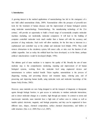
  • 7. 7 3.6 Diagnostic Imaging Techniques such as X-ray, computer tomography (CT), ultrasound (US), magnetic resonance imaging (MRI) and nuclear medicine (NM) are well established imaging techniques, widely used both in medicine and biochemical research. Originally, imaging techniques could only detect changes in the appearance of a tissue when the symptoms of the disease were relatively advanced. Later, targeting and contrast agents were introduced to mark the disease site at the tissue level, increasing imaging specificity and resolution. It is in this specific area that nanotechnology is making its highest contribution by developing better contrast agents for nearly all imaging techniques. The physiochemical characteristics of the nanoparticles (particle size, surface charge, surface coating and stability) allow the redirection and the concentration of the marker at the site of interest. An example of nanoparticles used in research for imaging are perfluorocarbon nanoparticles employed as contrast agents for nuclear imaging, magnetic resonance imaging and ultrasound, with application to the imaging of blood clots, angiogenesis, cancer metastases and other pathogenic changes in blood vessels. Gadolium complexes have been incorporated into emulsion nanoparticles for the molecular imaging of thrombus resulting in a dramatic enhancement of the signal compared to usual MRI contrast agents (Flacke et al., 2001). Figure 1. Low-resolution images (3D GraSE) of fibrin clots targeted with nanoparticles presenting a homogeneous, T1-weighted enhancement that improves with increasing gadolinium level (0, 2.5, and 20 mol%). (Flacke et al., 2001).
  • 8. 8 4. Nanotechnology and medical applications: 4.1 Liposomes Liposomes were first developed about 40 years ago. They are small artificial vesicles(50 – 100nm) developed from phospholipids such as phosphatidylcholine, phosphatidylglycerol,phosphatidylethanolamine and phosphatidylserine, which have been used in biology, biochemistry, medicine, food and cosmetics( Shafirovich et al., 2007; Patel-Predd , 2008;Hu et al., 2003;Heron et al., 2007).The characteristics of liposomes are determined by the choice of lipid, their composition, method of preparation, size and surface charge(Stylios et al., 2005). Liposomes have been applied as drug carriers due to their ability to prevent degradation of drugs, reduce side effects and target drugs to site of action (Cortie, 2004). However,limitations of liposomes include low encapsulation efficiency, rapid leakage of water-soluble drug in the presence of blood components and poor storage stability (Cortie , 2004;Cortie and van der Lingen , 2002). However, surface modification may confer stability and structure integrity against harsh bio-environment after oral or parenteral administration (Overbury, 2005). Surface modification can be achieved by attaching polymers such as poly (methacrylic acid-co-stearyl methacrylate) and polyethylene glycol units to improve the circulation time of liposomes in the blood; and by conjugation to antibodies or ligands such as lectins for target specific drug delivery and stability (Cortie and van der Lingen , 2002; Overbury , 2005; Jain et al., 2008). Applications of liposomes include transdermal drug delivery to enhance skin permeation of drugs with high molecular weight and poor water solubility (Roco , 1999); a carrier for delivery of drugs, such as gentamicin, in order to reduce toxicity( Yin , 2007); possible drug delivery to the lungs by nebulisation(Eijkel and van den Berg , 2006); ocular drug delivery ( Sheeparamatti et al.,2007) and in the treatment of parasitic infections. However, solid lipid nanoparticles (SLNs) provide an effective alternative due to their stability, ease of scalability and commercialisability (Smith, 2006).
  • 9. 9 Other vesicular structures include transferosomes, ethosomes, niosomes and marinosomes which are used mainly for transdermal delivery (Shafirovich et al., 2007; Ball , 2005;Bittner , 2005). Figure 2: Liposome for drug delivery (Smith, 2006). Liposomes discovered in mid 1960s were the original models of nanoscaled drug delivery devices.They are spherical nanoparticles made of lipid bilayer membranes with an aqueous interior but can be unilamellar with a single lamella of membrane or multilamellar with multiple membranes. They can be used as effective drug delivery systems. Cancer chemotherapeutic drugs and other toxic drugs like amphotericin and hamycin, when used as liposomal drugs produce much better efficacy and safety as compared to conventional preparations. These liposomes can be loaded with drugs either in the aqueous compartment or in the lipid membrane. Usually water soluble drugs are loaded in aqueous compartment and lipid soluble drugs are incorporated in the liposomal membrane (Gregoriadis and Ryman, 1972). The major limitation of liposome is its rapid degradation and clearance by the liver macrophages (McCormack and Gregoriadis, 1994),
  • 10. 10 thus reducing the duration of action of the drug it carries. This can be reduced to a certain extent with the advent of stealth liposomes where the liposomes are coated with materials like polyoxyethylene (Illum and Davis, 1984) which prevents opsonisation of the liposome and their uptake by macrophages (Senior et al., 1991). Other ways of prolonging the circulation time of liposomes are incorporation of substances like cholesterol (Kirby and Gregoriadis, 1983), polyvinylpyrollidone polyacrylamide lipids (Torchilin et al., 1994) and high transition temperature phospholipids distearoyl phosphatidylcholine (Forssen et al., 1992). 4.1.1 Targeting of liposomal drugs Immunoliposomes are liposomes conjugated with an antibody directed towards the tumour antigen. These immunoliposomes when injected into the body, reaches the target tissue and gets accumulated in its site of action. This reduces unwanted effects and also increases the drug delivery to the target tissue, thus enhancing its safety and efficacy (Surendiran et al., 2009). The targeted liposomal preparations are found to have a better efficacy than non targeted liposomes. 4.2 Nanopores Nanopores designed in 1997 by Desai and Ferrari (Desai et al., 1998), consist of wafers with high density of pores (20 nm in diameter). The pores allow entry of oxygen, glucose and other products like insulin to pass through. However, it does not allow immunoglobulin and cells to pass through them. Nanopores can be used as devices to protect transplanted tissues from the host immune system, at the same time, utilizing the benefit of transplantation. β cells of pancreas can be enclosed within the nanopore device and implanted in the recipient’s body. This tissue sample receives the nutrients from the surrounding tissues and at the same time remains undetected by the immune system and hence do not get rejected. This could serve as a newer modality of treatment for insulin dependent diabetes mellitus (Leoni and Desai, 2001).
  • 11. 11 Nanopores can also be employed in DNA sequencing. Nanopores are also being developed with ability to differentiate purines from pyrimidines. Further, incorporation of electricity conducting electrodes is being designed to improve longitudinal resolution for base pair identification ( Freitas , 2005).Such a method could possibly read a thousand bases per second per pore. These can be used for low cost high throughput genome sequencing (Deamer and Akeson , 2000)which would be of great benefit for application of pharmacogenomics in drug development process. 4.3 Fullerenes Fullerenes, a carbon allotrope, also called as “bucky balls” were discovered in 1985 (Thakral and Mehta, 2006).The buckminster fullerene is the most common form of fullerene measuring about 7 A in diameter with 60 carbon atoms arranged in a shape known as truncated icosahedrons (Kratschmer et al., 1990). It resembles a soccer ball with 20 hexagons and 12 pentagons and is highly symmetrical (Taylor et al., 1990). 4.3.1 Types of fullerenes Alkali doped fullerenes are structures with alkali metal atoms in between fullerenes Contributing valence electrons to neighbouring fullerenes (Chandrakumar and Ghosh , 2008).They occur because of the electronegative nature of the fullerenes. Endohedral fullerenes have another atom enclosed inside the buckyball. If a metallic atom is enclosed,these are called as metallofullerenes ( Fatouros et al., 2006; Komatsu et al., 2005). Due to the small size of C60 fullerene, it is difficult to synthesize endohedral C60 fullerenes. However, larger fullerenes such as C82 or C84 fullerenes are used for synthesizing endohedral fullerenes. Endohedral metallofullerenes can be used for diagnostic purposes as radio contrast media in magnetic resonance imaging and other imaging procedures. Since the radioactive metal is enclosed within the buckyball, these are less toxic and safer. This method can also be employed for imaging organs as radioactive tracers (Komatsu et al., 2005).
  • 12. 12 Exohedral fullerenes also called as fullerene derivatives are synthesized by chemical reaction between the fullerene and other chemical groups. These are also called as functionalized fullerenes. Such fullerenes can be used as photosensitizers in photodynamic therapy for malignancies. These generate reactive oxygen species when stimulated by light and kills the target cells. This method is now also being investigated for antimicrobial property as these cause cell membrane disruption especially in Gram positive bacteria and mycobacterium (Mroz et al., 2007; Tegos et al., 2005; Bosi et al., 2000). Heterofullerenes are fullerene compounds where one or more carbon atoms are replaced by other atoms like nitrogen or boron (Thakral and Mehta, 2006). Fullerenes are being investigated for drug transport of antiviral drugs, antibiotics and anticancer agents (Mroz et al., 2007; Tegos et al., 2005; Bosi et al., 2000; Ji et al., 2008). Fullerenes can also be used as free radical scavengers due to presence of high number of conjugated double bonds in the core structure. These are found to have a protective activity against mitochondrial injury induced by free radicals (Cai et al., 2008). However, fullerenes can also generate reactive oxygen species during photosensitization. This property can be used in cancer therapy (Markovic and Trajkovic, 2008). Fullerenes have the potential to stimulate host immune response and production of fullerene specific antibodies. Animal studies with C60 fullerene conjugated with thyroglobulin have produced a C60 specific immunological response which can be detected by ELISA with IgG specific antibodies. This can be used to design methods of estimation of fullerene levels in the body when used for therapeutic or diagnostic purposes (Chen et al., 1998).On intravenous injection, these get distributed to various parts of the body and get excreted unchanged through the kidney. Soluble derivates of fullerenes are more biocompatible compared to insoluble forms of fullerenes and have low toxic potential even at higher doses (Chen et al., 1998). Further, the degree of purification of fullerene determines its cost and highly purified fullerenes are expensive, restricting its application in medical field (Thakral and Mehta, 2006).
  • 13. 13 4.4 Biosensors A sensor is a device capable of recognizing a specific chemical species and ‘signalling’ the presence, activity or concentration of that species in solution through some chemical change. A ‘transducer’ converts the chemical signal (such as a catalytic activity of a specific biomolecule) into a quantifiable signal (such as a change in colour or intensity) with a defined sensitivity. When the sensing is based on biomolecular recognition it is called a biosensor. There are various types of biosensors, such as antibody/antigen based, nucleic acid based and enzyme based. Also, depending on the technique used in signal transduction, biosensors are classified as optical- detection biosensors (as in the example above), electrochemical biosensors, mass-sensitive biosensors and thermal biosensors (Kubik et al., 2005; Vo-Dinh and Cullum, 2000). One miniaturized biosensor concept makes use of small beams that bend when biological molecules become captured at the surface inducing a surface stress (see Figure 3). Figure 3.1 Schematic diagram of a cantilever based biosensor. The yellow molecules bind specifically to the red molecules on the right hand cantilever and are detected by the bending of the cantilever (Wang and Branton, 2001). There are numerous nanoparticles that can be used as biosensors components (Medina et al., 2007). These work as probes recognizing an analyte or differentiating between analytes of interest. In such applications some biological molecular species are attached to the surface of the nanoparticles to recognize, through a key -and-lock mechanism, the target of interest. The probes
  • 14. 14 then signal the presence of the target by a change in color, mass or other physical change. Nanoparticles that are used as elements for biosensors include quantum dots, metallic nanobeads, silica nanoparticles, magnetic beads, and fullerenes, which are hollow cages of carbon, shaped like soccer balls. Other biosensors use nanostructured particles as nano-sieves through which charged molecules are transported in an electric field. In this case particles with engineered nanopores are used (Wang and Branton, 2001). 4.5 Nanotubes Carbon nanotubes discovered in 1991 (Iijima, 1991) are tubular structures like a sheet of graphite rolled into a cylinder capped at one or both ends by a buckyball. Nanotubes can be single walled carbon nanotube (SWCNT) or multiwalled carbon nanotube (MWCNT) in concentric fashion. Single walled nanotube has an internal diameter of 1-2 nm and multiwalled nanotube has a diameter of 2-25 nm with 0.36 nm distance between layers of MWCNT. These vary in their length ranging from 1 μm to a few micrometers (Reilly, 2007). These are characterized by greater strength and stability hence can be used as stable drug carriers. Cell specificity can be achieved by conjugating antibodies to carbon nanotubes with fluorescent or radiolabelling (McDevitt et al., 2007). Entry of nanotubes into the cell may be mediated by endocytosis or by insertion through the cell membrane. Carbon nanotubes can be made more soluble by incorporation of carboxylic or ammonium groups to their structure and can be used for the transport of peptides, nucleic acids and other drug molecules. Indium-111 radionuclide labelled carbon nanotubes are being investigated for killing cancer cells selectively (Reilly, 2007). Amphotericin B nanotubes has shown increased drug delivery to the interior of cells compared to amphotericin B administration without nanotubes (Prato et al., 2008). The efficacy of amphotericin B nanotubes was greater as an antifungal agent compared to amphotericin B alone and it was effective on strains of fungi which are usually resistant to amphotericin B alone. Further, there was reduced toxicity to mammalian cells with amphotericin B nanotubes (Prato et al., 2008).The ability of nanotubes to transport DNA across cell membrane is used in studies
  • 15. 15 involving gene therapy. DNA can be attached to the tips of nanotubes or can be incorporated within the tubes (Prato et al., 2008).showed greater expression of the β galactosidase marker gene through nanotubes compared to transfer of naked DNA. This confers the advantage of non immunogenicity in contrast to viral vectors used for gene transfer. Gene silencing studies with small interfering RNA (siRNA) have been done as a modality of cancer therapy where tumour cells will be selectively modulated. Functionalized single walled carbon nanotubes can be used with siRNA to silence targeted gene expression (Zhang et al., 2006). It was observed that carbon nanotubes, except acetylated ones, when bonded with a peptide produce a higher immunological response compared to free peptides. This property can be used in vaccine production to enhance the efficacy of vaccines. Further, it was also found that compounds bound to nanotubes increase the efficacy of diagnostic methods like ELISA. These can also be used for designing of biosensors owing to property of functionalization and high length to diameter aspect ratio which provides a high surface to volume ratio (Pantarotto et al., 2003; Balasubramanian and Burghard, 2006). Water insoluble forms of nanotubes like pristine carbon nanotubes have high in vitro toxicity compared to modified water dispersible forms of nanotubes. It was also seen that the toxic potential decreases withfunctionalization. Further, functionalization also affects the elimination of the nanotube. SWCNTs without conjugation to monoclonal antibody have a high renal uptake and modest liver uptake as compared to SWCNTs with conjugation to monoclonal antibody having higher liver uptake and lower renal uptake (Reilly, 2007). Carbon nanotubes and nanowires are also employed for sensing (Kong et al., 2000; Collins et al., 2000; Cui et al., 2001). The latter can be fabricated out of a semiconductor material and their size tuned to have a specific conducting property. This, together with the ability to bind specific analytes on their surface, yields a direct, label-free electrical readout (Cui et al 2001; Patolsky et al., 2006). These nanowires biosensors allow detecting a wide range of chemical and biological species, including low concentration of protein and viruses and their application spans from the medical to the environmental sector. Figure 4 illustrates a silicon nanowires biosensor based on biorecognition.
  • 16. 16 Figure 3.2 Biorecognition on a silicon nanowire biosensor. The surface of the nanowire is modified with avidin molecules (purple stars) which can selectively bind a streptavidin- functionalised molecule or nanoparticle (Reilly, 2007). 4.6 Quantum dots Quantum dots are nanocrystals measuring around 2-10 nm which can be made to fluorescence when stimulated by light. Their structure consists of an inorganic core, the size of which determines the colour emitted an inorganic shell and an aqueous organic coating to which biomolecules are conjugated. The biomolecule conjugation of the quantum dots can be modulated to target various biomarkers (Iga et al., 2007). Quantum dots can be used for biomedical purposes as a diagnostic as well as therapeutic tool. These can be tagged with biomolecules and used as highly sensitive probes. A study done on prostate cancer developed in nude mice has shown accumulation of quantum dots probe by enhanced permeability and retention as well as by antibody directed targeting (Gao et al., 2004).The quantum dots conjugated with polyethylene glycol (PEG) and antibody to prostate specific membrane antigen (PSMA) were accumulated and retained in the grafted tumour tissue in the mouse (Gao et al., 2004). Quantum dots can also be used for imaging of sentinel node in cancer patients for tumour staging and planning of therapy. This method can be adopted for various malignancies like melanoma, breast, lung and gastrointestinal tumours (Iga et al., 2007). Quantum dot probes provide real time imaging of the sentinel node with Near Infra Red (NIR) fluorescence system. The NIR region of
  • 17. 17 the electromagnetic spectrum produces reduced background noise and deeper penetration of rays, of up to 2 to 5 cm into the biological sample. However, the traditional fluorescence dyes yield low signal intensity when used in NIR region. This limitation is overcome, by using NIR fluorescence system with quantum dot probes. The fluorescence produced by quantum dots is much brighter than those produced by conventional dyes when used with NIR fluorescence system (Amiot et al., 2008).However, the application of quantum dots in a clinical setting has limitations owing to its elimination factors. Functionalization of the quantum dots which protects from the toxic core, leads to increase in size of the nanoparticle greater than the pore size of endothelium and renal capillaries, thus reducing its elimination and resulting in toxicity. Also, in vivo studies are lacking on the metabolism and excretion of quantum dots (Iga et al., 2007). 4.7 Nanoshells Nanoshells were developed by West and Halas (West and Halas, 2000). At Rice University as a new modality of targeted therapy. Nanoshells consist of nanoparticles with a core of silica and a coating of thin metallic shell. These can be targeted to desired tissue by using immunological methods. This technology is being evaluated for cancer therapy. (Hirsh et al., 2003) used nanoshells which are tuned to absorb infra red rays when exposed from a source outside the body to demonstrate the thermo ablative property of nanoshells. The nanoshells when exposed to NIR region of the electromagnetic spectrum get heated and cause destruction of the tissue. This has been studied in both in vitro and in vivo experiments with HER 2 expressing SK-BR-3 human breast carcinoma cells. The control cells did not lose their viability even after treatment with nanoshells with non specific anti IgG or PEG and NIR ablation (Lowery et al., 2006). Nanoshells can also be embedded in a hydrogel polymer containing the drug. After directing the nanoshells to the tumour tissue by immunological methods, with an infrared laser, these can be made to get heated up, melting the polymer and releasing the drug at the tumour tissue. Targeting the drug release avoids the toxicity of cancer chemotherapy drugs. Nanoshells are currently being investigated for micro metastasis of tumours and also for treatment of diabetes (Freitas, 2005; Kherlopian., 2008).
  • 18. 18 Nanoshells are also useful for diagnostic purposes in whole blood immunoassays. Gold nanoshells can be coupled to antibodies and the size can be modulated so that it responds to NIR wavelength, which has the ability to penetrate whole blood specimens. With this method it is possible to detect immunoglobulins at a concentration range of nanograms per millilitre in plasma and whole blood (Freitas, 2005). 4.8 Nanobubbles Cancer therapeutic drugs can be incorporated into nanoscaled bubble like structures called as nanobubbles. These nanobubbles remain stable at room temperature and when heated to physiological temperature within the body coalesce to form microbubbles. These have the advantages of targeting the tumour tissue and delivering the drug selectively under the influence of ultrasound exposure. This results in increased intracellular uptake of the drug by the tumour cells. It also provides an additional advantage of enabling visualisation of the tumour by means of ultrasound methods (Klibanov, 2006; Gao et al., 2008). (Rapaport et al., 2007) have demonstrated the utility of nanobubbles in delivery of drugs like doxorubicin based on in vitro and in vivo experiments using breast cancer cells MDA MB231 and mice with breast cancer xenograft respectively. On administration of nanobubble loaded doxorubicin, these reach the tumour tissue through leaky vasculature and get accumulated at the site of tumour. This is followed by formation of microbubbles by coalescing of nanobubbles which can be visualized by ultrasound techniques. When the site is focused with high intensity focused ultrasound (HIFU), it causes disruption of the microbubbles resulting in release of the drug. The microbubbles retained the drug in a stable state until stimulated by HIFU. This results in attainment of higher levels of drug in the target cells and hence reduced toxicity and increased efficacy. This method needs further exploration for its utility in treatment of various malignancies. Liposomal nanobubbles and microbubbles are also being investigated for their role as effective non viral vectors for gene therapy. Nanobubbles combined with ultrasound exposure has shown improved transfer of gene in both in vitro and in vivo studies (Negishi et al., 2008; Suzuki et al., 2008). Nanobubbles are also being tried as a therapeutic measure for removal of clot in vascular system in combination with ultrasound, a process called as sonothrombolysis. This method has advantages of being non invasive and causing less damage to endothelium (Iverson et al., 2008).
  • 19. 19 4.9 Paramagnetic nanoparticles Paramagnetic nanoparticles are being tried for both diagnostic and therapeutic purposes. Diagnostically, paramagnetic iron oxide nanoparticles are used as contrast agents in magnetic resonance imaging. These have a greater magnetic susceptibility than conventional contrast agents. Targeting of these nanoparticles enables identification of specific organs and tissues (Cuenca et al., 2006).The use of iron oxide in MRI imaging faces limitations like specificity and internalization by macrophages (Peng et al., 2008). Paramagnetic nanoparticles conjugated with antibodies to HER-2/neu which are expressed on breast cancer cells have been used with MRI to detect breast cancer cells in vitro (Artemov et al., 2003).Study done by (Leuschner et al., 2005) has demonstrated the in vivo detection of breast cancer cells using paramagnetic nanoparticles conjugated with luteinizing hormone releasing hormone as breast cancer cells express LHRH receptors. Thus, use of antibodies to direct the nanoparticle to the target site helped to overcome problems with specificity of action. Internalization of the nanoparticles by macrophages can be reduced by treatment with drugs like lovastatin which reduce macrophage receptor expression for the nanoparticle by reducing the recycling of receptors (Peng et al., 2008). Further, injection of decoys of nanoparticle can be used to eliminate plasma opsonins and reduce uptake of the nanoparticles. Also, change of surface charge of the nanoparticle to neutral by covalent coupling to chemicals leads to an increase in circulation time (Peng et al., 2008). Monocrystalline iron oxide nanoparticles (MIONs) have been studied by (Knauth et al., 2001) in magnetic resonance imaging of brain. MIONs help in overcoming the disadvantage of surgically induced contrast enhancement with traditional contrast agents resulting in misinterpretation during intra-operative MR imaging of brain. Surgically induced contrast enhancement occurs in brain due to leak of contrast material from the cut end and oozing blood vessels in brain when MR imaging is done post- operatively.This is avoided when MIONs are used pre- operatively.These are rapidly taken up by the tumour cells (Moore et al., 1997), producing long lasting contrast enhancement of tumour and the remaining nanoparticles are removed from the circulation by reticuloendothelial system (Weissleder et al., 1989).
  • 20. 20 Magnetic microparticle probes with nanoparticle probes have been used for identification of proteins like prostate specific antigen. Here magnetic microparticles coated with antibodies together with nanoprobes with similar coating and a unique hybridized DNA barcode are used. The microparticle coated with antibody directed against prostate specific antigen combines with it to form a complex and can be separated by using magnetic separation. The presence of these separated complexes is determined by dehybridization of the complexed DNA barcode sequence and polymerase chain reaction for the oligonucleotides. This allows prostate specific antigen detection at 30 attomolar concentration (Nam et al., 2003). 4.10 Nanosomes Raoul Kopelman’s group at the University of Michigan, USA, has been working on nanosomes also called as PEBBLEs (Probes Encapsulated by Biologically Localized Embedding) which integrate various aspects of medical applications such as targeting, diagnosis and therapy. These nanosomes are being developed for treatment of various tumours, in particular CNS tumours. Silica coated iron oxide nanoparticles coated with polyethylene glycol (Xu et al., 2003) and affixed with targeting antibody and contrast elements like gadolinium are used to access specific areas of brain involved with tumour. Targeting aids in binding the nanoparticle specifically to the tumour cells and the contrast elements helps in better detection with magnetic resonance imaging. Subsequent treatment with laser can destroy the cells loaded with these nanoparticles by the heat generated by iron oxide particles by absorbing the infra red light. Nanosomes can also be integrated with a photocatalyst which produces reactive oxygen species when stimulated by light and destroy the target tissue. This method has advantage over conventional drugs in being much safer without the adverse effects of cancer chemotherapy drugs and also the absence of development of drug resistance. Nanosomes are being developed to integrate more and more components in it for flexibility of its applications (Freitas, 2005).
  • 21. 21 4.11 Dendrimers Dendrimers are nanostructures produced from macromolecules such as polyamidoamine (PAMAM), polypropyleneimine and polyaryl ether; and are highly branched with an inner core. The particle size range is between 1 to 100nm although their sizes are mostly less than 10nm. About 20 years ago, dendrimer studies centred on their synthesis, physical and chemical properties while exploration of their biological applications was initiated about thirteen years ago (Sahoo et al, 2007). The uniqueness of dendrimers is based on their series of branches,multivalency, well defined molecular weight and globular structure with controlled surface functionality, which enhances their potential as carriers for drug delivery (Sahoo et al, 2007). Their globular structures and the presence of internal cavities enable drugs to be encapsulated within the macromolecule interior. Dendrimers have been reported to provide controlled release from the inner core (Matija, 2004). Controlled multivalency of dendrimers enables attachment of several drug molecules, targeting groups and solubilising groups onto the surfaces of the dendrimers in a well defined manner (Sahoo et al., 2007) Dendrimers are employed due to their size (less than 10nm), ease of preparation, functionality and their ability to display multiple copies of surface groups for biological recognition process (Freitas , 2005). Water soluble dendrimers can bind and solubilise small molecules and can be used as coating agents to protect drugs and deliver to specific sites. Other applications of dendrimers include catalysis, gene and DNA delivery, biomimetics and as solution phase supports for combinatorial chemistry (Kostoff et al., 2007).Some of the drug delivery applications include therapeutic and diagnostic utilization for cancer treatment (Majumder et al., 2007) enhancement of drug solubility and permeability (dendrimer-drug conjugates)(Feynman,1960);and intracellular delivery (Smith, 2006). Dendrimers are also used as contrast agents for imaging.The 1, 4-diaminobutane (DAB) core dendrimer and the polyamidoamine (PAMAM) dendrimer are well studied commercially available dendrimers for imaging studies. Renal excretion is the main route of clearance and is dependent on the size of the particle and more than 60 per cent of injected DAB or PAMAM dendrimer is cleared from circulation within 15 min (Longmire et al., 2008). Smaller sized
  • 22. 22 dendrimers undergo rapid renal clearance whereas dendrimers with charged surface or hydrophobic surfaces are rapidly cleared by the liver. Those dendrimers with a hydrophilic surface escape renal clearance and have a greater circulation time (Duncan and Izzo, 2005). Cationic dendrimers have a greater potential to cause cytotoxicity compared to anionic dendrimer or PAMAM dendrimers. It is proposed to cause cell membrane instability and cell lysis. The toxicity of dendrimer is dependent on the size of the particle and increase with size. It can be reduced by means of surface modification of the dendrimers with incorporation of PEG or fatty acids (Svenson and Tomalia, 2005). 4.12 Polymeric nanoparticles Figure 4. Illustration of Nanospheres,Nanocapsules,Liposomes,Micelles (Svenson and Tomalia , 2005). Polymeric nanoparticles are colloidal solid particles with a size range of 10 to 1000nm (Stylios et al., 2005) and they can be spherical, branched or shell structures. The first fabrication of nanoparticles was about 35 years ago as carriers for vaccines and cancer chemotherapeutics. They are developed from non-biodegradable and biodegradable polymers. Their small sizes enable them to penetrate capillaries and to be taken up by cells, thereby increasing the accumulation of drugs at target sites. Drugs are incorporated into nanoparticles by dissolution, entrapment, adsorption, attachment or by encapsulation, and the nanoparticles provide sustained release of the drugs for longer periods, e.g., days and weeks (Roco, 2003). Nanoparticles
  • 23. 23 enhance immunization by prevention of degradation of the vaccine and increased uptake by immune cells (Singh et al., 2006). One of the determinants of the extent of uptake by immune cells is the type of polymer employed. In a study (Miyazaki and Islam, 2007) comparing poly-(_- caprolactone) (PCL), poly (lactide-coglycolide) (PLGA) and their blend, PCL nanoparticles were the most efficiently taken up by immune cells due to their hydrophobicity. However, all polymeric nanoparticles elicited vaccine (diphtheria toxoid) specific serum IgG antibody response significantly higher than free diphtheria toxoid. To target drugs to site of action, the drug can be conjugated to a tissue or cell specific ligand or coupled to macromolecules that reach the target organs. To target an anticancer agent to the liver, polymeric conjugate nanoparticles which comprised biotin and diamine-terminated poly (ethylene glycol) with a galactose moiety from lactobionic acid were prepared (Williams, 2008). Some other applications of nanoparticles include possible recognition of vascular endothelial dysfunction (Zong et al., 2005);oral deliveryof insulin (Gao et al., 2003); brain drug targeting for neurodegenerative disorders such as Alzheimer’s disease (Luo et al., 2005); topical administration to enhance penetration and distribution in and across the skin barrier(Tian et al., 2007); and pH-sensitive nanoparticles to improve oral bioavailability of drugs such as cyclosporine A (Shafirovich et al., 2006). 4.13 Solid lipid nanocarriers Solid lipid nanoparticles (SLN) are nanostructures made from solid lipids such as glyceryl behenate (Compritol), stearic triglyceride (tristearin), cetyl palmitate and glycerol tripalmitate (tripalmitin) with a size range of 50 and 1000 nm (Visakhapatnam, 2008). Research interest in SLN emerged about ten years ago due to their scalability potential. The lipids employed are well tolerated by the body; large scale production will be cost effective and simple by using high pressure homogenization. Some of the features of SLN include good tolerability, site-specific targeting, stability (stabilized by surfactants or polymers), controlled drug release and protection
  • 24. 24 of liable drugs from degradation. However, SLN are known for insufficient drug loading, drug expulsion after polymorphic transition on storage and relative high water content of the dispersions. SLN has been studied and developed for parenteral, dermal, ocular, oral, pulmonary and rectal routes of administration (Zsigmondy, 1926; Roco, 2000-2003; Horton and Khan, 2006;Guo , 2005; Liu et al., 2003). To overcome the limitations of SLN, nanostructured lipid carriers (NLC) were introduced. NLC is composed of solid lipids and a certain amount of liquid lipids with improved drug loading and increased stability on storage thereby reducing drug expulsion (Roco, 2000-2003). NLCs have been explored for dermal delivery in cosmetics and dermatological preparations (Roco, 2000- 2003). Lipid drug conjugate (LDC) nanoparticles were introduced to overcome the limitation of types of drugs incorporated in the solid lipid matrix. Lipophilic drugs are usually incorporated in SLN but due to partitioning effects during production, only highly potent hydrophilic drugs effective in low concentrations are incorporated in SLN (Wissing et al., 2004). LDC enables the incorporation of both hydrophilic (e.g., doxorubicin and tobramycin) and lipophilic (e.g., progesterone and cyclosporine A) drugs (Wissing et al., 2004). 4.14 Polymeric micelles Micelles are formed when amphiphilic surfactant or polymeric molecules spontaneously associate in aqueous medium to form core-shell structures or vesicles. Polymeric micelles are formed from amphiphilic block copolymers, such as poly(ethylene oxide)-poly(_-benzyl- Laspartate) and poly(N-isopropylacrylamide)- polystyrene, and are more stable than surfactant micelles in physiological solutions (Liu et al., 2003). They were first proposed as drug carriers about 24 years ago (Liu et al., 2003). The inner core of a micelle is hydrophobic which is surrounded by a shell of hydrophilic polymers such as poly (ethylene glycol) (Mansoori , 2002).Their hydrophobic core enables incorporation of poorly water soluble and amphiphilic drugs while their hydrophilic shell and size (<100nm) prolong their circulation time in the blood and increase accumulation in tumoural tissues (Liu et al., 2003).
  • 25. 25 Polymeric micelles are able to reach parts of the body that are poorly accessible to liposomes; accumulate more than free drugs in tumoural tissues due to increased vascular permeability (Liu et al., 2003). Thus, polymeric micelles can be employed to administer chemotherapeutics in a controlled and targeted manner with high concentration in the tumoural cells and reduced side effects. However, the targeting ability of polymeric micelles is limited due to low drug loading (Balzani, 2005; Teo and Sun, 2006). and low drug incorporation stability (Balzani, 2005). which cause the loaded drug to be released before getting to the site of action. Consequently, manipulation of the production parameters and the design of the inner core can improve drug loading and drug incorporation stability,respectively (Balzani , 2005; Teo and Sun, 2006). Lipid moieties, such as cholesterol and fatty acyl carnitines, can also be employed to impart good stability to the polymeric micelles. This is based on increased hydrophobic interaction between the polymeric chains in the inner core due to presence of fatty acid acyls (e.g.diacyllipid) (Liu et al., 2003). Polymeric micelles have been employed for targeted and intracellular delivery (Teo and Sun, 2006) sustained release and parenteral delivery (Liu et al., 2003). 4.15 Nanocapsules Nanocapsules are spherical hollow structures in which the drug is confined in the cavity and is surrounded by a polymer membrane (Romig et al., 2007).They were developed over 30 years ago. Sizes between 50 and 300nm are preferred for drug delivery and they may be filled with oil which can dissolve lipophilic drugs. They have low density, high loading capacity and are taken up by the mononuclear phagocyte system, and accumulate at target organs such as liver and spleen (Shea, 2005). Nanocapsules can be employed as confined reaction vessels, protective shell for cells or enzymes, transfection vectors in gene therapy, dye dispersants, carriers in heterogenous catalysis, imaging and drug carriers (Tratnyek and Johnson , 2006; Roco, 2001). They are known to
  • 26. 26 improve the oral bioavailability of protein and peptides which include insulin, elcatonin and salmon calcitonin (Romig et al., 2007; Salamanca-Buentello et al., 2005). Encapsulation of drugs such as ibuprofen(Shea , 2005). within nanocapsules protects liable drugs from degradation, reduces systemic toxicity, provide controlled release and mask unpleasant taste (Singh, 2007). Due to their high stability and low permeability, drugs may not be loaded into the capsules after formulation and also the release of the drug at target site may be difficult. To improve on their permeability, they are made responsive to physiological factors such as pH (Tegart, 2008). 4.16 Nanoemulsions Nanoemulsions are emulsions with droplet size below 1μ but usually between 20 and 200nm (Malsch, 1999; Renn and Roco, 2006). Unlike microemulsions which are white in colour due to their light scattering ability, nanoemulsions whose nanosize is often smaller than visible wavelength, are transparent (Malsch, 1999; Dunphy et al., 2000-2004). Nanoemulsions are biodegradable, biocompatible, easy to produce and used as carriers for lipophilic drugs which are prone to hydrolysis. They are employed as a sustained release delivery system for depot formation via subcutaneous injection (Renn and Roco , 2006). They enhance gastrointestinal absorption and reduce inter- and intra-subject variability for various drugs. Due to their very large interfacial area, they exhibit excellent drug release profile (Cobb and Macoubrice, 2004). Nanoemulsions have been studied and developed for parenteral, oral, ocular, pulmonary and dermal deliveries (Malsch, 1999). Stability against sedimentation is attained based on the nano size of the droplets because the sedimentation rate due to gravity is less than Brownian movement and diffusion (Malsch, 1999). Unlike microemulsions, nanoemulsions are metastable and can be destabilized by Ostwald ripening whereby the small droplets dissolve and their mass is taken up by the large droplets and depletion induced flocculation due to addition of thickening polymers. When this happens, the nanoemulsion becomes opaque and creaming will occur (Roco, 2003). However, addition of a small amount of a second oil with low solubility into the aqueous phase and addition of a second surfactant may reduce Ostwald ripening (Malsch, 1999). Also, a number of factors during production should be controlled (Dunphy et al., 2000-2004). These factors include selecting an
  • 27. 27 appropriate composition, controlling the order of addition of components, applying the shear in a manner that will effectively rupture the droplets, and ensuring that the dispersed phase molecules are insoluble in the continuous phase so that Ostwald ripening does not occur rapidly (Dunphy et al., 2000-2004). 4.17 Ceramic nanoparticles Ceramic nanoparticles are particles fabricated from inorganic compounds with porous characteristics such as silica, alumina and titania (Oberdörster et al., 2005; Nimesh et al., 2006; Soppimath et al., 2001). They can be prepared with the desired size, shape and porosity. Their sizes are less than 100nm and are able to avoid uptake by the reticulo-endothelial system as foreign bodies. Entrapped molecules such as drugs, proteins and enzymes are protected from denaturation at physiological pH and temperature as neither swelling nor change in porosity occurs (Soppimath et al., 2001).Hence, they are effective in delivering proteins and genes. However, these particles are not biodegradable and so there is concern that they may accumulate in the body and cause harmful effects (Nimesh et al., 2006). 4.18 Metallic nanoparticles Metallic nanoparticles include iron oxide, gold, silver, gadolinium and nickel which have been studied for targeted cellular delivery(Jung et al., 2000).Gold exhibits favourable optical and chemical properties at nanoscale for biomedical imaging and therapeutic applications (Nimesh et al., 2006). It can be manipulated to obtain the desired size in the range of 0.8 to 200nm. The surface can be modified with different functional groups for gene transfection, modified into gene delivery vector by conjugation and also modified to target proteins and peptides to the cell nucleus (Jung et al., 2000; Italia et al., 2007). Gadolinium has been studied for enhanced tumour targeted delivery by modification of the nanoparticles with folate, thiamine and poly (ethylene glycol). Modification with folate was reported to enhance the recognition, internalization and retention of gadolinium nanoparticles in tumour cells (Jung et al., 2000).
  • 28. 28 4.19 Carbon nanomaterials These include carbon nanotubes and fullerenes. Fullerenes are carbon allotrope made up of 60 or more carbon atoms with a polygonal structure. Nanotubes have been used for their high electrical conductivity and excellent strength (Nimesh et al., 2006). These materials are being studied for therapeutic applications. Fullerenes can be functionalized for delivery of drugs and biomolecules across cell membrane to the mitochondria (Jung et al., 2000). Carbon nanotubes’ unique properties including low cytotoxicity and good biocompatibility attract their use as vector system in target delivery of drugs, proteins and genes (Jung et al., 2000). However,toxicity of carbon nanotubes is of concern (Sahoo and Labhasetwar, 2003). Carbon nanotubes may cause inflammatory and fibrotic reactions. Figure 5: Illustration of some nanostructures A) Spherical polymeric nanoparticle; B) Liposome; C)Solid lipid nanoparticles – solid lipid enclosed within; D) Nanoemulsion – liquid enclosed within; E)Nanocapsule – hollow; F) Carbon nanotube; G) Dendrimer; I) Polymeric micelle. (Ochekpe et al., 2009).
  • 29. 29 5. Nanosurgery Present-day nanomedicine exploits carefully structured nanoparticles such as dendrimers (Borges and Schengrund, 2005), carbon fullerenes (buckyballs) (Mashino et al., 2005) and Nanoshells (O’Neal et al., 2004) to target specific tissues and organs. These nanoparticles may serve as diagnostic and therapeutic antiviral, antitumor or anticancer agents. But as this technology matures in the years ahead, complex nanodevices and even nanorobots will be fabricated, first of biological materials but later using more durable materials such as diamond to achieve the most powerful results. 5.1 Early vision The first and most famous scientist to voice this possibility was the late Nobel physicist Richard P. Feynman. In his remarkably prescient 1959 talk ‘‘There’s Plenty of Room at the Bottom,’’ Feynman proposed employing machine tools to make smaller machine tools, these are to be used in turn to make still smaller machine tools, and so on all the way down to the atomic level, noting that this is ‘‘a development which I think cannot be avoided.’’ (Feynman, 1960) Feynman was clearly aware of the potential medical applications of this new technology. He offered the first known proposal for a nanorobotic surgical procedure to cure heart disease: ‘‘a friend of mine (Albert R. Hibbs) suggests a very interesting possibility for relatively small machines. He says that, although it is a very wild idea, it would be interesting in surgery if you could swallow the surgeon. You put the mechanical surgeon inside the blood vessel and it goes into the heart and looks around. (Of course the information has to be fed out.) It finds out which valve is the faulty one and takes a little knife and slices it out. .[Imagine] that we can manufacture an object that maneuvers at that level!. Other small machines might be permanently incorporated in the body to assist some inadequately functioning organ.’’ (Feynman, 1960)
  • 30. 30 5.2 Medical microrobotics There are ongoing attempts to build microrobots for in vivo medical use. In 2002, at Tohoku University developed tiny magnetically driven spinning screws intended to swim along veins and carry drugs to infected tissues or even to burrow into tumors and kill them with heat (Freitas, 2005). In 2003, the ‘‘MR-Sub’’ project of Martel’s group at the NanoRobotics Laboratory of Ecole Polytechnique in Montreal tested using variable MRI magnetic fields to generate forces on an untethered microrobot containing ferromagnetic particles, developing sufficient propulsive power to direct the small device through the human body (Freitas, 2005). Brad Nelson’s team at the Swiss Federal Institute of Technology in Zurich continued this approach. In 2005, they reported the fabrication of a microscopic robot small enough (w200 mm) to be injected into the body through a syringe. They hope that this device or its descendants might someday be used to deliver drugs or perform minimally invasive eye surgery (Freitas, 2005). Nelson’s simple microrobot has successfully maneuvered through a watery maze using external energy from magnetic fields, with different frequencies that are able to vibrate different mechanical parts on the device to maintain selective control of different functions. Gordon’s group at the University of Manitoba has also proposed magnetically controlled ‘‘cytobots’’ and ‘‘karyobots’’ for performing wireless intracellular and intranuclear surgery (Freitas, 2005). 5.3 Manufacturing medical nanorobots The greatest power of nanomedicine will emerge, perhaps in the 2020s, when we can design and construct complete artificial nanorobots using rigid diamondoid nanometer-scale parts like molecular gears (Fig. 8) and bearings (Freitas , 2005).
  • 31. 31 Figure 6. A molecular planetary gear is a mechanical component that might be found inside a medical nanorobot. The gear converts shaft power from one angular frequency to another. The casing is a strained silicon shell with predominantly sulfur termination, with each of the nine planet gears attached to the planet carrier by a carbon-carbon single bond. The planetary gear shown here has not been built experimentally but has been modeled computationally. 5.4 Respirocytes and microbivores The ability to build complex diamondoid medical nanorobots to molecular precision, and then to build them cheaply enough in sufficiently large numbers to be useful therapeutically, will revolutionize the practice of medicine and surgery (Freitas, 2005).The first theoretical design study of a complete medical nanorobot ever published in a peer-reviewed journal (in 1998) described a hypothetica l artificial mechanical red blood cell or ‘‘respirocyte’’ made of 18 billion precisely arranged structural atoms (Freitas, 1998). The respirocyte is a bloodborne spherical 1- mm diamondoid 1000-atmosphere pressure vessel with reversible molecule-selective surface pumps powered by endogenous serum glucose. This nanorobot would deliver 236 times more oxygen to body tissues per unit volume than natural red cells and would manage carbonic acidity, controlled by gas concentration sensors and an onboard nanocomputer. A 5-cc
  • 32. 32 therapeutic dose of 50% respirocyte saline suspension containing 5 trillion nanorobots could exactly replace the gas carrying capacity of the patient’s entire 5.4 l of blood. Nanorobotic artificial phagocytes called ‘‘microbivores’’ (Fig. 9) Figure 7. Nanorobotic artificial phagocytes called ‘‘microbivores’’could patrol the bloodstream, seeking out and digesting unwanted pathogens (Freitas, 2005). Could patrol the bloodstream, seeking out and digesting unwanted pathogens including bacteria, viruses, or fungi (Freitas, 2005). 6. Nanotechnology in drug delivery Generally, nanostructures have the ability to protect drugs encapsulated within them from hydrolytic and enzymatic degradation in the gastrointestinal tract; target the delivery of a wide range of drugs to various areas of the body for sustained release and thus are able to deliver drugs, proteins and genes through the peroral route of administration (Nimesh et al., 2006; Soppimath et al., 2001; Jung et al., 2000). They deliver drugs that are highly water insoluble; can bypass the liver, thereby preventing the first pass metabolism of the incorporated drug (Italia et al., 2007; Sahoo and Labhasetwar , 2003). They increase oral bioavailability of drugs due to their specialized uptake mechanisms such as absorptive endocytosis and are able to remain in the
  • 33. 33 blood circulation for a longer time, releasing the incorporated drug in a sustained and continuous manner leading to less plasma fluctuations thereby minimizing side-effects caused by drugs (Italia et al., 2007). Due to the size of nanostructures, they are able to penetrate into tissues and are taken up by cells, allowing efficient delivery of drugs to sites of action. The uptake of nanostructures was found to be 15-250 times greater than that of microparticles in the 1-10μm range (Panyam and Labhasetwar , 2003). Through the manipulation of the characteristics of polymers, release of drug from nanostructures can be controlled to achieve the desired therapeutic concentration for the desired duration. For targeted delivery, nanostructures can be conjugated with targeting moieties such that the linkage between the polymer and the active substance can be manipulated to control the site and duration at which the drug is released. The linkage may be achieved by incorporation of amino acids, lipids, peptides or small chains as spacer molecules (Peppas, 2004). Drug targeting is crucial in chemotherapy, where a drug delivery system can target only the malignant tumour while shielding the healthy cells from uniform distribution of chemotherapeutics in the body and their harmful effects. The use of nanostructures such as polymeric nanoparticles is a non-invasive approach of penetrating the blood brain barrier for management of neurodegenerative disorders, cerebrovascular and inflammatory diseases (Garcia-Garcia et al., 2005; Ringe et al., 2004). Research and development of new drugs are capital- and time- intensive which requires that pharmaceutical companies, in addition,search for other means of meeting up with market demands. New drug delivery methods enable pharmaceutical companies reformulate existing drugs in the market. Nanotechnology is strategic in developing drug delivery systems which can expand drug markets. Nanotechnology can be applied to reformulate existing drugs thereby extending products’ lives, enhance their performance, improve their acceptability by increasing effectiveness, as well as increase safety and patient adherence, and ultimately reduce health care costs (Sahoo and Labhasetwar, 2003; Hughes, 2005). Nanotechnology may also enhance the performance of drugs that are unable to pass clinical trial phases (Hughes, 2005). It provides
  • 34. 34 drug delivery carriers, as well as treatment and management of chronic diseases which include cancer, HIV/AIDS and diabetes. Table 2 Examples of drug delivery technologies in relation to the current nanotechnology revolution (Hughes , 2005). Period Before Nanotechnology (Past) Transition Period (Present) Mature Nanotechnology (Future) Technology Emulsion-based preparation of nano/micro particles Nano/micro fabrication Nano/micro manufacturing Examples • - Liposomes • -Polymer micelles • - Dendrimers • - Nanoparticles • - Nanocrystals • - Microparticles • -Microchip systems • -Microneedle transdermal delivery systems • - Layer-by-layer assembled systems • - Microdispensed particles • -Nano/micro machines for scale-up production 7. Commercially available nano drug delivery systems Despite the challenges which include the huge volume of expenditure involved and the regulatory stages (preclinical and clinical stages – Phases 1 - 4) which are mandatory in order to obtain regulatory approval before a drug can get into the market, some nano drug delivery systems have made it to the market. Table 3 shows the list of some of nano drug delivery systems in the market:
  • 35. 35 Table 3: Nano drug delivery systems in the market(Wagner et al., 2006)
  • 36. 36 8. Conclusion The nanotechnology will help to improve health by enhancing the efficacy and safety of nanosystems and nanodevices. Moreover, early diagnosis, implants with improved properties, cancer treatment and minimum invasive treatments for heart disease, diabetes and other diseases are anticipated. In the coming years, nanotechnology will play a key role in the medicine of tomorrow providing revolutionary opportunities for early disease detection, diagnostic and therapeutic procedures to improving health and enhancing human physical abilities, and enabling precise and effective therapy tailored to the patient. Nanomedicine is in infancy, having the potential to change medical research dramatically in the 21st century. Nanomedical devices can be applied for analytical, imaging, detection, diagnostic and therapeutic purposes and procedures, such as targeting cancer, drug delivery, improving cell-material interactions, scaffolds for tissue engineering, and gene delivery systems, and provide innovative opportunities in the fight against incurable diseases. Many novel nanoparticles and nanodevices are expected to be used, with an enormous positive impact on human health. Over the next ten to twenty years nanotechnology may fundamentally transform science, technology, and society offering a significant opportunity to enhance human health in novel ways, especially by enabling early disease detection and diagnosis, as well as precise and effective therapy tailored to the patient.
  • 37. 37 References Admati R, Pfleiderer P. Robust financial contract and the role of venture capitalist. Journal of Finance, 1994; 2(4): 371–402. Alonso A, Goñi FM, Buckley JT. Lipids favoring inverted phase enhance the ability of aerolysm to permeabilize liposome bilayers. Biochemistry, 2000; 39 (46): 14019-14024. Alvarez-Román R, Naik A, Kalia YN, Guy RH, Fessi H. Skin penetration and distribution of polymeric nanoparticles. Journal of Controlled Release, 2004; 99(1): 53-62. Amiot CL, Xu S, Liang S, Pan L, Zhao JX. Near-infrared fluorescent materials for sensing of biological targets. Sensors 2008; 8: 3082-105. Arias JL, Ruiz MA, López-viota M, Delgado AV. Poly(alkylcyanoacrylate) colloidal particles as vehicles for antitumour drug delivery: a comparative study. Colloids and Surfaces B: Biointerfaces, 2008; 62(1): 64-70. Baker M, Gompers PA. The determinants of board structure at the initial public offering. Journal of Law and Economics, 2003; 46(2): 569–598. Bakowsky H, Richter T, Kneuer C, Hoekstra D, Rothe U, Bendas G, Ehrhardt C, Bakowsky U. Adhesion characteristics and stability assessment of lectin-modified liposomes for site-specific drug delivery. Biochimica et Biophysica Acta, 2008; 1778(1): 242-249. Balasubramanian K, Burghard M. Biosensors based on carbon nanotubes. Analytical & Bioanalytical Chemistry, 2006; 385(3) : 452-68. Banerjee R. Liposomes: applications in medicine. Journal of Biomaterials Applications, 2001; 16(1): 3-21. Barry BW. Is transdermal drug delivery research still important today.Drug Discovery Today, 2001; 6(19): 967-971.
  • 38. 38 Barry BW. Novel mechanisms and devices to enable successful transdermal drug delivery. European Journal of Pharmaceutical Sciences, 2001; 14(2): 101-114. Baysinger BD, Butler HN. Corporate governance and the board of directors: performance effects of changes in board composition. Journal of Law, Economics, and Organization, 1985; 1(1):101–124. Bharali DJ, Khalil M, Gurbuz M, Simone TM, Mousa SA. Nanoparticles and cancer therapy: a concise review with emphasis on dendrimers. International Journal of Nanomedicine 2009; 4(10): 1-7. Borges AR, Schengrund CL. Dendrimers and antivirals: a review. Current Drug Targets. Infectious Disorders, 2005; 5(3): 247-54. Bosi S, Da RT, Castellano S, Banfi E, Prato M. Antimycobacterial activity of ionic fullerene derivatives. Bioorganic and Medicinal Chemistry Letters, 2000; 10(10) : 1043-5. Brüsewitz C, Schendler A, Funke A, Wagner T, Lipp R. Novel poloxamer-based nanoemulsions to enhance the intestinal absorption of active compounds. International Journal of Pharmaceutics, 2007; 329(1-2): 1173-181. Budai L, Hajdu M, Budai M, Grof P, Béni S, Noszal B, Klebovich I, Antal I. Gels and liposomes in optimized ocular drug delivery: studies on ciprofloxacin formulations. International journal of Pharmaceutics, 2007; 343(1-2): 34-40. Cai X, Jia H, Liu Z, Hou B, Luo C, Feng Z et al. Polyhydroxylated fullerene derivative C(60)(OH)(24) prevents mitochondrial dysfunction and oxidative damage in an MPP(+)-induced cellular model of Parkinson’s disease. Journal of Neuroscience Research, 2008; 86(16) : 3622- 34. Casadei MA, Cerreto F, Cesa S, Giannuzzo M,Feeney M, Marianecci C, Paolicelli P. Solid lipid nanoparticles incorporated in dextran hydrogels: a new drug delivery system for oral formulations. International Journal of Pharmaceutics, 2006; 325(1-2):140-146.
  • 39. 39 Cavalli R, Gasco MR, Chetoni P, Burgalassi S,Saettone MF. Solid Lipid nanoparticles (SLN) as ocular delivery system for tobramycin. International Journal of Pharmaceutics, 2002; 238(1-2): 241-245. Chandrakumar KR, Ghosh SK. Alkali-metal-induced enhancement of hydrogen adsorption in C60 fullerene: an abInitio study. Nano Letters, 2008; 8(1): 13-9. Chen BX, Wilson SR, Das M, Coughlin DJ, Erlanger BF.Antigenicity of fullerenes : antibodies specific for fullerenes and their characteristics. Proceedings of the National Academy of the Sciences of the USA, 1998; 95(18) :10809-13. Cloninger MJ. Biological application of dendrimers. Current Opinion in Chemical Biology, 2002; 6(6): 742-748. Collins PG, Bradley K, Ishigami M, Zettl A. Extreme oxygen sensitivity of electronic properties of carbon nanotubes. Science, 2000; 287 (5459): 1801-1804. Cuenca AG, Jiang H, Hochwald SN, Delano M, Cance WG, Grobmyer SR. Emerging implications of nanotechnology on cancer diagnostics and therapeutics. Cancer, 2006; 107(3): 459-66. Cui Y, Wei Q,Park H, Lieber CM. Nanowire nanosensors for highly sensitive and selective detection of biological and chemical species. Science, 2001;293 (5533): 1289-1292. Dai J, Nagai T, Wang X, Zhang T, Meng M, Zhang Q. pH-sensitive nanoparticles for improving the oral bioavailability of cyclosporine A. International Journal of Pharmaceutics, 2004; 280(1- 2): 229-240. Damgé C, Maincent P, Ubrich N. Oral delivery of insulin associated to polymeric nanoparticles in diabetic rats. Journal of Controlled Release, 2007; 117(2): 163-170. Dat AA, Joshi MD, Patravale VB. Parasitic diseases: liposomes and polymeric nanoparticles versus lipid nanoparticles. Advanced Drug Delivery Reviews, 2007; 59(6): 505-521. Deamer DW, Akeson M. Nanopores and nucleic acids : prospects for ultrarapid sequencing. Trends in Biotechnology, 2000; 18(1) : 147-51.
  • 40. 40 Desai TA, Chu WH, Tu JK, Beattie GM, Hayek A, Ferrari M. Microfabricated immunoisolating biocapsules. Biotechnology and Bioengineering, 1998; 57(1) : 118-20. Duncan R, Izzo L. Dendrimer biocompatibility and toxicity. Advanced Drug Delivery Reviews, 2005; 57(15) : 2215-37. Edwin M, Schwartz M, Wagner S. Imitation costs and patents: an empirical study. Economic Journal, 1981; 91(3): 907–918. Fatouros PP, Corwin FD, Chen ZJ, Broaddus WC, Tatum JL, Kettenmann B et al. Invitro and invivo imaging studies of a new endohedral metallofullerene nanoparticle. Radiology 2006; 240(3): 756-64. Flacke S, Fischer S, Scott MJ, Fuhrhop RJ, Allen JS,McLean M,Winter P, Sicard GA, Gaffney PJ, Wickline SA, Lanza GM. Novel MRI Contrast Agent for Molecular Imaging of Fibrin Implications for Detecting Vulnerable Plaques. Circulation, 2001; 104(11): 1282-1285 Flynn T, Wei C. The pathway to commercialization for nanomedicine. Nanomedicine: Nanotechnology, Biology, and Medicine, 2005; 1(1): 47–51. Forssen EA, Coulter DM, Proffitt RT. Selective in vivo localization of daunorubicin small unilamellar vesicles in solid tumors. Cancer Research, 1992; 52(12) : 3255-61. Freitas Jr RA. Exploratory design in medical nanotechnology:a mechanical artificial red cell. Artificial Cells Blood Substitutes, and Immobilization Biotechnology, 1998; 26(4): 411-30. Freitas Jr RA. What is nanomedicine? . Nanomedicine: Nanotechnology, & Biological Medicine, 2005; 1(1): 2-9. Freitas Jr Robert A. Nanodentistry. Journal of the American Dental Association, 2000;131 (11):1559-65. Freitas RA Jr. Current Status of Nanomedicine and Medical Nanorobotics. Journal of. Computational and Theoretical Nanoscience, 2005; 2(1):1-25. Freitas RA Jr. Exploratory design in medical nanotechnology: a mechanical artificial red cell. Artificial Cells Blood Substitutes and Immobilization Biotechnology, 1998; 26(4): 411-430.
  • 41. 41 Freitas RA Jr. Nanodentistry. Journal of the American Dental Association, 2000; 131(11): 1559- 1565. Freitas RA Jr. Nanotechnology, nanomedicine and nanosurgery. International Journal of Surgery, 2005; 3(4): 243-6 Freitas RA Jr. The future of nanofabrication and molecular scale devices in nanomedicine. Studies Health Technology and Informatics, 2002; 80(2): 45-59. Freitas RA Jr., Christopher J. Phoenix. Vasculoid: A Personal Nanomedical Appliance to Replace Human Blood. Journal of Evolution and Technology, 2002; 11(3): 326-339 Gao X, Cui Y , Levenson RM, Chung LWK, Nie S. In-vivo cancer targeting and imaging with semiconductor quantum dots. Nature Biotechnology, 2004; 22(8) : 969-76. Gao Z, Kennedy AM, Christensen DA, Rapoport NY. Drugloaded nano/microbubbles for combining ultrasonography and targeted chemotherapy. Ultrasonics, 2008; 48(4) : 260-70. Garcia-Garcia E, Andrieux K, Gil S, Couvreur P.Colloidal carriers and blood-brain barrier (BBB) translocation: A way to deliver drugs to the brain. International Journal of Pharmaceutics, 2005; 298(2): 274-292. Gilles ER, Frechet JMJ. Dendrimers and dendritic polymers in drug delivery. Drug Discovery Today, 2005; 10(1): 35-43. Gregoriadis G, Ryman BE. Fate of protein-containing liposomes injected into rats. An approach to the treatment of storage diseases. European Journal of Biochemistry, 1972; 24(3) : 485-91. Gupta U, Agashe HB, Asthana A, Jain NK. A review of in vitro-in vivo investigations on dendrimers: the novel nanoscopic drug carriers. Nanomedicine:Nanotechnology, Biology,& Medicine, 2006; 2(2): 66-73. Härtig W, Paulke B-R, Varga C, Seeger J, Harkany T, Kacza J. Electron microscopic analysis of nanoparticles delivering thioflavin-T after intrahippocampal injection in mouse: implications for targeting B-amyloid in Alzheimer’s disease. Neuroscience Letters, 2003; 338(2): 174-176.
  • 42. 42 Hellmann T. The allocation of control rights in venture capital contracts. RAND Journal of Economics,1998; 29(1): 57–76, Hirsch LR, Gobin AM, Lowery AR, Tam F, Drezek RA, Halas NJ, West JL.Metal nanoshells. Annals Biomedical Engineering, 2006; 34(1):15-22. Hirsch LR, Stafford RJ, Bankson JA, Sershen SR, Rivera B, Price RE, et al. Nanoshell-mediated near-infrared thermal therapy of tumors under magnetic resonance guidance. Proceedings of the National Academy of Sciences of the USA 2003; 100(23) : 13549-54. Iga AM, Robertson JH, Winslet MC, Seifalian AM. Clinical potential of quantum dots. Journal of Biomedicine & Biotechnology, 2007; 2007(10) :76087-97. Iijima S. Helical microtubules of graphitic carbon. Nature biotechnology,1991; 354 : 56-8. Ikuta K, Mori T, Yamamoto T, Niidome T,Shimokawa H, Katayama Y. Development of polymeric drug delivery system for recognizing vascular endothelial dysfunction. Bioorganic and Medicinal Chemistry, 16(6): 2811-2818. Illum L, Davis SS. The organ uptake of intravenously administered colloidal particles can be altered using a nonionic surfactant (Poloxamer 338). FEBS Letters, 1984; 167(1) : 79-82. Italia JL, Bhatt DK, Bhardwaj V, Tikoo K, Ravi Kumar MNV. PLGA nanoparticles for oral delivery of cyclosporine: nephrotoxicity and pharmacokinetic studies in comparison to sandimmune neural. Journal of Controlled Release, 2007; 119(2): 197-206. Iverson N, Plourde N, Chnari E, Nackman GB, Moghe PV.Convergence of nanotechnology and cardiovascular medicine: progress and emerging prospects. BioDrugs 2008; 22(1):1-10. Ji H, Yang Z, Jiang W, Geng C, Gong M, Xiao H, et al.Antiviral activity of nano carbon fullerene lipidosome against influenza virus in vitro. Journal of Huazhong University of Science & Technolog. Medical Sciences, 2008; 28(3) : 243-6. Jia Y, Joly H, Omri H. Liposomes as a carrier for gentamicin delivery: development and evaluation of the physicochemical properties. International Journal of Pharmaceutics, 2008; 359(1-2): 254-263.
  • 43. 43 Jiang B, Hu L, Gao C, Shen J. Crosslinked polysaccharide nanocapsules: preparation and drug release properties. Acta Biomaterialia,2006; 2(1): 9-18. Jones M-C, Leroux J-C. Polymeric micelles-a new generation of colloidal drug carriers. European Journal of Pharmaceutics & Biopharmaceutics, 1999; 48(2): 101-111. Jung T, Kamm W, Breitenbach A, Kaiserling E, Xiao JX, Kissel T. Biodegradable nanoparticles for oral delivery of peptides: is there a role for polymers to affect mucosal uptake. European Journal of Pharmaceutics & Biopharmaceutics, 2000; 50(1): 147-160. Kherlopian AR, Song T, Duan Q, Neimark MA, Po MJ, Gohagan JK, et al. A review of imaging techniques for systems biology. BMC Systems of Biology, 2008; 2(5) : 74-92. Kim BYS, Rutka JT, Chan WCW. Current Concepts Nanomedicine. The New England Journal of Medicine, 2010; 363(25): 2434-43. Kim IS, Kim S-H. Development of polymeric nanoparticulate drug delivery systems: evaluation of nanoparticles based on biotinylated poly (ethylene glycol) with sugar moiety. International Journal of Pharmaceutics, 2003; 257(1-2): 195-203. Kingsley JD, Dou H, Morehead J, Rabinow B, Gendelman HE, Destache CJ. Nanotechnology: a focus on nanoparticles as drug delivery system. Journal of Neuroimmune Pharmacology, 2006; 1(3): 340-35. Kirby C, Gregoriadis G. The effect of lipid composition of small unilamellar liposomes containing melphalan and vincristine on drug clearance after injection in mice. Biochemical Pharmacology, 1983; 32(4): 609-15. Klibanov AL. Microbubble contrast agents : targeted ultrasound imaging and ultrasound-assisted drug-delivery applications. Investigative Radiology, 2006; 41(3) : 354-62. Knauth M, Egelhof T, Roth SU, Wirtz CR, Sartor K.Monocrystalline iron oxide nanoparticles : possible solution to the problem of surgically induced intracranial contrast enhancement in intraoperative MR imaging. AJNR .American Journal of Neuroradiology, 2001; 22(1) : 99-102.
  • 44. 44 Komatsu K, Murata M, Murata Y . Encapsulation of molecular hydrogen in fullerene C60 by organic synthesis. Science, 2005; 307(5707) : 238-40. Kong J, Franklin NR, Zhou C, Chapline MG, Peng S, Cjo K,Dailt H. Nanotube molecular wires as chemical sensors. Science, 2000; 287 (5453): 622-625. Kostoff RN, Koytcheff RG, Lau CGY.Global nanotechnology research literature overview. Technological forecasting & Social Change, 2007; 74(9):1733-1747. Kratschmer W, Lamb LD, Fostiropoulos K, Hoffman DR.Solid C60 : a new form of carbon. Nature ,1990; 347 : 354-8. Kubik T, Bogunia-Kubik K, Sugisaka M. Nanotechnology on duty in medical applications, Current Pharmaceutical Biotechnology, 2005; 6(1): 17-33. Kubik T, Bogunia-Kubik K, Sugisaka M. Nanotechnology on duty in medical applications, Current Pharmaceutical Biotechnology, 2005; 6(1):17-33. Leoni L, Desai TA. Nanoporous biocapsules for the encapsulation of insulinoma cells: biotransport and biocompatibility considerations. IEEE Transaction on Biomedical Engineering, 2001; 48(11) : 1335-41. Leuschner C, Kumar C, Urbina MO. The use of ligand conjugated supraparamagnetic iron oxide nanoparticles for early detection of metastasis. NSTI Nanotechnol 2005; 1(12):5-6. Lim HJ, Cho EC, Shim J, Kim D-H, An EJ, Kim J.Polymer-associated liposomes as a novel delivery system for cyclodextrin-bound drugs.Journal of Colloid and Interface Science, 2008; 320(2): 460-468. Liu J, Gong T, Fu H, Wang C, Wang X, Chien Q,Zhang Q, He Q, Zhang Z. Solid lipid nanoparticles for pulmonary delivery of insulin.International Journal of Pharmaceutics, 2008; 356(1-2): 333-344. Longmire M, Choyke PL, Kobayashi H. Clearance properties of nano-sized particles and molecules as imaging agents: considerations and caveats. Nanomedicine, 2008; 3(5) : 703-17.
  • 45. 45 Lowery AR, Gobin AM, Day ES, Halas NJ, West JL.Immunonanoshells for targeted photothermal ablation of tumor cells. International Journal of Nanomedicine, 2006; 1(2) : 149- 54. Markovic Z, Trajkovic V. Biomedical potential of the reactive oxygen species generation and quenching by fullerenes (C60). Biomaterials, 2008; 29(26) : 3561-73. McCormack B, Gregoriadis G. Drugs-in-cyclodextrins-inliposomes – a novel concept in drug- delivery. International Journal of Pharmaceutics, 1994; 112(5) : 249-58. McDevitt MR, Chattopadhyay D, Kappel BJ, Jaggi JS, Schiffman SR, Antczak C, et al. Tumor targeting with antibody-functionalized, radiolabeled carbon nanotubes. Journal of Nuclear Medicine, 2007; 48(7) : 1180-9. Medina C, Santos-Martinez MJ, Radomski A, Corrigan OI, Radomski MW. Nanoparticles : pharmacological and toxicological significance. British Journal of Pharmacology 2007; 150(5): 552-8. Medina C, Santos-Martinez MJ, Radomski A,Corrigan OI, Radomski MW. Nanoparticles: Pharmacological and toxicological significance. British Journal of Pharmacology, 2007; 150(5): 552-558. Medina C,Santos-Martinez MJ, Radomski A, Corrigan OI, Radomski MW. Nanoparticles: pharmacological and toxicological significance. British Journal of Pharmacology, 2007; 150(5): 1-7 Miyazaki K, Islam N. Nanotechnology systems of innovation – An analysis of industry and academia research activities. Technovation, 2007; 27 (11): 661-671. Moore A, Weissleder R, Bogdanov A Jr. Uptake of dextrancoated monocrystalline iron oxides in tumor cells and macrophages. Journal of Magnetic Resonance and Imaging, 1997; 7(6) : 1140-5. Morigi V, Tocchio A, Pellegrini CB, Sakamoto JH, Arnone M, Tasciotti E. Review Article Nanotechnology in Medicine: From Inception to Market Domination. Journal of Drug Delivery, 2012; 2012(6): 389485-7
  • 46. 46 Moussaoui N, Cansell M, Denizot A. Marinosomes, marine lipid-based liposomes: physical characterization and potential application in cosmetics. International Journal of Pharmaceutics, 2002; 242 (1-2): 361-385. Mroz P, Pawlak A, Satti M, Lee H, Wharton T, Gali H, et al. Functionalized fullerenes mediate photodynamic killing of cancer cells: Type I versus Type II photochemical mechanism. Free Radical Biology and Medicine, 2007; 43(5): 711-9. Müller RH, Mäder K, Gohla S. Solid lipid nanoparticles (SLN) for controlled drug delivery- a review of the state of the art. European Journal of Pharmaceutics & Biopharmaceutics, 2000; 50(1): 161-177. Najlah M, D’Emanuele A. Crossing cellular barriers using dendrimer nanotechnologies. Current Opinion in Pharmacology, 2006; 6(5): 522-527. Najlah M, Freeman S, Attwood D, D’Emanuele A. In vitro evaluation of dendrimer prodrug for oral drug delivery. International Journal of Pharmaceutics, 2007; 336(1): 183-190. Nam JM, Thaxton CS, Mirkin CA. Nanoparticle-based biobar codes for the ultrasensitive detection of proteins. Science, 2003; 301(5641) : 1884-6. Negishi Y , Endo Y , Fukuyama T, Suzuki R, Takizawa T,Omata D, et al. Delivery of siRNA into the cytoplasm by liposomal bubbles and ultrasound. Journal of Controlled Release, 2008; 132(2) : 124-30. Nimesh S, Manchanda R, Kumar R, Saxena A,Chaudhary P, Yadav V, Mozumdar S, Chandra R. Preparation, characterization and in vitro drug release studies of novel polymeric nanoparticles. International Journal of Pharmaceutics, 2006; 323(1-2): 146-152. Nishiyama N, Kataoka K. Current state achievements and future prospects of polymeric micelles as nanocarriers for drug and gene delivery. Pharmacology and Therapeutics, 2006; 112(3): 630- 648. O’Neal DP, Hirsch LR, Halas NJ, Payne JD, West JL. Photothermal tumor ablation in mice using near infrared-absorbing nanoparticles. Cancer Letters, 2004; 209(2):171-6.
  • 47. 47 Ochekpe NA, Olorunfemi PO, Ngwuluka NC. Nanotechnology and Drug Delivery Part 1: Background and Applications. Tropical Journal of Pharmaceutical Research, 2009; 8 (3): 265- 274 Ochekpe NA, Olorunfemi PO, Ngwuluka NC.Nanotechnology and Drug Delivery Part 2: Nanostructures for Drug Delivery. Tropical Journal of Pharmaceutical Research, 2009; 8 (3): 275-287 Pantarotto D, Partidos CD, Graff R, Hoebeke J, Briand JP,Prato M, et al. Synthesis, structural characterization, and immunological properties of carbon nanotubes functionalized with peptides. Journal of American Chemical Society, 2003; 125(20) : 6160-4. Panyam J, Labhasetwar V. Biodegradable nanoparticles for drug and gene delivery to cells and tissue. Advanced Drug Delivery Reviews, 2003; 55(3):329-347. Park JW. Liposome-based drug delivery in breast cancer treatment. Breast Cancer Research 2002; 4(3): 95-99. Partha R, Conyers JL. Biomedical applications of functionalized fullerene-based nanomaterials. International Journal of Nanomedicine, 2009; 4(14): 261-275. Patolsky F,Zheng G,Lieber CM. Nanowire-based biosensors. Analytical Chemistry, 2006; 78(13): 4261-4269 Peer D, Karp JM, Hong S, Farokhzad OC, Margalit R, Langer R. Nanocarriers as an emerging platform for cancer therapy. Nature Nanotechnology, 2007; 2(12): 751-60. Peng XH, Qian X, Mao H, Wang AY, Chen Z, Nie S, et al. Targeted magnetic iron oxide nanoparticles for tumor imaging and therapy. International Journal of Nanomedicine , 2008; 3(3) : 311-21. Penn SC, He I, Natan MJ. Nanoparticle for bioanalysis. Current Opinion in Chemical Biology, 2003; 7(5): 609-615. Peppas NA. Intelligent therapeutics: biomimetic systems and nanotechnology in drug delivery.Advanced Drug Delivery Reviews, 2004; 56(11): 1529-1531.
  • 48. 48 Prato M, Kostarelos K, Bianco A. Functionalized carbon nanotubes in drug design and discovery. Accounts of Chemical Research, 2008; 41(1) : 60-8. Prego C, Torres D, Fernandez-Megia E, Novoa- Carballal R, Quinoa E, Alonso MJ. Chitosan - Peg nanocapsules as new carriers for oral peptide delivery: effect of chitosan pegylation degree. Journal of Controlled Release, 2006; 111(3): 299-308. Puglia C, Blasi P, Rizza I, Schoubben A, Bonina F,Rossi C, Ricc M. Lipid nanoparticles for prolonged topical delivery: an in vitro and in vivo investigation. International Journal of Pharmaceutics, 2008; 357(1-2): 295-304. Qiu Y, Gao Y, Hu K, Li F. Enhancement of skin permeation of docetaxel: a novel approach combining microneedle and elastic liposomes.Journal of Controlled Release, 2008; 129(2): 144 - 150. Rapoport N, Gao Z, Kennedy A. Multifunctional nanoparticles for combining ultrasonic tumor imaging and targeted chemotherapy. Journal of the National Cancer Institutes, 2007; 99(14): 1095-106. Rawat M, Singh D, Saraf S, Saraf S. Nanocarriers: promising vehicle for bioactive drugs. Biological & Pharmaceutical Bulletin, 2006; 29(9): 1790-1798. Reilly RM. Carbon nanotubes : potential benefits and risks of nanotechnology in nuclear medicine. Journal of Nuclear Medicine, 2007; 48(7) :1039-42. Reilly RM. Carbon nanotubes : potential benefits and risks of nanotechnology in nuclear medicine. Journal of Nuclear Medicine, 2007; 48(7) :1039-42. Renn O, Roco MC. Nanotechnology and the need to risk governance. Journal of Nanoparticle Researches, 2006; 8(2):153-191. Roco MC. Nanotechnology: Convergence with modern biology and medicine. Current Opinion in Biotechnology, 2003; 14(3): 337-346. Saha M. Nanomedicine: Promising Tiny machine for the healthcare in future-a review . Oman Medical Journal, 2009; 24( 4): 242-7
  • 49. 49 Sahoo SK, Labhasetwar V. Nanotech approaches to drug delivery and imaging. Drug Discovery Today, 2003; 8(24): 1112-1120. Santos-Magalhães NS, Pontes A, Pereira VMW, Caetano MNP. Colloidal carriers for benzathin penicillin G: nanoemulsions and nanocapsules. International Journal of Pharmaceutics, 2000; 208 (1-2):71-80. Senior J, Delgado C, Fisher D, Tilcock C, Gregoriadis G.Influence of surface hydrophilicity of liposomes on their interaction with plasma-protein and clearance from the circulation – studies with poly (ethylene glycol)-coated vesicles. Biochimica et Biophysica Acta, 1991; 1062(1) : 77- 82. Seow WY, Xue JM, Yang Y-Y. Targeted and intracellular delivery of paclitaxel using multifunctional polymeric micelles. Biomaterials, 2007; 28(9): 1730-1740. Sihorkar V, Vyas SP. Potential of polysaccharide anchored liposomes in drugs delivery, targeting and immunization. Journal of Pharmacy & Pharmaceutical Sciences, 2001; 4(2): 138- 158. Singh J, Pandit S, Bramwell VW, Alpar OH.Diphtheria toxoid loaded-(-caprolactone) nanoparticles as mucosal vaccine delivery systems. Methods, 2006; 38(2): 96-106. Sonneville-Aubrun O, Simonnet J-T, L’Alloret F.Nanoemulsions: a new vehicle for skin care products. Advances in Colloid and interface Science, 2004; 108(1): 145-149. Sonneville-Aubrun O, Simonnet J-T, L’Alloret F.Nanoemulsions: a new vehicle for skin care products. Advances in Colloid and interface Science, 2004; 108(1): 145-149. Soppimath K, Aminabhavi TM, Kulkarni AR,Rudzinski WE. Biodegradable polymeric nanoparticles as drug delivery devices. Journal of Controlled Release, 2001; 70(1-2): 1-20. Stya KS, Srinivasa BP. Nanotechnology: A new approach in cancer therapy. Pharm Times 2006; 39(13):17-20. Stylios GK, Giannoudis PV, Wan T. Applications of nanotechnologies in medical practice. Injury, 2005; 36 (4): 6-13.
  • 50. 50 Surendiran A, Sandhiya S,Pradhan SC ,Adithan C. Novel applications of nanotechnology in medicine. The Indian Journal of Medical Research, 2009;130(6): 689-701 Suzuki R, Takizawa T, Negishi Y , Utoguchi N, Maruyama K. Effective gene delivery with novel liposomal bubbles and ultrasonic destruction technology. International Journal of Pharmaceutics, 2008; 354(1- 2): 49-55. Svenson S, Tomalia DA. Dendrimers in biomedical applications-reflections on the field. Advanced Drug Delivery Reviews, 2005; 57(15) : 2106-29. Taylor R, Hare JP, Abdul-Sada AK, Kroto HW. Isolation,separation and characterisation of the fullerenes C60 and C70: the third form of carbon. Journal of the Chemical Society, Chemical Communications 1990; 20:1423-5. Tegos GP, Demidova TN, Arcila-Lopez D, Lee H, Wharton T, Gali H, et al. Cationic fullerenes are effective and selective antimicrobial photosensitizers. Chemistry and Biology, 2005; 12(10) : 1127-35. Thakral S, Mehta RM. Fullerenes : an introduction and overview of their biological properties. Indian Journal of Pharmaceutical Sciences, 2006; 68(6) : 13-9. Thorek DL, Chen AK, Czupryna J, Tsourkas A. Superparamagnetic iron oxide nanoparticle probes for molecular imaging. Annals of Biomedical Engineering, 2006; 34(1): 23-38. Torchilin VP, Shtilman MI, Trubetskoy VS, Whiteman K,Milstein AM. Amphiphilic vinyl polymers effectively prolong liposome circulation time in vivo. Biochimica et Biophysica Acta, 1994; 1195(1) : 181-4. Vo-Dinh T,Cullum B. Biosensors and biochips:advances in biological and medical diagnostics (Review), Fresenius Journal of Analytical Chemistry, 2000; 366(6-7): 540-551. Wagner V, Dullaart A, Bock A-K, Zweck A. The emerging nanomedicine landscape. Nature Biotechnology, 2006; 24(10): 1211-1217. Wang H, Branton D. Nanopores with a spark for single-molecule detection. Nature Biotechnology, 2001; 19(7): 622-623.
  • 51. 51 Weissleder R, Stark DD, Engelstad BL, Bacon BR, Compton CC, White DL, et al. Superparamagnetic iron oxide : pharmacokinetics and toxicity. AJR. American Journal of Roentgenology, 1989; 152(1): 167-73. West JL, Halas NJ. Applications of nanotechnology to biotechnology commentary. Current Opinion in Biotechnology, 2000; 11(2): 215-7. Whelan J. Nanocapsules for controlled delivery.Drug Discovery Today, 2001; 6(23): 1183-1184. Williams D. The relationship between biomaterials and nanotechnology. Biomaterials, 2008; 29(12):1737-8. Wissing SA, Kayser O, Müller RH. Solid Lipid nanoparticles for parenteral drug delivery. Advanced Drug Delivery Reviews, 2004; 56(9): 1257-1272. Wissing SA, Kayser O, Muller RH. Solid Lipid nanoparticles for parenteral drug delivery. Advanced Drug Delivery Reviews, 2004; 56(9): 1257-1272. Wolinsky JB, Grinstaff MW. Therapeutic and diagnostic applications of dendrimers for cancer treatment. Advanced Drug Delivery Reviews, 2008; 60(9):1037-1055. Xia Y, Xiong YJ, Lim B, Skrabalak SE. Shape-controlled synthesis of metal nanocrystals: simple chemistry meets complex physics. Angewandte Chemie( International ed. in English), 2009; 48(1):60-103. Xu H, Y an F, Monson EE, Kopelman R. Room-temperature preparation and characterization of poly (ethylene glycol) coated silica nanoparticles for biomedical applications. Journal of Biomedical Materials Research.Part A , 2003; 66(4) : 870-9. Yamamoto T, Yokoyam M, Opanasopit P, Hayama A, Kawano K, Maitani Y. What are determining factors for stable drug incorporation into polymeric micelle carriers? Consideration on physical and chemical characters of the micelle inner core. Journal of Controlled Release, 2007; 123(1): 11-18. Yih TC, Al-Fandi M. Engineered nanoparticles as precise drug delivery systems. Journal of Cellular Biochemistry, 2006; 97(6): 1184-1190.
  • 52. 52 Zaru M, Mourtas S, Klepetsanis P, Fadda AM,Antimisiaris SG. Liposomes for drug delivery to the lungs by nebulisation. European Journal of Pharmaceutics and Biopharmaceutics, 2007; 67(3): 655-666. Zhang Z, Yang X, Zhang Y , Zeng B, Wang S, Zhu T. Delivery of telomerase reverse transcriptase small interfering RNA in complex with positively charged single-walled carbon nanotubes suppresses tumor growth. Clinical Cancer Research, 2006; 12(16): 4933-9.