Hematology Summary Guide
•Download as DOCX, PDF•
1 like•319 views

1. The document provides information on various hematological conditions including indications for blood transfusion, iron and vitamin deficiencies, hemolytic anemias, leukemias, lymphomas, and bleeding disorders. 2. Key points covered include diagnostic criteria and testing, typical clinical presentations, complications, and treatment approaches for conditions like iron deficiency anemia, thalassemia, vitamin B12 and folate deficiency, hemolytic anemias, sickle cell disease, leukemias, lymphomas, immune thrombocytopenia, hemophilia, and von Willebrand disease. 3. Treatment recommendations are provided for issues such as indications for blood transfusion in sickle cell disease, chemotherapy regimens for

Recommended
More Related Content
What's hot
What's hot (20)
Viewers also liked
Viewers also liked (19)
Similar to Hematology Summary Guide
Similar to Hematology Summary Guide (20)
Recently uploaded
Recently uploaded (20)
Hematology Summary Guide
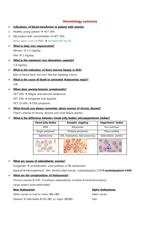
- 1. Hematology summary Indications of blood transfusion in patient with anemia: 1. Healthy young patient HCT 20% 2. Old patient with comorbidities HCT 30% Note: every I unit of PRBC increase HCT by 3% What is daily iron requirements? Woman 2-3 mg/day Man 1 mg/day What is the maximum iron absorption capacity? 3-4 mg/day What is the indication of bone marrow biopsy in IDA? IDA on blood tests, but can’t find the bleeding source. What is the cause of death in untreated thalassemia major? CHF When does anemia become symptomatic? HCT 30% fatigue and exercise intolerance HCT 25% tachypnea and dyspnea HCT 15-20% CNS symptoms What should you always remember about anemia of chronic disease? There’s anemia of chronic disease and renal failure anemia What is the difference between howel jolly bodies and pappinhemir bodies? Howel jolly bodies Basopilic stippling Pappinhemir bodies DNA Ribosomes Iron overload Single peripheral Multiple peripheral Many variable Splenectomy IDA, thalassemia, lead poisoning Sideroblastic anemia What are causes of sideroblastic anemia? Congenital aminolevulinic acid synthase or B6 metabolism Aquiredchloramphinicol, INH, alcohol, lead toxicity, myelodysplasia, CTDmyelodysplasiaAML What are the complications of thalassemia? Chronic anemia & CHF. Transfusion dependence; cirrhosis & hemochromatosis. Large spleen, bone deformities. Beta thalassemia: Alpha thalassemia: Silent carrier or trait or minor B(B+/B0) Silent carrier Disease intermedia B+(B+/B0) or major (B0/B0) trait
- 2. Disease 1 gene deleted 2 genes deleted 3 genes deleted Alpha thal Normal clinical & labs Mild microcytic anemia; HCT 30-40 Moderate anemia; HCT 20-30 Beta thal Normal clincal & labs Severe microcytic anemia st 6 months with growth failure -- What parasite causes B12 defeciency? Tapewarm; diphyllobothrium latum What are the causes of folate deficiency? Eczema, dialysis, anticonvulsants, alcohol. How long does the liver store B12 and folate? B12 3 yrs, folate 3 months At what levels of the spinal cord does B12 damage? Posterior and lateral spinocortical, and spinocerebellar. But can cause anything. Note: methylmalonate & homocystine elevated in B12 def. only homocystine elevated in folate def. What are the causes of hemolytic anemia? Acute medication, autoimmune, G6PD Chronic RBC abnormalities, PNH Anemia Picture Diagnosis Treatment IDA Glossitis, brittle nails, poikilonychia, pica Iron studies; blood smear Bone marrow biopsy Oral ferrous sulfate Parenteral; Best: BT (not used) Chronic dx Same as any anemia Iron studies Iron and erythropiotin only if renal dx or cancer Tx sideroblastic Same as any anemia Iron studies, prussian blue; pappinhemir bodies, BM RE iron high Tx cause, B6 2-4 mg, iron chelation Thalassemia Same as any anemia Iron studies, electropheresis: ↑H, A2 in beta thalassemia, target cells. Crew-cut on skull xray B major BT 1-2/month. Splenectomy +/- BMT B12 defect Neurologic signs, glossitis, diarrhea, abd pain Blood smear, ↓retic, hypercellular BM B12 and methylmallonate & homocystine levels. Schilling test B12 + Folate therapy; watch for HYPOKALEMIA Folate defect Same as any anemia Same as B12 defeciency Folate replacement Hemolysis Same as anemia, jaundice & dark urine Rapid fever, chills, chest & back pain, ↑HR Labs, blood smear, urine has hemosiderin not bilirubin Hydration to protect kidneys
- 3. Sickle cell Painful ↓O2, dehydration, ↓pH, fever, infection Aplasticspleen sequester, G6PD, B19, folate def Hemolytic, Vasoocclusive, sequestration. Affects all symptoms ↑retic unless folate defec/B19 Electropheresis, blood in urine ↑WBC, sickle prep Fluid, analgesics, O2, ceftriaxone Severe BT / exchange transfusion Folate + vaccine pneumo+influenza Prophylactic penicillin 4 mnth -6 yrs. Hydroxyurea. Autoimmune Fever, syncope, CHF, hemoglobinuria, cyanosis Coombs, cold agglutinin, spherocyte Steroid splenectomy immunomodulators If CD20 rituximab Spherocytosi s Splenomegaly, cholecystitis in young Osmotic fragility test Splenectomy PNH Venous thrombosis (budd-chiari); early morning Sugar-water &Ham test, ↓DAF Most accurate flow cytometry for CD55,59 Iron, steroid, anticoagulation, eculizumab, BMT G6PD Jaundice, dark urine, weakness, tachycardia Heinz bodies; bite cells. G6PD level Hydration and transfusion Aplastic anemia Pancytopenia Pancytopenia+empty bone marrow BMT, if not possible immunosupressio antithymocyte globn+sterod+cyclosporin Aplastic crisis Blood transfusion Recovery in a week Painful crisis Self-limited Recovery in a week Dactylitis in children -- -- Acute chest sx -- -- Splenic infarcts Splenomegaly in childhood. Not palbable at 4 yrs Avascular necrosis -- -- Priapism Nifidipine/hydralazine Self limited erection for 30 min to 3 hr after cold, exercise, void Sustained priapism Medical ER Erection > 3 hrs Papillary necrosis Sel-limited -- What are the causes of autoimmune hemolytic anemia? 1. Blooc cancer lymphoma, CLL, leukemia 2. Viral infections 3. CTDs 4. Medications antibiotics, thiazide, methyldopa 5. UC Which type of AIHA is more common? Warm >> cold Warm AIHA Cold AIHA medications, CTDs, cancer (lympphoma, CLL) Mycoplasma & IM Intravascular Extravascular Splenomegaly Liver IgG IgM
- 4. Direct coombs +ve & spherocytes Complement-coated RBCs OR cold agglutinin titer Steroid & splenectomy Avoid cold & chemotherapy What are the causes of cold-agglutnin hemolytic anemia? 1. Mycoplasma, mononucleosis 2. Lymphoma, waldenstorm macroglubinemia What are the complications of PNH? Aplastic anemia and leukemia What is the cause of death in PNH? Budd-chiari syndrome What are the excerbation factors of G6PD? Infections, medications (sulfa, nitrofurantoin, malaria, dapsone), fava beans What is the typical picture of aplastic anemia? Pancytopenia, empty bone marrow What is the benefit of hydroxyurea in SCD? Reduce recurrence of painful crises, and accelerated leg ulcers healing What are the indications of blood transfusion in SCD? CNS, LUNG, HEART, PRIAPISM What are the causes of spherocytosis? Congenital, G6PD, fever, autoimmune hemolytic anemia, ABO incompatibility What are the causes of aplastic anemia? 1. Radiation and chemotherapy 2. Benzene 3. NSAIDs, chloramphinocol 4. Infections CMV, EBV, B19, CMV, hepatits, TB 5. Cancer infiltration What are the causes of acute leukemia? 1. Aplastic anemia 2. Down and klenfilter syndrome 3. Myleodysplasia 4. Sideroblastic anemia 5. Retroviruses What is the pathogenesis of acute leukemia? Normal blood cells number, non functional (functional pancytopenia) What translocation is associated with APL? t(15:17) pancytopenia start ATRA immidiately (very sick patient) What are the poor prognostic factors in ALL? 2<age<9. WBC>10^5. CCNS involvement.
- 5. ALL AML Children Adults Can’t ddx clinically Extramedullary infiltration Extramedullary infiltration Leucostasis causes vasocclusion pheresis+chemo Leucostasis causes vasocclusion Pancytopenia Pancytopenia Leukemic blasts on smear Leukemic blasts on smear BM > 20% blasts BM > 20% blasts CALLA, TdT Auer bodies, myeloperoxidase, esterase Remission induction consolidation Relapse BMT (the leukemia most responsive to Tx) Remission induction consolidation Relapse BMT Cystosine arabinoside+daunorubicin/idarubicin Daunorubicin, vincristine, prednisone Intrathecal methotrexate to CNS prevention Testicular disease Skin nodules CML CLL Functional myeloid cells Mature B cells Philadelphia chromosome CD19; smudge cells neutrophilia (left shift) + B symptom + massive splenomegaly + bone No LN, no bleeding or infection Lymphocytosis/LN/splenomegaly/anemia/↑platelets No bleeding or infections; autoimmune hemolysis Leukocostasis 200-500.000 vaso-occlusion -- Imatinib Stage 0-1 nothing Stage 3-4 fludarabine +/- rituximab ITP/hemolysis Basophilia is present in all myeloproliferative disorders What chromosome is associated with MDS? 5q What is MDS picture? Pancytopenia + blasts 1-20% Leads to AML Bi-lobed neutrophil (pulger-heut) Tx: peroidic transfusion TNF inhibitors: lenalidomide/thalidomide 5q: azacitidine/decitabine Young patient BMT What is multiple myeloma picture? Picture Bone pain + infections + renal failure Hypercalcemia + anemia + leucostasis Diagnosis protein electropheresis + skeletal xray + B2 microglobulin + calcium level + KFT + urinanalysis in acid urine + bone marrow biopsy (>10% plasma cells)
- 6. ** note: albumin level is low in multiple myeloma Criteria: plasma cells > 10% + (M protein in plasma/urine/lytic bone lesions) Tx a. <70 yrs BMT + thalidomide + dexamethasone (DON’T GIVE CHEMO TX if considering BMT) b. > 70 yrs mephalan + prednisone c. Hypercalcemia fluid + duiretics + bisphosphonates DDx : MGUS bone marrow plasma cells < 10% protein electopheresis IgG spike <3 g bence jones protein < 1 g/24 hr no end-organ damage What is the most common cause of death in MM? Infections (lung or UTI) What complication of MM is a medical emergency? Cord compression by fracture or plasmacytoma immidiate steroid MM CRAB Skeletal xray+plasma cells>10%+bencejones+electropheresis IgG>3 BMT/chemo+steroid Waldenstorm LN/splenomegaly/hyperviscosity/neuro No bone lesions+IgM>5+bence jones Chemo+plasmapheresis MGUS Asymptomatic plasmacytosis No end-organ+bence<1g/d+IgG<3+plasma cells<10 -- Hodgkin’s Non-hodgkin’s Cancer of lymphocytes in LN; centripetal Cancer of lymphocytes in LN and extranodal Reed-sternberg No reed-sternberg Idiopathic Infections; HIV, EBV, HTLV-1, Hep C, h.pylori +immunosupression+CTDs Palbable LN + B symtoms + Extranodal + pruritis Palbable LN + B symtoms + Extranodal + pruritis -- HIV, EBV burkitt, HIV immunoblastic lymphoma + CNS involvment Excisional biopsy + CT for staging + BM biopsy Same (BM biopsy is more important than in HL) Stage 1 & 2 radiation Stage 3 & 4 OR B symptoms ABVD >> MOPP Stage 1 & 2 radiation Stage 3 & 4 OR B symptoms R-CHOP (1st test for hep B & C) CNS radiation + CHOP Multiple myeloma Pancytopenia No LN No splenomegaly Hodgkin No pancytopenia LN Splenomegaly NHL Pancytopenia LN Splenomegaly Acute leukemia Pancytopenia LN Splenomegaly CML Pancytopenia LN Splenomegaly CLL Pancytopenia LN Splenomegaly What are the causes of ITP? 1. Blood cancer Lymphoma & CLL 2. Infections HIV
- 7. 3. CTDs How is ITP diagnosed? 1. Thrombocytopenia 2. Antiplatelet antibodies 3. Bone marrow full of platelets & exclude other causes What is the treatment of ITP? Prednisone splenectomy thrombopoietin agents ( romiplostim/eltrombopag) PLT < 10.000 IVIG in Rh +ve patients When type of thrombi are found in TTP? Hyaline thrombi of smallest vessels fatal if not plasmapheresis done What is the main complication of HIT? DVT How is HIT diagnosed? Anti-platelet factor IV or serotonin release assay What are the clues to diagnosis of BSS? Adherance by GPIb few large platelets. What are the clues to diagnosis of glanzman? Aggregation by GPIIb/IIIa normal platelet count What are the types of vWF disease? Type 1 Decreased count Desmopressin Factor 8 concentrate after surgery or trauma Type 2 Qualitative +/- desmopressin Factor 8 concentrate after surgery or trauma Type 3 Absent -- Factor 8 concentrate after surgery or trauma What are the sites of bleeding in hemophilia? 1. Hemarthrosis 2. Intracranial 3. Retroperitoneal 4. Hematuria & hemospermia 5. Intramuscular hematoma What is the treatment of hemophilia? Hemophilia A Factor 8 concentrate + desmopressin Hemophilia B Factor 9 concentrate What lab tests are used in hemophilia diagnosis? 1. PTT prolonged 2. Factor 8 or 9 level 3. vWF level 4. detection for factor inhibitor mix patient plasma with normal plasma check for PTT correction What are the normal ranges of coagulation studies? 1. PT 11-15 sec
- 8. 2. PTT 25-40 sec 3. BT 2-7 min How to ddx by labs between liver and vit K problems? 1. Liver disease PT and PTT prolonged 2. Vit K def. PT only Note: normal or elevated fibrinogen R/O DIC Antithrombin III def. AD Thrombosis Anti-phospholipid Aquired Arterial & venous thrombosis + thrombocytopenia Protein C AD Factor 5 & 8 Protein S - Cofactor for protein C Factor V leiden - -- Prothrombin mutation - -- hyperhomocysteinemia - -- When to suspect inherited hypercoagulable state? 1. Family Hx 2. Recurrent DVT 3. First episode before 40 4. DVT in unusual site (cerebral/mesentric/renal/IVC) Low grade Small lymphocytic B cell, resembles CLL Mycosis fungoides T cell, Eczema, cribriform lymphocytes Radio Tx Follicular small cleaved T(14,18). BCL2, may transform to diffuse large cell Radio Tx Extranodal MALT intermediate Follicular large cell -- Diffuse large cell B cell, T cell, mixed CHOP High grade Lymphoblastic T cell, children, ALL, CNS & skin Chemo Tx Adult T cell lymphoma HTLV-1, ↑Ca+2 Sezary syndrome T cell, blood (burkit or diffuse-large cell) Small non-cleaved (Burkit) t(8,14), c-myc, EBV Chemo Tx immunoblastic T or B cell