SlideShare a Scribd company logo
1 of 17
NEURON AND MUSCLE POTENTIALS 
BY 
UMAR SHU’AIBU 
12/15/2014
INTRODUCTION 
Muscle is a soft tissue found in most animals. they , produced a contraction that changes 
both the length and the shape of the cell. Muscles function to produce force 
and motion. They are primarily responsible for maintaining and 
changing posture, locomotion as well as movement of internal organs, such as the 
contraction of the heart and the movement of food through the digestive system. The 
Movement of the muscle generate potential known as muscle potential. 
The neuron also known as a nerve cell is an electrically excitable cell that processes and 
transmits information through electrical and chemical signals. neurons are the core 
components of the nervous system. neuron respond to touch, sound, light and all other 
stimuli affecting the cells of the sensory organs that then send signals to the spinal cord 
and brain.
NEURON POTENTIAL 
The neuron potential result from the electrochemical activity of excitable cells 
also known as neuron. These cells are surrounded by body fluids having a high 
Cl concentration. Neuron acts as a constant current source when stimulated, 
and creates an ionic current within the body fluid. This current induces 
electrical potentials within the human body. This potential decrease in 
amplitude with increasing distance from the excitable cell. 
Resting neurons maintain a difference in charge across their cell membrane with 
negative charges inside, and a positive charges outside. When neuron is 
stimulated this polarity is reversed. The neuron potential is divided into 
resting potential and action potential which can be explain as follow
NEURON RESTING POTENTIAL 
Resting membrane potential is the difference in voltage of the fluids inside a cell 
and outside a cell, which is usually between -70 to -80 millivolts (mV). All 
cells have this difference, but it is particularly important in relation to nerve and 
muscle cells, since any stimulus that changes the voltage and makes it different 
from the resting membrane potential is what allows the cells to transmit 
electrical signals. If resting potential rises above threshold, an action potential 
starts to travel from cell body down the axon
5 
RESTING POTENTIAL 
OUTSIDE 
Cl- 
+ + + + + + + + + + + + + + + + + + + + + + + + + + + + + + + + + + + + + + + + + + + 
- - - - - - - - - - - - - - - - - - - - - - - - - - - - - - - - - - - - - - - - - - - - - - - - - - - - -- - - - - - - 
Force of Diffusion 
INSIDE 
Na+ 
Na 
+ 
K+ 
K+ 
K+ = Potassium; Na+ = Sodium; Cl- = Chloride; 
Force of Diffusion 
Cl- 
Closed 
channel 
open 
channel 
open 
channel 
no 
channel 
3Na/2K 
pump 
- 65 mV
INITIALIZATION OF ACTION POTENTIAL 
Stimulation of a neuron opens some of the membrane proteins (a.k.a. Na+gates) 
allows to pass freely into the cells free flow of Na+ into the cell causes a 
reversal of membrane polarity polarity reversal is called the action 
potential Stimulation of neuron due to environmental changes opens some of 
the membrane protein there by allowing Na+ to pass feely into the cell. The 
free flow of into the cell causes a reversal of membrane polarity. This 
polarity reversal is called action potential. 
This result to the changes in potential from -70mV to about 40mV. spike in voltage 
causes the K+ gate open and Na+ gate to close . This make K+ ions rush out of 
the neuron. The inside becomes negative again. This is repolarization. 
So many K+ ions get out that the charge goes below the resting potential. While the 
neuron is in this state it cannot react to additional stimuli. This is termed as 
refractory period which last for about 0.5.
The sequence of depolarization and repolarization generates a small electrical current 
in this localized area. The current affects the nearby protein channels for Na+ and 
causes them to open. When the adjacent channels open, Na+ions flood into that area 
of the neuron and an action potential occurs. This in turn will affect the areas next 
to it and the impulse passes along the entire neuron. The electric current pas.ses 
outward over the membrane in all directions.
MUSCLE POTENTIAL 
The action potential will continue to flow along the axon until it reaches 
neuromuscular junction as can be shown below
The neuromuscular junction consist of two part. Ie presynaptic terminal on the side of 
the axon, and the postsynaptic terminal on the side of the muscle, between them 
existed a gap called synaptic cleft. To this end, a chemical known as acetylcholine 
will be released( also known as neurotransmitter) this will allow the action potential 
to cross the gap and move to the target muscle fibre.
MUSCLE FIBRE EXCITATION 
Each muscle fibre has one neuromuscular junction, receiving input from just 
one efferent neuron . The neuro transmitter allows Na+ ions to enter the 
cell, causing a depolarizing excitatory postsynaptic potential (EPSP) that is 
above the threshold potential, this triggered an action potential in the muscle 
fibre. the EPSP is always well above threshold. This means that under normal 
circumstances, an action potential in a somatic efferent neuron always elicits an 
action potential in the muscle fibre. Consider the diagram below.
The above figure shows a muscle cell EPSP in response to a single action 
potential in a somatic efferent neuron . The amount of Ach(neuro transmitter) 
released with one neuronal action potential is enough to depolarize the muscle 
fibre well above the threshold for eliciting an action potential. The degree that 
the EPSP exceeds threshold is known as the safety factor. 
The term 'safety factor' refers to the ability of neuromuscular transmission to 
remain effective under various physiological conditions and stresses.
This is a result of the amount of transmitter released per nerve impulse being 
greater than that required to trigger an action potential in the muscle fibre. 
The safety factor is a measure of this excess of released transmitter. This 
means that any action potential in the neuron will be enough to triggered 
action potential in the muscle fibre(also known as sarcolemma). 
a single action potential in a motor neuron can activate hundreds of muscle 
fibers in synchrony, the resulting currents sum to generate a potential known 
as the compound action potential, which is the summed action potentials 
of all the muscle fibers in the motor unit. This is also termed as the muscle 
potential as can be shown in the figure below.
The action potential in the muscle will cause the muscle contraction( also known as 
twitch). The twitch is divided into three period. 
 1. Latent period 
brief delay between the stimulus and the muscle contraction The latent period is 
less than 2milliseconds in humans
2. Period of contraction 
3. Period of relaxation 
This can be as shown in the figure below
If the muscle is allowed to relax completely before each stimulus than the muscle 
will contract with the same force. 
 If the muscle is stimulated again before it has completely relaxed, then the force 
of the next contraction increases. 
i.e. stimulating the muscle at a rapid frequency increases the force of contraction. 
This is called summation
Neuron and muscle potential

More Related Content

What's hot

Kinetics and kinematics of gait
Kinetics and kinematics of gaitKinetics and kinematics of gait
Kinetics and kinematics of gait
Athul Soman
 
Biomechanics of knee complex 6 tibiofemoral jt function
Biomechanics of knee complex 6 tibiofemoral jt functionBiomechanics of knee complex 6 tibiofemoral jt function
Biomechanics of knee complex 6 tibiofemoral jt function
Dibyendunarayan Bid
 

What's hot (20)

Bpo(1)
Bpo(1)Bpo(1)
Bpo(1)
 
Hand splinting in common orthopedic & neurological condition 1
Hand splinting in common orthopedic & neurological condition 1Hand splinting in common orthopedic & neurological condition 1
Hand splinting in common orthopedic & neurological condition 1
 
Posture Analysis In Biomechanics
Posture Analysis In BiomechanicsPosture Analysis In Biomechanics
Posture Analysis In Biomechanics
 
Biomechanics of hip complex 2
Biomechanics of hip complex 2Biomechanics of hip complex 2
Biomechanics of hip complex 2
 
Kinship, marriage and the household
Kinship, marriage and the householdKinship, marriage and the household
Kinship, marriage and the household
 
Pictorial essay ni caren ramos
Pictorial essay ni caren ramosPictorial essay ni caren ramos
Pictorial essay ni caren ramos
 
Kinetics and kinematics of gait
Kinetics and kinematics of gaitKinetics and kinematics of gait
Kinetics and kinematics of gait
 
CERVICAL BIOMECHANICS.pptx
CERVICAL BIOMECHANICS.pptxCERVICAL BIOMECHANICS.pptx
CERVICAL BIOMECHANICS.pptx
 
Human cultural variation
Human cultural variationHuman cultural variation
Human cultural variation
 
Biomechanics of knee complex 6 tibiofemoral jt function
Biomechanics of knee complex 6 tibiofemoral jt functionBiomechanics of knee complex 6 tibiofemoral jt function
Biomechanics of knee complex 6 tibiofemoral jt function
 
motor point
 motor point motor point
motor point
 
Iba’t Ibang Uri ng Akademikong Sulatin.pptx
Iba’t Ibang Uri ng Akademikong Sulatin.pptxIba’t Ibang Uri ng Akademikong Sulatin.pptx
Iba’t Ibang Uri ng Akademikong Sulatin.pptx
 
Antas ng Pagbasa
Antas ng PagbasaAntas ng Pagbasa
Antas ng Pagbasa
 
Biomech lumbar spine
Biomech lumbar spineBiomech lumbar spine
Biomech lumbar spine
 
Orthoses for shoulder, elbow and forearm
Orthoses for shoulder, elbow and forearmOrthoses for shoulder, elbow and forearm
Orthoses for shoulder, elbow and forearm
 
Principles of Biomechanics and Exercise
Principles of Biomechanics and ExercisePrinciples of Biomechanics and Exercise
Principles of Biomechanics and Exercise
 
Prehension
PrehensionPrehension
Prehension
 
Biomechanics of wrist joint
Biomechanics of wrist joint Biomechanics of wrist joint
Biomechanics of wrist joint
 
Sitting to Standing Mechanism and Osteoarthritis
Sitting to Standing Mechanism and OsteoarthritisSitting to Standing Mechanism and Osteoarthritis
Sitting to Standing Mechanism and Osteoarthritis
 
Joint lubrication by dr debasis mukherjee
Joint lubrication by dr debasis mukherjeeJoint lubrication by dr debasis mukherjee
Joint lubrication by dr debasis mukherjee
 

Similar to Neuron and muscle potential

Lab Report- Neurophysiology Of Nerve Impulses Essay
Lab Report- Neurophysiology Of Nerve Impulses EssayLab Report- Neurophysiology Of Nerve Impulses Essay
Lab Report- Neurophysiology Of Nerve Impulses Essay
Stephanie King
 
HAP 1 anatomy physiology and pathophysio
HAP 1 anatomy physiology and pathophysioHAP 1 anatomy physiology and pathophysio
HAP 1 anatomy physiology and pathophysio
christinajohn24
 
cape biology unit 2-_fundamentals_of_the_nervous_system
cape biology unit 2-_fundamentals_of_the_nervous_systemcape biology unit 2-_fundamentals_of_the_nervous_system
cape biology unit 2-_fundamentals_of_the_nervous_system
Hilton Ritch
 
Nervous system review
Nervous system reviewNervous system review
Nervous system review
eruder
 
Homeostasis Topic 6.5
Homeostasis Topic 6.5 Homeostasis Topic 6.5
Homeostasis Topic 6.5
Bob Smullen
 

Similar to Neuron and muscle potential (20)

Presentation EXCITABLE TISSUES.pptx
Presentation EXCITABLE TISSUES.pptxPresentation EXCITABLE TISSUES.pptx
Presentation EXCITABLE TISSUES.pptx
 
Lab Report- Neurophysiology Of Nerve Impulses Essay
Lab Report- Neurophysiology Of Nerve Impulses EssayLab Report- Neurophysiology Of Nerve Impulses Essay
Lab Report- Neurophysiology Of Nerve Impulses Essay
 
Biology notes - topic 8
Biology notes -  topic 8Biology notes -  topic 8
Biology notes - topic 8
 
HAP 1 anatomy physiology and pathophysio
HAP 1 anatomy physiology and pathophysioHAP 1 anatomy physiology and pathophysio
HAP 1 anatomy physiology and pathophysio
 
Nerve physiology
Nerve physiologyNerve physiology
Nerve physiology
 
cape biology unit 2-_fundamentals_of_the_nervous_system
cape biology unit 2-_fundamentals_of_the_nervous_systemcape biology unit 2-_fundamentals_of_the_nervous_system
cape biology unit 2-_fundamentals_of_the_nervous_system
 
Nerve Muscle Physiology
Nerve Muscle PhysiologyNerve Muscle Physiology
Nerve Muscle Physiology
 
Neuron communication
Neuron communicationNeuron communication
Neuron communication
 
lecturei-150109042144-conversion-gate01.pdf
lecturei-150109042144-conversion-gate01.pdflecturei-150109042144-conversion-gate01.pdf
lecturei-150109042144-conversion-gate01.pdf
 
Nervous system review
Nervous system reviewNervous system review
Nervous system review
 
Neurophysiology kritika
Neurophysiology kritikaNeurophysiology kritika
Neurophysiology kritika
 
Neural control and coordination
Neural control and coordinationNeural control and coordination
Neural control and coordination
 
Types of communication
Types of communicationTypes of communication
Types of communication
 
Neuro physiology notes
Neuro physiology notesNeuro physiology notes
Neuro physiology notes
 
GROUP6.pptx
GROUP6.pptxGROUP6.pptx
GROUP6.pptx
 
types of action-pot-B.Sc 18 - Copy (2).pptx
types of action-pot-B.Sc 18 - Copy (2).pptxtypes of action-pot-B.Sc 18 - Copy (2).pptx
types of action-pot-B.Sc 18 - Copy (2).pptx
 
Nerve
NerveNerve
Nerve
 
rafractory period.pptx
rafractory period.pptxrafractory period.pptx
rafractory period.pptx
 
conduction of nerve impulse & synaps
conduction of nerve impulse & synapsconduction of nerve impulse & synaps
conduction of nerve impulse & synaps
 
Homeostasis Topic 6.5
Homeostasis Topic 6.5 Homeostasis Topic 6.5
Homeostasis Topic 6.5
 

More from Umar Shuaib (8)

Intelligent travelling and home automation aid, for visually impaired
Intelligent travelling and home  automation aid, for visually impairedIntelligent travelling and home  automation aid, for visually impaired
Intelligent travelling and home automation aid, for visually impaired
 
Zigbee based dc motors remote monitoring and control
Zigbee based dc motors remote monitoring and controlZigbee based dc motors remote monitoring and control
Zigbee based dc motors remote monitoring and control
 
Assistive technologies and devices for visually impaired
Assistive technologies and devices for visually impairedAssistive technologies and devices for visually impaired
Assistive technologies and devices for visually impaired
 
Analog to digital conversion technique
Analog to digital conversion techniqueAnalog to digital conversion technique
Analog to digital conversion technique
 
Passive infrared sensor technology(pir)
Passive infrared sensor technology(pir)Passive infrared sensor technology(pir)
Passive infrared sensor technology(pir)
 
Dc motor interfacing with 8051 microcontroller
Dc motor interfacing with 8051 microcontrollerDc motor interfacing with 8051 microcontroller
Dc motor interfacing with 8051 microcontroller
 
Biopotential generation
Biopotential generationBiopotential generation
Biopotential generation
 
Serial buses
Serial busesSerial buses
Serial buses
 

Neuron and muscle potential

  • 1. NEURON AND MUSCLE POTENTIALS BY UMAR SHU’AIBU 12/15/2014
  • 2. INTRODUCTION Muscle is a soft tissue found in most animals. they , produced a contraction that changes both the length and the shape of the cell. Muscles function to produce force and motion. They are primarily responsible for maintaining and changing posture, locomotion as well as movement of internal organs, such as the contraction of the heart and the movement of food through the digestive system. The Movement of the muscle generate potential known as muscle potential. The neuron also known as a nerve cell is an electrically excitable cell that processes and transmits information through electrical and chemical signals. neurons are the core components of the nervous system. neuron respond to touch, sound, light and all other stimuli affecting the cells of the sensory organs that then send signals to the spinal cord and brain.
  • 3. NEURON POTENTIAL The neuron potential result from the electrochemical activity of excitable cells also known as neuron. These cells are surrounded by body fluids having a high Cl concentration. Neuron acts as a constant current source when stimulated, and creates an ionic current within the body fluid. This current induces electrical potentials within the human body. This potential decrease in amplitude with increasing distance from the excitable cell. Resting neurons maintain a difference in charge across their cell membrane with negative charges inside, and a positive charges outside. When neuron is stimulated this polarity is reversed. The neuron potential is divided into resting potential and action potential which can be explain as follow
  • 4. NEURON RESTING POTENTIAL Resting membrane potential is the difference in voltage of the fluids inside a cell and outside a cell, which is usually between -70 to -80 millivolts (mV). All cells have this difference, but it is particularly important in relation to nerve and muscle cells, since any stimulus that changes the voltage and makes it different from the resting membrane potential is what allows the cells to transmit electrical signals. If resting potential rises above threshold, an action potential starts to travel from cell body down the axon
  • 5. 5 RESTING POTENTIAL OUTSIDE Cl- + + + + + + + + + + + + + + + + + + + + + + + + + + + + + + + + + + + + + + + + + + + - - - - - - - - - - - - - - - - - - - - - - - - - - - - - - - - - - - - - - - - - - - - - - - - - - - - -- - - - - - - Force of Diffusion INSIDE Na+ Na + K+ K+ K+ = Potassium; Na+ = Sodium; Cl- = Chloride; Force of Diffusion Cl- Closed channel open channel open channel no channel 3Na/2K pump - 65 mV
  • 6. INITIALIZATION OF ACTION POTENTIAL Stimulation of a neuron opens some of the membrane proteins (a.k.a. Na+gates) allows to pass freely into the cells free flow of Na+ into the cell causes a reversal of membrane polarity polarity reversal is called the action potential Stimulation of neuron due to environmental changes opens some of the membrane protein there by allowing Na+ to pass feely into the cell. The free flow of into the cell causes a reversal of membrane polarity. This polarity reversal is called action potential. This result to the changes in potential from -70mV to about 40mV. spike in voltage causes the K+ gate open and Na+ gate to close . This make K+ ions rush out of the neuron. The inside becomes negative again. This is repolarization. So many K+ ions get out that the charge goes below the resting potential. While the neuron is in this state it cannot react to additional stimuli. This is termed as refractory period which last for about 0.5.
  • 7. The sequence of depolarization and repolarization generates a small electrical current in this localized area. The current affects the nearby protein channels for Na+ and causes them to open. When the adjacent channels open, Na+ions flood into that area of the neuron and an action potential occurs. This in turn will affect the areas next to it and the impulse passes along the entire neuron. The electric current pas.ses outward over the membrane in all directions.
  • 8.
  • 9. MUSCLE POTENTIAL The action potential will continue to flow along the axon until it reaches neuromuscular junction as can be shown below
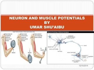
  • 10. The neuromuscular junction consist of two part. Ie presynaptic terminal on the side of the axon, and the postsynaptic terminal on the side of the muscle, between them existed a gap called synaptic cleft. To this end, a chemical known as acetylcholine will be released( also known as neurotransmitter) this will allow the action potential to cross the gap and move to the target muscle fibre.
  • 11. MUSCLE FIBRE EXCITATION Each muscle fibre has one neuromuscular junction, receiving input from just one efferent neuron . The neuro transmitter allows Na+ ions to enter the cell, causing a depolarizing excitatory postsynaptic potential (EPSP) that is above the threshold potential, this triggered an action potential in the muscle fibre. the EPSP is always well above threshold. This means that under normal circumstances, an action potential in a somatic efferent neuron always elicits an action potential in the muscle fibre. Consider the diagram below.
  • 12. The above figure shows a muscle cell EPSP in response to a single action potential in a somatic efferent neuron . The amount of Ach(neuro transmitter) released with one neuronal action potential is enough to depolarize the muscle fibre well above the threshold for eliciting an action potential. The degree that the EPSP exceeds threshold is known as the safety factor. The term 'safety factor' refers to the ability of neuromuscular transmission to remain effective under various physiological conditions and stresses.
  • 13. This is a result of the amount of transmitter released per nerve impulse being greater than that required to trigger an action potential in the muscle fibre. The safety factor is a measure of this excess of released transmitter. This means that any action potential in the neuron will be enough to triggered action potential in the muscle fibre(also known as sarcolemma). a single action potential in a motor neuron can activate hundreds of muscle fibers in synchrony, the resulting currents sum to generate a potential known as the compound action potential, which is the summed action potentials of all the muscle fibers in the motor unit. This is also termed as the muscle potential as can be shown in the figure below.
  • 14. The action potential in the muscle will cause the muscle contraction( also known as twitch). The twitch is divided into three period.  1. Latent period brief delay between the stimulus and the muscle contraction The latent period is less than 2milliseconds in humans
  • 15. 2. Period of contraction 3. Period of relaxation This can be as shown in the figure below
  • 16. If the muscle is allowed to relax completely before each stimulus than the muscle will contract with the same force.  If the muscle is stimulated again before it has completely relaxed, then the force of the next contraction increases. i.e. stimulating the muscle at a rapid frequency increases the force of contraction. This is called summation