SlideShare a Scribd company logo
1 of 10
Download to read offline
Section IX – Nuclear Radiology


                       Figure 1A                                              Figure 1B


366. A 34-year-old male presented with symptoms of irregular heartbeat and tremor. Thyroid function tests
     revealed an elevated serum T3, normal T4 and reduced TSH. The 24 hour I-123 thyroid uptake was < 1%.
     Based on these results and the accompanying I-123 thyroid images (Figure 1A: anterior image of neck with
     radioactive markers around the chin and at the sternal notch, and Figure 1B: anterior image of the neck
     without markers), which one of the following is the MOST likely diagnosis?
      A. Graves’ disease
      B.   Acute bacterial thyroiditis
      C. Plummer’s disease
      D. Painless thyroiditis
      E.   Recent iodinated contrast administration


                                   Diagnostic In-Training Exam 2002                                             1
Section IX – Nuclear Radiology
Question #366
Findings: There is near-complete non-visualization of the thyroid on I-123 imaging (Figures 1A & 1B), with only
faint visualization of the gland, which appears grossly normal in size. Thyroid morphology cannot accurately be
assessed.

Rationales:
A) Incorrect. While the patient’s symptoms and laboratory findings are consistent with hyperthyroidism, the
   markedly reduced I-123 thyroid uptake and near non-visualization of the thyroid on I-123 imaging are not
   consistent with Graves’ disease, in which an elevated uptake and an enlarged thyroid with diffusely increased
   uptake on thyroid imaging would be expected.
B) Incorrect. Acute bacterial thyroiditis presents with fever, elevated white blood cell count and focal tenderness
   over a portion of the gland. The thyroid uptake is variable, and thyroid imaging would most likely demonstrate
   a focal hypofunctioning (“cold”) nodule, with normal visualization of the remainder of the gland.
C) Incorrect. Plummer’s disease is toxic nodular goiter. The thyroid uptake may be normal or mildly increased in
   this disorder, but would not be decreased. I-123 thyroid imaging in Plummer’s disease demonstrates one or
   more focal areas of increased tracer uptake, with associated areas of suppression of uptake in other parts of the
   gland, findings which are not present in this case.
D) Correct. Subacute or painless thyroiditis is the most likely etiology for these findings. In this viral disorder,
   there is diffuse or focal inflammation of the gland, with release of pre-formed thyroid hormone into the
   circulation during the acute phase, resulting in signs and symptoms of hyperthyroidism and elevated thyroid
   function tests, as in this case. There is markedly reduced synthesis of new thyroid hormone by the gland, with
   associated decreased thyroid uptake and poor visualization of the thyroid on I-123 or Tc-99m pertechnetate
   thyroid imaging. These findings may also be seen in thyrotoxicosis factitia (intake of exogenous thyroid
   hormone) or in patients with ectopic thyroid hormone production (eg. struma ovarii), but these conditions
   were not listed as possible answers.
E) Incorrect. Recent iodinated contrast administration may result in a falsely low I-123 thyroid uptake
   measurement, secondary to flooding of the extracellular iodine pool with non-radioactive iodine. In turn, poor
   visualization or non-visualization of the thyroid on thyroid scintigraphy may result. However, in most cases,
   these findings would not be associated with clinical evidence of hyperthyroidism, as is present in this case.

Citations:
Lazarus JH: Silent Thyroiditis and Subacute Thyroiditis. in Werner and Ingbar’s The Thyroid, a Fundamental and Clinical Text,
7th Edition, (Braverman LE and Utiger RD, eds.), Lippincott-Raven, Philadelphia, 1996, pp. 577-591.
Mettler FA, Jr. and Guiberteau MJ: Essentials of Nuclear Medicine Imaging, 4th edition, WB Saunders Co., Philadelphia, 1998,
pp. 117-118


2                                          American College of Radiology
Section IX – Nuclear Radiology


                                                 Figure 2A


                    Figure 2B                                                 Figure 2C

367. A 57-year-old male presented with a 4 cm right upper lobe pulmonary mass on an outside chest radiograph
     and chest CT scan. The CT study demonstrated no evidence of mediastinal or hilar adenopathy. You are
     shown representative coronal, transaxial and sagittal images from an F-18 fluorodeoxyglucose (FDG) PET
     scan (Figure 2A) and the complete series of coronal images (Figures 2B and 2C). The lesion demonstrated a
     standardized uptake value (SUV) of 7.2. Based upon these findings, which one of the following is the MOST
     likely diagnosis?
      A. Stage IIIA squamous cell bronchogenic carcinoma
      B.   Stage IB squamous cell bronchogenic carcinoma
      C. MALT (mucosa-associated lymphoid tissue) lymphoma
      D. Hamartoma
      E.   Bronchoalveolar carcinoma


                                 Diagnostic In-Training Exam 2002                                            3
Section IX – Nuclear Radiology
Question #367
Findings: There is a large focal area of markedly increased FDG uptake in the right upper lobe, corresponding
to the site of the patient’s known pulmonary nodule on prior imaging studies. There is no evidence of mediastinal
or hilar lymphadenopathy. No additional pulmonary nodules are identified. The remaining areas of FDG uptake
(eg. liver, heart, bowel, kidneys, bone marrow) represent normal sites of uptake.

Rationales:
A) Incorrect. The right upper lobe lesion is highly suspicious for bronchogenic carcinoma, especially in light of the
   SUV value of 7.2. However, in the absence of mediastinal or hilar adenopathy, this lesion is not consistent with
   Stage IIIA disease, which includes patients with primary lesions of any size, but only those with ipsilateral
   mediastinal or hilar adenopathy.
B) Correct. On the basis of this PET scan, this patient has a T2 N0 M0 lesion, which is consistent with Stage 1B
   involvement. Despite its 4 cm size, only a primary lesion with evidence of invasion of the mediastinum, heart,
   great vessels, trachea, esophagus, vertebral body, carina or lesions associated with additional tumor nodules or
   malignant pleural effusions can be placed into the category of Stage III disease without evidence of adenopathy.
C) Incorrect. Lymphomas are in general very FDG-avid. However, the MALT type lesion has been shown to be
   much less FDG-avid than other cell types, and FDG PET imaging is not recommended for patients with this
   disorder. Furthermore, a large focal lung nodule would represent an unusual manifestation of lymphoma in any
   case, even for FDG-avid cell types.
D) Incorrect. Mildly increased FDG uptake may occur in infectious and granulomatous processes, including
   histoplasmosis, tuberculosis and others. Such occurrences may result in false positive PET scans for
   bronchogenic carcinoma. However, FDG uptake in such lesions is usually mild, typically with SUV values
   < 2.5, and in any event, such a diagnosis is much less likely than bronchogenic carcinoma, given the findings
   in this case.
E) Incorrect. Bronchoalveolar carcinoma demonstrates variable FDG uptake, and has been reported to be
   a common cause of a false negative FDG PET scan in bronchogenic carcinoma. The presentation of a
   solitary, large focal pulmonary nodule is not the most common appearance for bronchoalveolar carcinoma.
   Furthermore, it is less common than other cell types of bronchogenic carcinoma, and this diagnosis would
   therefore be less likely than other cells types of non-small cell or small cell carcinoma.

Citations:
Bar-Shalom R, et. al.: PET Imaging in Oncology. Semin Nucl Med 2000; 30:150-185.
Hoffmann M. et. al.: Positron emission tomography with fluorine-18-2-fluoro-deoxy-D-glucose (F18-FDG) does not visualize
extranodal B-cell lymphoma of the mucosa-associated lymphoid tissue (MALT)-type. Annals Oncol 1999; 10:1185-1189.


4                                         American College of Radiology
Section IX – Nuclear Radiology


                   Figure 3A                                                  Figure 3B


                                                                            Figure 3D
                        Figure 3C

368. A 43-year-old female with a history of ovarian carcinoma presented with shortness of breath and low-grade
     fever following a long airplane ride. You are shown posterior Xe-133 ventilation images (Figure 3A), Tc-99m
     MAA perfusion images (Figure 3B) and a concurrent PA and lateral chest radiograph (Figures 3C and 3D).
     Which one of the following BEST characterizes the overall findings in this case?
      A. High probability for pulmonary embolism
      B.   Intermediate probability for pulmonary embolism
      C. Low probability for pulmonary embolism
      D. Normal study
      E.   Lymphangitic carcinomatosis

                                  Diagnostic In-Training Exam 2002                                             5
Section IX – Nuclear Radiology
Question #368
Findings: The Xe-133 ventilation study (Figure 3A) demonstrates only mild xenon retention bilaterally, most
striking at the lung bases. The Tc-99m MAA perfusion images (Figure 3B) demonstrate a single moderate
subsegmental perfusion defect in the lateral basal segment of the left lower lobe, which does not correspond to a
focal ventilatory defect on the ventilation study. The chest radiograph demonstrates no focal infiltrates or pleural
effusions.

Rationales:
A) Incorrect. The V/Q scan findings of a single moderate ventilation-perfusion mismatch are not consistent with
   a high probability for pulmonary embolism, using any of the commonly employed criteria for interpretation
   of these studies. Both the modified PIOPED criteria and modified Biello criteria categorize such cases as
   intermediate probability for pulmonary embolism.
B) Correct. As discussed in item A above, a single moderate V/Q mismatch is consistent with an intermediate
   probability for pulmonary embolism using the two most prevalent sets of diagnostic criteria currently in use.
   In published retrospective and prospective studies, approximately one-third of such cases have been associated
   with pulmonary embolism on pulmonary angiography.
C) Incorrect. Again, a single moderate subsegmental V/Q matching defect is consistent with an intermediate
   probability for pulmonary embolism, both in the modified PIOPED and modified Biello diagnostic criteria.
   The original PIOPED study incorrectly classified these cases as low probability, an error which was corrected
   when the criteria were subsequently modified following the study.
D) Incorrect. This ventilation-perfusion lung scan demonstrates abnormalities on both the ventilation and
   perfusion portions of the exam. It is not a normal study.
E) Incorrect. The pattern of perfusion abnormality in this case is not suggestive of lymphangitic carcinomatosis.
   In that entity, the perfusion images typically produce a diffuse pattern of irregularity, with numerous small
   subsegmental perfusion defects, corresponding to the interlobular septae. This pattern of abnormality has
   been termed “contour mapping”. It is not present in this case, nor is there an interstitial pattern of pulmonary
   parenchymal abnormality on the chest radiography to suggest this entity.

Citations:
Biello DR, et. al.: Ventilation-perfusion studies in suspected pulmonary embolism. Amer J Roentgenol 1979; 133:1033-1037.
Gottschalk A, et. al.: Ventilation-perfusion scintigraphy in the PIOPED study. Part ii. Evaluation of the scintigraphic criteria and
interpretations. J Nucl Med 1993; 1119-1126


6                                            American College of Radiology
Section IX – Nuclear Radiology


                                                                                 Figure 4C


      Figure 4A                       Figure 4B
                                                                                 Figure 4D


                        Figure 4E


                                                                                  Figure 4F

369. A 15-year-old female volleyball player presented with low back pain, without a specific incident of trauma.
     You are presented with AP and lateral radiographs of the lumbar spine (Figures 4A and 4B), anterior and
     posterior planar spot images (Figure 4C) and coronal, transaxial and sagittal SPECT images from a Tc-99m
     MDP radionuclide bone scan (Figures 4D - 4F). No other lesions were present elsewhere in the skeleton.
     Which one of the following is the MOST likely diagnosis?
      A. Stress fracture of the pars interarticularis
      B.   Osteoid osteoma
      C. Ewing’s sarcoma
      D. Vertebral osteomyelitis
      E.   Ankylosing spondylitis

                                    Diagnostic In-Training Exam 2002                                               7
Section IX – Nuclear Radiology
Question #369
Findings: The AP and lateral radiographs of the lumbar spine (Figures 4A and 4B) demonstrate very mild scoliosis
of the lower lumbar spine, possibly positional. Anterior and posterior planar Tc-99m MDP bone images (Figure
4C) demonstrate a focal area of increased tracer uptake at L5 on the left, best seen on the posterior image. The
coronal, transaxial and sagittal SPECT images (Figures 4D-4F) localize this finding to the region of the lamina, in
the expected location of the pars interarticularis.

Rationales:
A) Correct. The findings described above are typical for a stress fracture of the left pars interarticularis of L5,
   a common injury occurring in children involved in athletic activities, such as gymnastics, volleyball, etc.
   Radiographs may or may not demonstrate a pars defect, and in the case of bilateral pars defects, spondylolysis
   may also be present. SPECT imaging is useful in detecting subtle lesions not evident on planar views, as well
   as localizing the lesion to the region of the pars. Increased bone turnover in the site of the lesion suggests that
   it is the etiology for the patient’s symptoms of localized back pain.
B) Incorrect. Osteoid osteoma can occur in the spine, and may be associated with focal areas of increased uptake
   on bone scintigraphy. However, it is less likely to be associated with normal radiographs, usually presenting
   with a focal area of sclerosis, sometimes with a visible central radiolucent nidus. It commonly presents with
   rigid, painful scoliosis, and may have the classic presentation of pain, worse at night and relieved by aspirin.
   It is less common than a stress fracture of the pars, especially in the present clinical setting.
C) Incorrect. Ewing’s sarcoma is uncommon relative to stress fractures, and the vertebral column is an uncommon
   site of involvement. When arising in the vertebral column, the sacrum is the most common site. It is often
   associated with a long history of pain, a soft tissue mass and in many cases, associated systemic symptoms.
   Vertebral involvement most often involves the vertebral bodies primarily, may be associated with vertebral
   collapse, and is not usually associated with normal radiographic findings. Ewing’s sarcoma is associated with
   markedly increased uptake of Tc-99m MDP, although usually the lesion is much larger at presentation.
D) Incorrect. Vertebral osteomyelitis is usually located in the vertebral bodies, and may also spread to the
   intervertebral disc spaces. Radiographic findings include areas of bone destruction and/or sclerosis, similar to
   the findings of osteomyelitis in other regions of the skeleton. Fever, leukocytosis and systemic symptoms are
   often present. Vertebral osteomyelitis is less common than stress fractures, and does not fit the present clinical
   setting well. Again, normal radiographic findings would not be expected with vertebral osteomyelitis.
E) Incorrect. Findings that may be seen in ankylosing spondylitis include increased uptake in the sacro-iliac joints,
   focal areas of increased uptake in the apophyseal joints, increased uptake in the intervertebral disc spaces and
   other sites of arthritic involvement in the peripheral joints and costovertebral and sternoclavicular joints. These
   findings are not present in this case. While this patient is at the lower end of the typical age range for this
   disorder, ankylosing spondylitis is more common in males, and is far less common than post-traumatic injuries.

Citations:
Mettler FA, Jr. and Guiberteau MJ. Essentials of Nuclear Medicine Imaging, 4th edition, WB Saunders Co., Philadelphia, 1998,
pp. 285-333.
Greenfield GB. Radiology of Bone Diseases. 2nd edition, J.B. Lippincott Co., Philadelphia, 1975.
Resnick D. Bone and Joint Imaging. W.B. Saunders Co., Philadelphia, 1989.


8                                          American College of Radiology
Section IX – Nuclear Radiology


                                                       Figure 5A


                                                   Figure 5B

370. A 46-year-old female underwent a living-related donor renal transplant placed in the left iliac fossa, and
     presented 5 days post-operatively with the acute onset of markedly reduced urine output and elevated serum
     creatinine. You are presented with an anterior flow study (Figure 5A) and serial static anterior images of the
     abdomen and pelvis (Figure 5B) from a Tc-99m MAG-3 renal scan. Which one of the following is the BEST
     interpretation for these findings?
      A. Post-operative acute tubular necrosis (ATN)
      B.   Renal artery stenosis
      C. Immunosuppressive drug toxicity
      D. Ureteral obstruction
      E.   Accelerated rejection

                                   Diagnostic In-Training Exam 2002                                                   9
Section IX – Nuclear Radiology
Question #370
Findings: The flow study (Figure 5A) demonstrates no evidence of perfusion to the transplanted kidney in the left
iliac fossa. The delayed static images (Figure 5B) demonstrate increased background activity and photopenic areas
in the configuration of bowel loops, without visualization of the renal transplant.

Rationales:
A) Incorrect. Post-operative ATN is present immediately after transplantation, and a 5 day delay before the onset
   of symptoms would not be expected. In addition, ATN is usually associated with disproportionate impairment
   of renal tubular function, with relatively preserved perfusion, which is not present in this case. It is also usually
   less striking in the case of living-related donor transplantation, in which cases the ischemic time of the donor
   kidney is minimized.
B) Incorrect. Renal artery stenosis is a later complication of transplantation, and would not be expected within
   the first post-operative week. The high-grade degree of impaired perfusion in this case is also not typical for
   renal artery stenosis.
C) Incorrect. Immunosuppressive drug toxicity, similar to ATN, is most often associated with a disproportionate
   impairment of renal tubular function relative to perfusion, which is not the case in this instance. Complete
   lack of perfusion to the renal transplant is not an expected finding with drug toxicity.
D) Incorrect. Complete obstruction of the transplanted kidney could conceivably produce these findings. However,
   it would be unusual to see complete non-perfusion and non-function of the transplant, as is present in this case.
   Acute rejection is a more likely etiology for these findings.
E) Correct. Severe acute or accelerated rejection, is the most likely explanation for these findings. Rejection is
   typically associated with impairment both of perfusion and tubular function of the transplanted kidney, and is
   consistent with the time course in this case. While much less common in living-related donor transplants, it can
   occur even in this setting. The most likely differential diagnostic possibility would be acute occlusion or
   disruption of the renal vessels, which is not one of the options listed.

Citations:
Mettler FA, Jr. and Guiberteau MJ. Essentials of Nuclear Medicine Imaging, 4th edition, WB Saunders Co., Philadelphia, 1998,
pp. 335-361.


10                                         American College of Radiology

More Related Content

What's hot

Presentation1, pet ct, functional mri and ct angiography in bone tumour.
Presentation1, pet ct, functional mri and ct angiography in bone tumour.Presentation1, pet ct, functional mri and ct angiography in bone tumour.
Presentation1, pet ct, functional mri and ct angiography in bone tumour.
Abdellah Nazeer
 
Presentation1, role of mri imaging in pulmonary nodules.
Presentation1, role of mri imaging in pulmonary nodules.Presentation1, role of mri imaging in pulmonary nodules.
Presentation1, role of mri imaging in pulmonary nodules.
Abdellah Nazeer
 
Presentation1.pptx, radiological imaging of the nasopharyngeal diseases.
Presentation1.pptx, radiological imaging of the nasopharyngeal diseases.Presentation1.pptx, radiological imaging of the nasopharyngeal diseases.
Presentation1.pptx, radiological imaging of the nasopharyngeal diseases.
Abdellah Nazeer
 
Presentation2, radiological imaging of gastrointestinal schwannoma.
Presentation2, radiological imaging of gastrointestinal schwannoma.Presentation2, radiological imaging of gastrointestinal schwannoma.
Presentation2, radiological imaging of gastrointestinal schwannoma.
Abdellah Nazeer
 
Pediatric orbit tumors and tumorlike
Pediatric orbit tumors and tumorlikePediatric orbit tumors and tumorlike
Pediatric orbit tumors and tumorlike
Sidra Afzal
 
Presentation1, radiological application of diffusion weighted mri in neck mas...
Presentation1, radiological application of diffusion weighted mri in neck mas...Presentation1, radiological application of diffusion weighted mri in neck mas...
Presentation1, radiological application of diffusion weighted mri in neck mas...
Abdellah Nazeer
 
Presentation1.pptx, radiological imaging of scrotal diseases.
Presentation1.pptx, radiological imaging of scrotal diseases.Presentation1.pptx, radiological imaging of scrotal diseases.
Presentation1.pptx, radiological imaging of scrotal diseases.
Abdellah Nazeer
 
Presentation2, radiological imaging of intra cranial meningioma.
Presentation2, radiological imaging of intra cranial meningioma.Presentation2, radiological imaging of intra cranial meningioma.
Presentation2, radiological imaging of intra cranial meningioma.
Abdellah Nazeer
 

What's hot (20)

23204977
2320497723204977
23204977
 
23205036
2320503623205036
23205036
 
23204958
2320495823204958
23204958
 
23205045
2320504523205045
23205045
 
23204973
2320497323204973
23204973
 
23204964
2320496423204964
23204964
 
23205022
2320502223205022
23205022
 
23204919
2320491923204919
23204919
 
Presentation1, pet ct, functional mri and ct angiography in bone tumour.
Presentation1, pet ct, functional mri and ct angiography in bone tumour.Presentation1, pet ct, functional mri and ct angiography in bone tumour.
Presentation1, pet ct, functional mri and ct angiography in bone tumour.
 
Presentation1, role of mri imaging in pulmonary nodules.
Presentation1, role of mri imaging in pulmonary nodules.Presentation1, role of mri imaging in pulmonary nodules.
Presentation1, role of mri imaging in pulmonary nodules.
 
Presentation1.pptx, radiological imaging of the nasopharyngeal diseases.
Presentation1.pptx, radiological imaging of the nasopharyngeal diseases.Presentation1.pptx, radiological imaging of the nasopharyngeal diseases.
Presentation1.pptx, radiological imaging of the nasopharyngeal diseases.
 
Presentation2, radiological imaging of gastrointestinal schwannoma.
Presentation2, radiological imaging of gastrointestinal schwannoma.Presentation2, radiological imaging of gastrointestinal schwannoma.
Presentation2, radiological imaging of gastrointestinal schwannoma.
 
Pediatric orbit tumors and tumorlike
Pediatric orbit tumors and tumorlikePediatric orbit tumors and tumorlike
Pediatric orbit tumors and tumorlike
 
Presentation1, mri imaging of the prostate
Presentation1, mri imaging of the prostatePresentation1, mri imaging of the prostate
Presentation1, mri imaging of the prostate
 
Presentation1, radiological application of diffusion weighted mri in neck mas...
Presentation1, radiological application of diffusion weighted mri in neck mas...Presentation1, radiological application of diffusion weighted mri in neck mas...
Presentation1, radiological application of diffusion weighted mri in neck mas...
 
SCROTAL ULTRASOUND
SCROTAL ULTRASOUNDSCROTAL ULTRASOUND
SCROTAL ULTRASOUND
 
Presentation1.pptx, radiological imaging of scrotal diseases.
Presentation1.pptx, radiological imaging of scrotal diseases.Presentation1.pptx, radiological imaging of scrotal diseases.
Presentation1.pptx, radiological imaging of scrotal diseases.
 
Presentation2, radiological imaging of intra cranial meningioma.
Presentation2, radiological imaging of intra cranial meningioma.Presentation2, radiological imaging of intra cranial meningioma.
Presentation2, radiological imaging of intra cranial meningioma.
 
Case presentation on orbital Rhabdomyosarcoma
Case presentation on  orbital RhabdomyosarcomaCase presentation on  orbital Rhabdomyosarcoma
Case presentation on orbital Rhabdomyosarcoma
 
Imaging in musculoskeletal complications of AIDS
Imaging in musculoskeletal complications of AIDSImaging in musculoskeletal complications of AIDS
Imaging in musculoskeletal complications of AIDS
 

Viewers also liked

Socialroi altimiter group
Socialroi altimiter groupSocialroi altimiter group
Socialroi altimiter group
Timothy Hooker
 
Diff Calc Third Partial review
Diff Calc Third Partial reviewDiff Calc Third Partial review
Diff Calc Third Partial review
Carlos Vázquez
 
Sdlp Conference Obama's Organisations - ideas for the SDLP to make a change
Sdlp Conference Obama's Organisations - ideas for the SDLP to make a changeSdlp Conference Obama's Organisations - ideas for the SDLP to make a change
Sdlp Conference Obama's Organisations - ideas for the SDLP to make a change
Neil Ward
 
Challenges of AJAX Development for Smartphone Platforms
Challenges of AJAX Development for Smartphone PlatformsChallenges of AJAX Development for Smartphone Platforms
Challenges of AJAX Development for Smartphone Platforms
MaxMotovilov
 

Viewers also liked (20)

23205052
2320505223205052
23205052
 
Socialroi altimiter group
Socialroi altimiter groupSocialroi altimiter group
Socialroi altimiter group
 
Transit of Venus Presentation
Transit of Venus PresentationTransit of Venus Presentation
Transit of Venus Presentation
 
Diff Calc Third Partial review
Diff Calc Third Partial reviewDiff Calc Third Partial review
Diff Calc Third Partial review
 
USA TRIP
USA TRIPUSA TRIP
USA TRIP
 
Achieve and Maintain 5-Star Ratings for Health Peformance
Achieve and Maintain 5-Star Ratings for Health PeformanceAchieve and Maintain 5-Star Ratings for Health Peformance
Achieve and Maintain 5-Star Ratings for Health Peformance
 
Voluntary Benefits Portfolio 1.09
Voluntary Benefits Portfolio 1.09Voluntary Benefits Portfolio 1.09
Voluntary Benefits Portfolio 1.09
 
Horizon Report Presentation Innovation Day 2014
Horizon Report Presentation Innovation Day 2014Horizon Report Presentation Innovation Day 2014
Horizon Report Presentation Innovation Day 2014
 
Sdlp Conference Obama's Organisations - ideas for the SDLP to make a change
Sdlp Conference Obama's Organisations - ideas for the SDLP to make a changeSdlp Conference Obama's Organisations - ideas for the SDLP to make a change
Sdlp Conference Obama's Organisations - ideas for the SDLP to make a change
 
從A到A+ Chap3
從A到A+ Chap3從A到A+ Chap3
從A到A+ Chap3
 
Annuja Jain Photo Profile
Annuja  Jain  Photo  ProfileAnnuja  Jain  Photo  Profile
Annuja Jain Photo Profile
 
Challenges of AJAX Development for Smartphone Platforms
Challenges of AJAX Development for Smartphone PlatformsChallenges of AJAX Development for Smartphone Platforms
Challenges of AJAX Development for Smartphone Platforms
 
Gala Final Presentation
Gala Final PresentationGala Final Presentation
Gala Final Presentation
 
KPOEs Constellation Contemplation
KPOEs Constellation ContemplationKPOEs Constellation Contemplation
KPOEs Constellation Contemplation
 
Karen Glover 2012 V2 Resume2
Karen Glover 2012 V2 Resume2Karen Glover 2012 V2 Resume2
Karen Glover 2012 V2 Resume2
 
Not just 4_astro_anymore_(webchat_animations)
Not just 4_astro_anymore_(webchat_animations)Not just 4_astro_anymore_(webchat_animations)
Not just 4_astro_anymore_(webchat_animations)
 
DiffCalculus August 7, 2012
DiffCalculus August 7, 2012DiffCalculus August 7, 2012
DiffCalculus August 7, 2012
 
Meet yolanda
Meet yolandaMeet yolanda
Meet yolanda
 
Wsi Franchise Webinar Presentation
Wsi Franchise Webinar PresentationWsi Franchise Webinar Presentation
Wsi Franchise Webinar Presentation
 
EdTechProgramNationalUniversity
EdTechProgramNationalUniversityEdTechProgramNationalUniversity
EdTechProgramNationalUniversity
 

Similar to 23204916

Disseminated lung metastases in thyroid cancer
Disseminated lung metastases in thyroid cancerDisseminated lung metastases in thyroid cancer
Disseminated lung metastases in thyroid cancer
Jacoblix
 
(FULL) CME Glass (of milk) Half Full copy.pdf
(FULL) CME Glass (of milk) Half Full copy.pdf(FULL) CME Glass (of milk) Half Full copy.pdf
(FULL) CME Glass (of milk) Half Full copy.pdf
gdbnqj7b9v
 
Superior vena cava syndrome
Superior vena cava syndromeSuperior vena cava syndrome
Superior vena cava syndrome
Ranjita Pallavi
 
Evaluation of lung ultrasound for the diagnosis of pneumonia in the ED
Evaluation of lung ultrasound for the diagnosis of pneumonia in the EDEvaluation of lung ultrasound for the diagnosis of pneumonia in the ED
Evaluation of lung ultrasound for the diagnosis of pneumonia in the ED
Mario Robusti
 

Similar to 23204916 (20)

23204992
2320499223204992
23204992
 
Disseminated lung metastases in thyroid cancer
Disseminated lung metastases in thyroid cancerDisseminated lung metastases in thyroid cancer
Disseminated lung metastases in thyroid cancer
 
Role of Radiology in Pulmonary Tuberculosis
Role of Radiology in Pulmonary TuberculosisRole of Radiology in Pulmonary Tuberculosis
Role of Radiology in Pulmonary Tuberculosis
 
Role of Radiology in Pulmonary Tuberculosis
Role of Radiology in Pulmonary TuberculosisRole of Radiology in Pulmonary Tuberculosis
Role of Radiology in Pulmonary Tuberculosis
 
(FULL) CME Glass (of milk) Half Full copy.pdf
(FULL) CME Glass (of milk) Half Full copy.pdf(FULL) CME Glass (of milk) Half Full copy.pdf
(FULL) CME Glass (of milk) Half Full copy.pdf
 
Caso clinico 37 2015 nejm
Caso clinico 37 2015 nejmCaso clinico 37 2015 nejm
Caso clinico 37 2015 nejm
 
23204970
2320497023204970
23204970
 
Austin Journal of Radiology
Austin Journal of Radiology Austin Journal of Radiology
Austin Journal of Radiology
 
Case report :systemic torulopsis after gastric bypass operation
Case report :systemic torulopsis after gastric bypass operationCase report :systemic torulopsis after gastric bypass operation
Case report :systemic torulopsis after gastric bypass operation
 
The Great Masquerader: Pulmonary Embolism_Crimson Publishers
The Great Masquerader: Pulmonary Embolism_Crimson PublishersThe Great Masquerader: Pulmonary Embolism_Crimson Publishers
The Great Masquerader: Pulmonary Embolism_Crimson Publishers
 
Radiology most important signs sample questions based on neet pg , usmle, pla...
Radiology most important signs sample questions based on neet pg , usmle, pla...Radiology most important signs sample questions based on neet pg , usmle, pla...
Radiology most important signs sample questions based on neet pg , usmle, pla...
 
Superior vena cava syndrome
Superior vena cava syndromeSuperior vena cava syndrome
Superior vena cava syndrome
 
Sarcoidosis from head to toe: What the radiologist needs to know
Sarcoidosis from head to toe: What the radiologist needs to knowSarcoidosis from head to toe: What the radiologist needs to know
Sarcoidosis from head to toe: What the radiologist needs to know
 
cardiology template.pptx
cardiology template.pptxcardiology template.pptx
cardiology template.pptx
 
Evaluation of lung ultrasound for the diagnosis of pneumonia in the ED
Evaluation of lung ultrasound for the diagnosis of pneumonia in the EDEvaluation of lung ultrasound for the diagnosis of pneumonia in the ED
Evaluation of lung ultrasound for the diagnosis of pneumonia in the ED
 
Pulmonary nocardiosis
Pulmonary nocardiosisPulmonary nocardiosis
Pulmonary nocardiosis
 
COVID 19 radiology.ppt human education system
COVID 19 radiology.ppt human education systemCOVID 19 radiology.ppt human education system
COVID 19 radiology.ppt human education system
 
23205059
2320505923205059
23205059
 
Solitary Thyroid Nodule
Solitary Thyroid NoduleSolitary Thyroid Nodule
Solitary Thyroid Nodule
 
10 pulmonary
10 pulmonary10 pulmonary
10 pulmonary
 

More from radgirl (16)

23205056
2320505623205056
23205056
 
23205042
2320504223205042
23205042
 
23205048
2320504823205048
23205048
 
23205032
2320503223205032
23205032
 
23205025
2320502523205025
23205025
 
23205019
2320501923205019
23205019
 
23205011
2320501123205011
23205011
 
23204998
2320499823204998
23204998
 
23205008
2320500823205008
23205008
 
23205004
2320500423205004
23205004
 
23204995
2320499523204995
23204995
 
23204989
2320498923204989
23204989
 
23204986
2320498623204986
23204986
 
23204980
2320498023204980
23204980
 
23204983
2320498323204983
23204983
 
23204967
2320496723204967
23204967
 

Recently uploaded

College Call Girls in Haridwar 9667172968 Short 4000 Night 10000 Best call gi...
College Call Girls in Haridwar 9667172968 Short 4000 Night 10000 Best call gi...College Call Girls in Haridwar 9667172968 Short 4000 Night 10000 Best call gi...
College Call Girls in Haridwar 9667172968 Short 4000 Night 10000 Best call gi...
perfect solution
 
Call Girls in Gagan Vihar (delhi) call me [🔝 9953056974 🔝] escort service 24X7
Call Girls in Gagan Vihar (delhi) call me [🔝  9953056974 🔝] escort service 24X7Call Girls in Gagan Vihar (delhi) call me [🔝  9953056974 🔝] escort service 24X7
Call Girls in Gagan Vihar (delhi) call me [🔝 9953056974 🔝] escort service 24X7
9953056974 Low Rate Call Girls In Saket, Delhi NCR
 
Call Girls Aurangabad Just Call 8250077686 Top Class Call Girl Service Available
Call Girls Aurangabad Just Call 8250077686 Top Class Call Girl Service AvailableCall Girls Aurangabad Just Call 8250077686 Top Class Call Girl Service Available
Call Girls Aurangabad Just Call 8250077686 Top Class Call Girl Service Available
Dipal Arora
 

Recently uploaded (20)

Call Girls Agra Just Call 8250077686 Top Class Call Girl Service Available
Call Girls Agra Just Call 8250077686 Top Class Call Girl Service AvailableCall Girls Agra Just Call 8250077686 Top Class Call Girl Service Available
Call Girls Agra Just Call 8250077686 Top Class Call Girl Service Available
 
The Most Attractive Hyderabad Call Girls Kothapet 𖠋 9332606886 𖠋 Will You Mis...
The Most Attractive Hyderabad Call Girls Kothapet 𖠋 9332606886 𖠋 Will You Mis...The Most Attractive Hyderabad Call Girls Kothapet 𖠋 9332606886 𖠋 Will You Mis...
The Most Attractive Hyderabad Call Girls Kothapet 𖠋 9332606886 𖠋 Will You Mis...
 
Top Rated Bangalore Call Girls Ramamurthy Nagar ⟟ 9332606886 ⟟ Call Me For G...
Top Rated Bangalore Call Girls Ramamurthy Nagar ⟟  9332606886 ⟟ Call Me For G...Top Rated Bangalore Call Girls Ramamurthy Nagar ⟟  9332606886 ⟟ Call Me For G...
Top Rated Bangalore Call Girls Ramamurthy Nagar ⟟ 9332606886 ⟟ Call Me For G...
 
All Time Service Available Call Girls Marine Drive 📳 9820252231 For 18+ VIP C...
All Time Service Available Call Girls Marine Drive 📳 9820252231 For 18+ VIP C...All Time Service Available Call Girls Marine Drive 📳 9820252231 For 18+ VIP C...
All Time Service Available Call Girls Marine Drive 📳 9820252231 For 18+ VIP C...
 
College Call Girls in Haridwar 9667172968 Short 4000 Night 10000 Best call gi...
College Call Girls in Haridwar 9667172968 Short 4000 Night 10000 Best call gi...College Call Girls in Haridwar 9667172968 Short 4000 Night 10000 Best call gi...
College Call Girls in Haridwar 9667172968 Short 4000 Night 10000 Best call gi...
 
Call Girls Jabalpur Just Call 8250077686 Top Class Call Girl Service Available
Call Girls Jabalpur Just Call 8250077686 Top Class Call Girl Service AvailableCall Girls Jabalpur Just Call 8250077686 Top Class Call Girl Service Available
Call Girls Jabalpur Just Call 8250077686 Top Class Call Girl Service Available
 
Call Girls Dehradun Just Call 9907093804 Top Class Call Girl Service Available
Call Girls Dehradun Just Call 9907093804 Top Class Call Girl Service AvailableCall Girls Dehradun Just Call 9907093804 Top Class Call Girl Service Available
Call Girls Dehradun Just Call 9907093804 Top Class Call Girl Service Available
 
(Low Rate RASHMI ) Rate Of Call Girls Jaipur ❣ 8445551418 ❣ Elite Models & Ce...
(Low Rate RASHMI ) Rate Of Call Girls Jaipur ❣ 8445551418 ❣ Elite Models & Ce...(Low Rate RASHMI ) Rate Of Call Girls Jaipur ❣ 8445551418 ❣ Elite Models & Ce...
(Low Rate RASHMI ) Rate Of Call Girls Jaipur ❣ 8445551418 ❣ Elite Models & Ce...
 
Pondicherry Call Girls Book Now 9630942363 Top Class Pondicherry Escort Servi...
Pondicherry Call Girls Book Now 9630942363 Top Class Pondicherry Escort Servi...Pondicherry Call Girls Book Now 9630942363 Top Class Pondicherry Escort Servi...
Pondicherry Call Girls Book Now 9630942363 Top Class Pondicherry Escort Servi...
 
Call Girls Ooty Just Call 8250077686 Top Class Call Girl Service Available
Call Girls Ooty Just Call 8250077686 Top Class Call Girl Service AvailableCall Girls Ooty Just Call 8250077686 Top Class Call Girl Service Available
Call Girls Ooty Just Call 8250077686 Top Class Call Girl Service Available
 
Call Girls Kochi Just Call 8250077686 Top Class Call Girl Service Available
Call Girls Kochi Just Call 8250077686 Top Class Call Girl Service AvailableCall Girls Kochi Just Call 8250077686 Top Class Call Girl Service Available
Call Girls Kochi Just Call 8250077686 Top Class Call Girl Service Available
 
Call Girls Visakhapatnam Just Call 9907093804 Top Class Call Girl Service Ava...
Call Girls Visakhapatnam Just Call 9907093804 Top Class Call Girl Service Ava...Call Girls Visakhapatnam Just Call 9907093804 Top Class Call Girl Service Ava...
Call Girls Visakhapatnam Just Call 9907093804 Top Class Call Girl Service Ava...
 
VIP Hyderabad Call Girls Bahadurpally 7877925207 ₹5000 To 25K With AC Room 💚😋
VIP Hyderabad Call Girls Bahadurpally 7877925207 ₹5000 To 25K With AC Room 💚😋VIP Hyderabad Call Girls Bahadurpally 7877925207 ₹5000 To 25K With AC Room 💚😋
VIP Hyderabad Call Girls Bahadurpally 7877925207 ₹5000 To 25K With AC Room 💚😋
 
Night 7k to 12k Chennai City Center Call Girls 👉👉 7427069034⭐⭐ 100% Genuine E...
Night 7k to 12k Chennai City Center Call Girls 👉👉 7427069034⭐⭐ 100% Genuine E...Night 7k to 12k Chennai City Center Call Girls 👉👉 7427069034⭐⭐ 100% Genuine E...
Night 7k to 12k Chennai City Center Call Girls 👉👉 7427069034⭐⭐ 100% Genuine E...
 
Best Rate (Hyderabad) Call Girls Jahanuma ⟟ 8250192130 ⟟ High Class Call Girl...
Best Rate (Hyderabad) Call Girls Jahanuma ⟟ 8250192130 ⟟ High Class Call Girl...Best Rate (Hyderabad) Call Girls Jahanuma ⟟ 8250192130 ⟟ High Class Call Girl...
Best Rate (Hyderabad) Call Girls Jahanuma ⟟ 8250192130 ⟟ High Class Call Girl...
 
Call Girls in Gagan Vihar (delhi) call me [🔝 9953056974 🔝] escort service 24X7
Call Girls in Gagan Vihar (delhi) call me [🔝  9953056974 🔝] escort service 24X7Call Girls in Gagan Vihar (delhi) call me [🔝  9953056974 🔝] escort service 24X7
Call Girls in Gagan Vihar (delhi) call me [🔝 9953056974 🔝] escort service 24X7
 
Call Girls Aurangabad Just Call 8250077686 Top Class Call Girl Service Available
Call Girls Aurangabad Just Call 8250077686 Top Class Call Girl Service AvailableCall Girls Aurangabad Just Call 8250077686 Top Class Call Girl Service Available
Call Girls Aurangabad Just Call 8250077686 Top Class Call Girl Service Available
 
VIP Service Call Girls Sindhi Colony 📳 7877925207 For 18+ VIP Call Girl At Th...
VIP Service Call Girls Sindhi Colony 📳 7877925207 For 18+ VIP Call Girl At Th...VIP Service Call Girls Sindhi Colony 📳 7877925207 For 18+ VIP Call Girl At Th...
VIP Service Call Girls Sindhi Colony 📳 7877925207 For 18+ VIP Call Girl At Th...
 
Best Rate (Patna ) Call Girls Patna ⟟ 8617370543 ⟟ High Class Call Girl In 5 ...
Best Rate (Patna ) Call Girls Patna ⟟ 8617370543 ⟟ High Class Call Girl In 5 ...Best Rate (Patna ) Call Girls Patna ⟟ 8617370543 ⟟ High Class Call Girl In 5 ...
Best Rate (Patna ) Call Girls Patna ⟟ 8617370543 ⟟ High Class Call Girl In 5 ...
 
Call Girls in Delhi Triveni Complex Escort Service(🔝))/WhatsApp 97111⇛47426
Call Girls in Delhi Triveni Complex Escort Service(🔝))/WhatsApp 97111⇛47426Call Girls in Delhi Triveni Complex Escort Service(🔝))/WhatsApp 97111⇛47426
Call Girls in Delhi Triveni Complex Escort Service(🔝))/WhatsApp 97111⇛47426
 

23204916

  • 1. Section IX – Nuclear Radiology
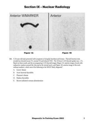
 Figure 1A Figure 1B 366. A 34-year-old male presented with symptoms of irregular heartbeat and tremor. Thyroid function tests revealed an elevated serum T3, normal T4 and reduced TSH. The 24 hour I-123 thyroid uptake was < 1%. Based on these results and the accompanying I-123 thyroid images (Figure 1A: anterior image of neck with radioactive markers around the chin and at the sternal notch, and Figure 1B: anterior image of the neck without markers), which one of the following is the MOST likely diagnosis? A. Graves’ disease B. Acute bacterial thyroiditis C. Plummer’s disease D. Painless thyroiditis E. Recent iodinated contrast administration Diagnostic In-Training Exam 2002 1
  • 2. Section IX – Nuclear Radiology Question #366 Findings: There is near-complete non-visualization of the thyroid on I-123 imaging (Figures 1A & 1B), with only faint visualization of the gland, which appears grossly normal in size. Thyroid morphology cannot accurately be assessed. Rationales: A) Incorrect. While the patient’s symptoms and laboratory findings are consistent with hyperthyroidism, the markedly reduced I-123 thyroid uptake and near non-visualization of the thyroid on I-123 imaging are not consistent with Graves’ disease, in which an elevated uptake and an enlarged thyroid with diffusely increased uptake on thyroid imaging would be expected. B) Incorrect. Acute bacterial thyroiditis presents with fever, elevated white blood cell count and focal tenderness over a portion of the gland. The thyroid uptake is variable, and thyroid imaging would most likely demonstrate a focal hypofunctioning (“cold”) nodule, with normal visualization of the remainder of the gland. C) Incorrect. Plummer’s disease is toxic nodular goiter. The thyroid uptake may be normal or mildly increased in this disorder, but would not be decreased. I-123 thyroid imaging in Plummer’s disease demonstrates one or more focal areas of increased tracer uptake, with associated areas of suppression of uptake in other parts of the gland, findings which are not present in this case. D) Correct. Subacute or painless thyroiditis is the most likely etiology for these findings. In this viral disorder, there is diffuse or focal inflammation of the gland, with release of pre-formed thyroid hormone into the circulation during the acute phase, resulting in signs and symptoms of hyperthyroidism and elevated thyroid function tests, as in this case. There is markedly reduced synthesis of new thyroid hormone by the gland, with associated decreased thyroid uptake and poor visualization of the thyroid on I-123 or Tc-99m pertechnetate thyroid imaging. These findings may also be seen in thyrotoxicosis factitia (intake of exogenous thyroid hormone) or in patients with ectopic thyroid hormone production (eg. struma ovarii), but these conditions were not listed as possible answers. E) Incorrect. Recent iodinated contrast administration may result in a falsely low I-123 thyroid uptake measurement, secondary to flooding of the extracellular iodine pool with non-radioactive iodine. In turn, poor visualization or non-visualization of the thyroid on thyroid scintigraphy may result. However, in most cases, these findings would not be associated with clinical evidence of hyperthyroidism, as is present in this case. Citations: Lazarus JH: Silent Thyroiditis and Subacute Thyroiditis. in Werner and Ingbar’s The Thyroid, a Fundamental and Clinical Text, 7th Edition, (Braverman LE and Utiger RD, eds.), Lippincott-Raven, Philadelphia, 1996, pp. 577-591. Mettler FA, Jr. and Guiberteau MJ: Essentials of Nuclear Medicine Imaging, 4th edition, WB Saunders Co., Philadelphia, 1998, pp. 117-118 2 American College of Radiology
  • 3. Section IX – Nuclear Radiology Figure 2A Figure 2B Figure 2C 367. A 57-year-old male presented with a 4 cm right upper lobe pulmonary mass on an outside chest radiograph and chest CT scan. The CT study demonstrated no evidence of mediastinal or hilar adenopathy. You are shown representative coronal, transaxial and sagittal images from an F-18 fluorodeoxyglucose (FDG) PET scan (Figure 2A) and the complete series of coronal images (Figures 2B and 2C). The lesion demonstrated a standardized uptake value (SUV) of 7.2. Based upon these findings, which one of the following is the MOST likely diagnosis? A. Stage IIIA squamous cell bronchogenic carcinoma B. Stage IB squamous cell bronchogenic carcinoma C. MALT (mucosa-associated lymphoid tissue) lymphoma D. Hamartoma E. Bronchoalveolar carcinoma Diagnostic In-Training Exam 2002 3
  • 4. Section IX – Nuclear Radiology Question #367 Findings: There is a large focal area of markedly increased FDG uptake in the right upper lobe, corresponding to the site of the patient’s known pulmonary nodule on prior imaging studies. There is no evidence of mediastinal or hilar lymphadenopathy. No additional pulmonary nodules are identified. The remaining areas of FDG uptake (eg. liver, heart, bowel, kidneys, bone marrow) represent normal sites of uptake. Rationales: A) Incorrect. The right upper lobe lesion is highly suspicious for bronchogenic carcinoma, especially in light of the SUV value of 7.2. However, in the absence of mediastinal or hilar adenopathy, this lesion is not consistent with Stage IIIA disease, which includes patients with primary lesions of any size, but only those with ipsilateral mediastinal or hilar adenopathy. B) Correct. On the basis of this PET scan, this patient has a T2 N0 M0 lesion, which is consistent with Stage 1B involvement. Despite its 4 cm size, only a primary lesion with evidence of invasion of the mediastinum, heart, great vessels, trachea, esophagus, vertebral body, carina or lesions associated with additional tumor nodules or malignant pleural effusions can be placed into the category of Stage III disease without evidence of adenopathy. C) Incorrect. Lymphomas are in general very FDG-avid. However, the MALT type lesion has been shown to be much less FDG-avid than other cell types, and FDG PET imaging is not recommended for patients with this disorder. Furthermore, a large focal lung nodule would represent an unusual manifestation of lymphoma in any case, even for FDG-avid cell types. D) Incorrect. Mildly increased FDG uptake may occur in infectious and granulomatous processes, including histoplasmosis, tuberculosis and others. Such occurrences may result in false positive PET scans for bronchogenic carcinoma. However, FDG uptake in such lesions is usually mild, typically with SUV values < 2.5, and in any event, such a diagnosis is much less likely than bronchogenic carcinoma, given the findings in this case. E) Incorrect. Bronchoalveolar carcinoma demonstrates variable FDG uptake, and has been reported to be a common cause of a false negative FDG PET scan in bronchogenic carcinoma. The presentation of a solitary, large focal pulmonary nodule is not the most common appearance for bronchoalveolar carcinoma. Furthermore, it is less common than other cell types of bronchogenic carcinoma, and this diagnosis would therefore be less likely than other cells types of non-small cell or small cell carcinoma. Citations: Bar-Shalom R, et. al.: PET Imaging in Oncology. Semin Nucl Med 2000; 30:150-185. Hoffmann M. et. al.: Positron emission tomography with fluorine-18-2-fluoro-deoxy-D-glucose (F18-FDG) does not visualize extranodal B-cell lymphoma of the mucosa-associated lymphoid tissue (MALT)-type. Annals Oncol 1999; 10:1185-1189. 4 American College of Radiology
  • 5. Section IX – Nuclear Radiology Figure 3A Figure 3B Figure 3D Figure 3C 368. A 43-year-old female with a history of ovarian carcinoma presented with shortness of breath and low-grade fever following a long airplane ride. You are shown posterior Xe-133 ventilation images (Figure 3A), Tc-99m MAA perfusion images (Figure 3B) and a concurrent PA and lateral chest radiograph (Figures 3C and 3D). Which one of the following BEST characterizes the overall findings in this case? A. High probability for pulmonary embolism B. Intermediate probability for pulmonary embolism C. Low probability for pulmonary embolism D. Normal study E. Lymphangitic carcinomatosis Diagnostic In-Training Exam 2002 5
  • 6. Section IX – Nuclear Radiology Question #368 Findings: The Xe-133 ventilation study (Figure 3A) demonstrates only mild xenon retention bilaterally, most striking at the lung bases. The Tc-99m MAA perfusion images (Figure 3B) demonstrate a single moderate subsegmental perfusion defect in the lateral basal segment of the left lower lobe, which does not correspond to a focal ventilatory defect on the ventilation study. The chest radiograph demonstrates no focal infiltrates or pleural effusions. Rationales: A) Incorrect. The V/Q scan findings of a single moderate ventilation-perfusion mismatch are not consistent with a high probability for pulmonary embolism, using any of the commonly employed criteria for interpretation of these studies. Both the modified PIOPED criteria and modified Biello criteria categorize such cases as intermediate probability for pulmonary embolism. B) Correct. As discussed in item A above, a single moderate V/Q mismatch is consistent with an intermediate probability for pulmonary embolism using the two most prevalent sets of diagnostic criteria currently in use. In published retrospective and prospective studies, approximately one-third of such cases have been associated with pulmonary embolism on pulmonary angiography. C) Incorrect. Again, a single moderate subsegmental V/Q matching defect is consistent with an intermediate probability for pulmonary embolism, both in the modified PIOPED and modified Biello diagnostic criteria. The original PIOPED study incorrectly classified these cases as low probability, an error which was corrected when the criteria were subsequently modified following the study. D) Incorrect. This ventilation-perfusion lung scan demonstrates abnormalities on both the ventilation and perfusion portions of the exam. It is not a normal study. E) Incorrect. The pattern of perfusion abnormality in this case is not suggestive of lymphangitic carcinomatosis. In that entity, the perfusion images typically produce a diffuse pattern of irregularity, with numerous small subsegmental perfusion defects, corresponding to the interlobular septae. This pattern of abnormality has been termed “contour mapping”. It is not present in this case, nor is there an interstitial pattern of pulmonary parenchymal abnormality on the chest radiography to suggest this entity. Citations: Biello DR, et. al.: Ventilation-perfusion studies in suspected pulmonary embolism. Amer J Roentgenol 1979; 133:1033-1037. Gottschalk A, et. al.: Ventilation-perfusion scintigraphy in the PIOPED study. Part ii. Evaluation of the scintigraphic criteria and interpretations. J Nucl Med 1993; 1119-1126 6 American College of Radiology
  • 7. Section IX – Nuclear Radiology Figure 4C Figure 4A Figure 4B Figure 4D Figure 4E Figure 4F 369. A 15-year-old female volleyball player presented with low back pain, without a specific incident of trauma. You are presented with AP and lateral radiographs of the lumbar spine (Figures 4A and 4B), anterior and posterior planar spot images (Figure 4C) and coronal, transaxial and sagittal SPECT images from a Tc-99m MDP radionuclide bone scan (Figures 4D - 4F). No other lesions were present elsewhere in the skeleton. Which one of the following is the MOST likely diagnosis? A. Stress fracture of the pars interarticularis B. Osteoid osteoma C. Ewing’s sarcoma D. Vertebral osteomyelitis E. Ankylosing spondylitis Diagnostic In-Training Exam 2002 7
  • 8. Section IX – Nuclear Radiology Question #369 Findings: The AP and lateral radiographs of the lumbar spine (Figures 4A and 4B) demonstrate very mild scoliosis of the lower lumbar spine, possibly positional. Anterior and posterior planar Tc-99m MDP bone images (Figure 4C) demonstrate a focal area of increased tracer uptake at L5 on the left, best seen on the posterior image. The coronal, transaxial and sagittal SPECT images (Figures 4D-4F) localize this finding to the region of the lamina, in the expected location of the pars interarticularis. Rationales: A) Correct. The findings described above are typical for a stress fracture of the left pars interarticularis of L5, a common injury occurring in children involved in athletic activities, such as gymnastics, volleyball, etc. Radiographs may or may not demonstrate a pars defect, and in the case of bilateral pars defects, spondylolysis may also be present. SPECT imaging is useful in detecting subtle lesions not evident on planar views, as well as localizing the lesion to the region of the pars. Increased bone turnover in the site of the lesion suggests that it is the etiology for the patient’s symptoms of localized back pain. B) Incorrect. Osteoid osteoma can occur in the spine, and may be associated with focal areas of increased uptake on bone scintigraphy. However, it is less likely to be associated with normal radiographs, usually presenting with a focal area of sclerosis, sometimes with a visible central radiolucent nidus. It commonly presents with rigid, painful scoliosis, and may have the classic presentation of pain, worse at night and relieved by aspirin. It is less common than a stress fracture of the pars, especially in the present clinical setting. C) Incorrect. Ewing’s sarcoma is uncommon relative to stress fractures, and the vertebral column is an uncommon site of involvement. When arising in the vertebral column, the sacrum is the most common site. It is often associated with a long history of pain, a soft tissue mass and in many cases, associated systemic symptoms. Vertebral involvement most often involves the vertebral bodies primarily, may be associated with vertebral collapse, and is not usually associated with normal radiographic findings. Ewing’s sarcoma is associated with markedly increased uptake of Tc-99m MDP, although usually the lesion is much larger at presentation. D) Incorrect. Vertebral osteomyelitis is usually located in the vertebral bodies, and may also spread to the intervertebral disc spaces. Radiographic findings include areas of bone destruction and/or sclerosis, similar to the findings of osteomyelitis in other regions of the skeleton. Fever, leukocytosis and systemic symptoms are often present. Vertebral osteomyelitis is less common than stress fractures, and does not fit the present clinical setting well. Again, normal radiographic findings would not be expected with vertebral osteomyelitis. E) Incorrect. Findings that may be seen in ankylosing spondylitis include increased uptake in the sacro-iliac joints, focal areas of increased uptake in the apophyseal joints, increased uptake in the intervertebral disc spaces and other sites of arthritic involvement in the peripheral joints and costovertebral and sternoclavicular joints. These findings are not present in this case. While this patient is at the lower end of the typical age range for this disorder, ankylosing spondylitis is more common in males, and is far less common than post-traumatic injuries. Citations: Mettler FA, Jr. and Guiberteau MJ. Essentials of Nuclear Medicine Imaging, 4th edition, WB Saunders Co., Philadelphia, 1998, pp. 285-333. Greenfield GB. Radiology of Bone Diseases. 2nd edition, J.B. Lippincott Co., Philadelphia, 1975. Resnick D. Bone and Joint Imaging. W.B. Saunders Co., Philadelphia, 1989. 8 American College of Radiology
  • 9. Section IX – Nuclear Radiology Figure 5A Figure 5B 370. A 46-year-old female underwent a living-related donor renal transplant placed in the left iliac fossa, and presented 5 days post-operatively with the acute onset of markedly reduced urine output and elevated serum creatinine. You are presented with an anterior flow study (Figure 5A) and serial static anterior images of the abdomen and pelvis (Figure 5B) from a Tc-99m MAG-3 renal scan. Which one of the following is the BEST interpretation for these findings? A. Post-operative acute tubular necrosis (ATN) B. Renal artery stenosis C. Immunosuppressive drug toxicity D. Ureteral obstruction E. Accelerated rejection Diagnostic In-Training Exam 2002 9
  • 10. Section IX – Nuclear Radiology Question #370 Findings: The flow study (Figure 5A) demonstrates no evidence of perfusion to the transplanted kidney in the left iliac fossa. The delayed static images (Figure 5B) demonstrate increased background activity and photopenic areas in the configuration of bowel loops, without visualization of the renal transplant. Rationales: A) Incorrect. Post-operative ATN is present immediately after transplantation, and a 5 day delay before the onset of symptoms would not be expected. In addition, ATN is usually associated with disproportionate impairment of renal tubular function, with relatively preserved perfusion, which is not present in this case. It is also usually less striking in the case of living-related donor transplantation, in which cases the ischemic time of the donor kidney is minimized. B) Incorrect. Renal artery stenosis is a later complication of transplantation, and would not be expected within the first post-operative week. The high-grade degree of impaired perfusion in this case is also not typical for renal artery stenosis. C) Incorrect. Immunosuppressive drug toxicity, similar to ATN, is most often associated with a disproportionate impairment of renal tubular function relative to perfusion, which is not the case in this instance. Complete lack of perfusion to the renal transplant is not an expected finding with drug toxicity. D) Incorrect. Complete obstruction of the transplanted kidney could conceivably produce these findings. However, it would be unusual to see complete non-perfusion and non-function of the transplant, as is present in this case. Acute rejection is a more likely etiology for these findings. E) Correct. Severe acute or accelerated rejection, is the most likely explanation for these findings. Rejection is typically associated with impairment both of perfusion and tubular function of the transplanted kidney, and is consistent with the time course in this case. While much less common in living-related donor transplants, it can occur even in this setting. The most likely differential diagnostic possibility would be acute occlusion or disruption of the renal vessels, which is not one of the options listed. Citations: Mettler FA, Jr. and Guiberteau MJ. Essentials of Nuclear Medicine Imaging, 4th edition, WB Saunders Co., Philadelphia, 1998, pp. 335-361. 10 American College of Radiology