
Recommended
More Related Content
What's hot
What's hot (20)
Viewers also liked
Viewers also liked (17)
Similar to Struttura_dna
Similar to Struttura_dna (20)
Recently uploaded
Recently uploaded (20)
Struttura_dna
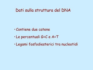
- 1. Dati sulla struttura del DNA •Contiene due catene •Le percentuali G=C e A=T •Legami fosfodiesterici tra nucleotidi
- 2. Lewin, IL GENE VIII, Zanichelli editore S.p.A. Copyright © 2006
- 3. Lewin, IL GENE VIII, Zanichelli editore S.p.A. Copyright © 2006
- 4. N N N N NN O H H O N sugar H sugar CH3 timina adenina N N N N O N H N N N H O sugar H H H sugar citosina guanina L’appaiamento delle basi implica la formazione di legami idrogeno
- 5. Lewin, IL GENE VIII, Zanichelli editore S.p.A. Copyright © 2006
- 6. Figura riportata nella pubblicazione originale di Watson e Crick Nature, Aprile 1953
- 7. Lewin, IL GENE VIII, Zanichelli editore S.p.A. Copyright © 2006 20
- 8. La struttura del DNA (B) •Il DNA ha una forma ad elica regolare, diametro 20Å, passo 34Å •Legami idrogeno tra le basi •L’impilamento delle basi è determinato da interazioni idrofobiche •Ogni coppia è ruotata di 36° •Solchi maggiore (22Å) e minore (12Å) •Avvolgimento in senso orario (elica destrorsa)
- 9. Nature, Vol. 171, p.737, April 25, 1953 MOLECULAR STRUCTURE OF NUCLEIC ACIDS A Structure for Deoxyribose Nucleic Acid We wish to s uggest a structure for the salt of deoxyribose nucleic acid (D.N.A.). This structure has novel features which are of considerable biological interest. A structure for nucleic acid has already been proposed by Pauling and Corey (1). They kindly made their manuscript available to us in advance of publication. Their model consists of three intertwined chains, with the phosphates near the fibre axis, and the bases on the outside. In our opinion, this structure is unsatisfactory for two reasons: (1) We believe that the material which gives the X-ray diagrams is the salt, not the free acid. Without the acidic hydrogen atoms it is not clear what forces would hold the structure together, especially as the negatively charged phosphates near the axis will repel each other. (2) Some of the van der Waals distances appear to be too small. Another three-chain structure has also been suggested by F raser (in the press). In his model the phosphates are on the outside and the bases on the inside, linked together by hyd rogen bonds. This structure as described is rather ill-defined, and for this reason we shall not comment on it. We wish to put forward a ra dically different structure for the salt of deoxyribose nucleic acid. This structure has two helical chains each coiled round the same axis (see diagram). We have made the usual chemical assumptions, namely, that each chain consists of phosphate diester groups joining ß-D- deoxyribofuranose residues with 3',5' linkages. The two chains (but not their bases) are related by a dyad perpendicular to the fibre axis. Both chains follow right- handed helices, but owing to the dyad the sequences of the atoms in t he two chains run in opposite directions. Each chain loosely resembles Furberg's2 model No. 1; that is, the bases are on the inside of the helix and the phosphates on the outside. The configuration of the sugar and the atoms near it is close to Furberg's 'standard configuration', the sugar being roughly perpendicular to the attached base. There is a residue on each every 3.4 A. in the z-direction. We have assumed an angle of 36° between adjacent residues in the same chain, so that the structure repeats after 10 residues on each chain, that is, after 34 A. The distance of a phosphorus atom from the fibre axis is 10 A. A s the phosphates are on the outside, cations have easy access to them. The structure is an open one, and its water content is rather high. At lower water contents we would expect the bases to tilt so that the structure could become more compact. The novel feature of the structure is the manner in which the two chains are held together by the purine and pyrimidine bases. The planes of the bases are perpendicular to the fibre axis. The are joined together in pairs, a single base from the other chain, so that the two lie side by side with identical z-co-ordinates. One of the pair must be a purine and the other a pyrimidine for bonding to occur. The hydrogen bonds are made as follows : purine position 1 t o pyrimidine position 1 ; purine position 6 to pyrimidine position 6. If it is assumed that the bases only occur in the structure in the most plausible tautomeric forms (that is, with the keto rather than the enol configurations) it is found that only specific pairs of bases can bond together. These pairs are : adenine (purine) with thymine (pyrimidine), and guanine (purine) with cytosine (pyrimidine). In other words, if an adenine forms one member of a pair, on either chain, then on these assumptions the other member must be thymine ; similarly for guanine and cytosine. The sequence of bases on a single chain does not appear to be restricted in any way. However, if only specific pairs of bases can be formed, it follows that if the sequence of bases on one chain is given, then the sequence on t he other chain is automatically determined. It has been found experimentally (3,4) that the ratio of the amounts of adenine to thymine, and the ration of guanine to cytosine, are always bery close to unity for deoxyribose nucleic acid. It is probably impossible to build this structure with a ribose sugar in place of the deoxyribose, as the extra oxygen atom would make too close a van der Waals contact. The previously published X-ray data (5,6) on deoxyribose nucleic acid are insufficient for a rigorous test of our structure. So far as we can tell, it is roughly compatible with the experimental data, but it must be regarded as unproved until it has been checked against more exact results. Some of these are given in the following communications. We were not aware of the details of the results presented there when we devised our structure, which rests mainly though not entirely on published experimental data and stereochemical arguments. It has not escaped our notice that the specific pairing we have postulated immediately suggests a possible copying mechanism for the genetic material. Full details of the structure, including the conditions assumed in building it, together with a set of co-ordinates for the atoms, will be published elsewhere. We are much indebted to Dr. Jerry Donohue for constant advice and criticism, especially on interatomic distances. We have also been stimulated by a knowledge of the general nature of the unpublished experimental results and ideas of Dr. M. H. F. Wilkins, Dr. R. E . Franklin and their co-workers at King's College, London. One of us (J. D. W.) has been aided by a fellowship from the National Foundation for Infantile Paralysis. J. D. WATSON F. H. C. CRICK Medical Research Council Unit for the Study of Molecular Structure of Biological Systems, Cavendish Laboratory, Cambridge. 1. Pauling, L., and Corey, R. B., Nature, 171, 346 (1953); Proc. U.S. Nat. Acad. Sci., 39, 84 (1953). 2. Furberg, S., Acta Chem. Scand., 6, 634 (1952). 3. Ch argaff, E., for references see Zamenhof, S., Brawerman, G., and Chargaff, E., Biochim. et Biophys. Acta, 9, 402 (1952). 4. Wyatt, G. R., J. Gen. Physiol., 36, 201 (1952). 5. A stbury, W. T., Symp. Soc. Exp. Biol. 1, Nucleic Acid, 66 (Camb. Univ. Press, 1947). 6. W ilkins, M. H . F., and Randall, J. T., Biochim. et Biophys. Acta, 10, 192 (1953).
- 10. It has not escaped our notice that the specific pairing we have postulated immediately suggests a possible copying mechanism for the genetic material.
- 11. Il modello a doppia elica di Crick e Watson suggerisce un meccanismo di replicazione OLD NEW OLD NEW NEW NEW OLD OLD
- 12. Lewin, IL GENE VIII, Zanichelli editore S.p.A. Copyright © 2006
- 13. Lewin, IL GENE VIII, Zanichelli editore S.p.A. Copyright © 2006
- 14. Lewin, IL GENE VIII, Zanichelli editore S.p.A. Copyright © 2006
- 15. Lewin, IL GENE VIII, Zanichelli editore S.p.A. Copyright © 2006
- 16. Strutture alternative del DNA
- 17. Gli acidi nucleici possono formare vari tipi di doppia elica
- 18. Gli acidi nucleici possono formare vari tipi di doppia elica Elica Coppie di basi per giro Rotazione tra due basi Diametro B 10 (10,4) 36 (34,6) 20 (19) A 11 32,7 23 Z 12 -30 18
- 19. Strutture superiori del DNA
- 20. DNA circolare con superavvolgimento = 0 DNA superavvolto negativamente
- 21. Lewin, IL GENE VIII, Zanichelli editore S.p.A. Copyright © 2006
- 22. Lewin, IL GENE VIII, Zanichelli editore S.p.A. Copyright © 2006
- 23. Lewin, IL GENE VIII, Zanichelli editore S.p.A. Copyright © 2006
- 24. Il superavvolgimento negativo può convertirsi nella separazione locale dei due filamenti DNA superavvolto negativamente
- 25. separazione dei filamenti (denaturazione di regioni ricche in A+T) strutture cruciformi (in corrispondenza di palindromi) struttura Z (in corrispondenza di regioni di alternanza Pu/Py)
- 26. Superavvolgimento del DNA Positivo = DNA superspiralizzato Negativo = DNA sottospiralizzato
- 27. Enzimi che alterano la topologia del DNA Topoisomerasi: tipo I tipo II (girasi)
- 28. Lewin, IL GENE VIII, Zanichelli editore S.p.A. Copyright © 2006
- 29. Lewin, IL GENE VIII, Zanichelli editore S.p.A. Copyright © 2006
- 30. Enzimi per gli acidi nucleici •Polimerasi: DNA polimerasi RNA polimerasi •Nucleasi: esonucleasi endonucleasi
- 31. Lewin, IL GENE VIII, Zanichelli editore S.p.A. Copyright © 2006
- 32. Lewin, IL GENE VIII, Zanichelli editore S.p.A. Copyright © 2006