BAG TECHNIQUE Bag technique-a tool making use of public health bag through wh...
A new ultrasound marker of tetralogy of fallot
1. Ultrasound Obstet Gynecol 2010; 36: 556–560
Published online 6 October 2010 in Wiley Online Library (wileyonlinelibrary.com). DOI: 10.1002/uog.7614
The ‘question mark’ sign as a new ultrasound marker
of tetralogy of Fallot in the fetus
J. M. MART´INEZ, O. G ´OMEZ, M. BENNASAR, A. OLIVELLA, F. CRISPI, B. PUERTO
and E. GRATAC ´OS
Fetal Cardiology Unit, Maternal-Fetal Medicine Department, Institut Cl´ınic de Ginecologia, Obstetr´ıcia I Neonatologia (ICGON), Hospital
Cl´ınic, Institut d’Investigacions Biom`ediques Augusto Pi i Sunyer (IDIBAPS), University of Barcelona and Centro de Investigaci´on
Biom´edica en Red de Enfermedades Raras (CIBERER), Barcelona, Spain
KEYWORDS: congenital heart defects; pulmonary atresia with ventricular septal defect; tetralogy of Fallot; ultrasonographic
marker; ultrasound screening
ABSTRACT
Objective To describe a new ultrasonographic marker,
the ‘question-mark’ sign, to assist in the diagnosis of
tetralogy of Fallot (TOF) in the fetus, and to evaluate
its prevalence in TOF as compared with other cardiac
defects.
Methods A prospective evaluation over a 5-year period of
a consecutive series of 3998 pregnant women undergoing
fetal echocardiography from 12 to 40 weeks’ gestation
due to high risk for congenital heart disease (CHD).
Standard echocardiographic planes with color Doppler
assessment and evaluation of the whole aortic arch, from
the left ventricular outflow tract to the descending aorta in
the axial upper mediastinum views, were performed. The
question-mark sign corresponded with an enlarged and
dilated ascending aorta and aortic arch in the three-vessel
view of the upper fetal mediastinum. The frequency of
this sign was evaluated in cases with TOF and in other
cases of cardiac defects, as well as in fetuses with normal
cardiac scans in this series.
Results CHD was diagnosed in a total of 447 (11.2%)
fetuses at a median gestational age of 24 (range, 12–40)
weeks. Forty-two of the 447 (9.4%) had TOF, of which
29 cases (69.0%) had classical TOF (pulmonary stenosis),
nine (21.4%) pulmonary atresia and four (9.5%) absent
pulmonary valve syndrome. A question-mark sign was
observed in 16/29 (55.2%) cases of classical TOF and in
8/9 (88.9%) cases of TOF with pulmonary atresia. The
sign was never observed in any of the cases of TOF with a
right-sided aortic arch. Likewise, the sign was observed in
1/405 (0.2%) cases with other cardiac anomalies (a fetus
with a complex cardiac defect) and in none of the fetuses
with normal hearts.
Conclusions The finding of an enlarged aorta with a
question-mark shape should raise a strong suspicion
of tetralogy of Fallot, in particular the variant with
pulmonary atresia. This sign may be useful in screening
considering that prenatal diagnosis of TOF by routine
ultrasonography remains a challenge. Copyright 2010
ISUOG. Published by John Wiley & Sons, Ltd.
INTRODUCTION
Tetralogy of Fallot (TOF) occurs in approximately
8–12% of infants with CHD1–3
. TOF is frequently asso-
ciated with chromosomal and extracardiac anomalies,
and cases with severe obstruction of the pulmonary tract
very often require assessment and treatment immediately
after birth. Therefore, prenatal detection of TOF is criti-
cally important4–8. Unfortunately, the prevalence of TOF
found in fetal series is much lower than that found in
infant series3,9
, which indicates that TOF remains among
the least diagnosed CHD during the prenatal period2,4,10
.
The use of key signs may be of great help in assisting
in the identification of cardiac defects in fetal ultrasound
screening. In our clinical experience, we have observed
that fetuses with TOF often present a characteristic
appearance of the ascending aorta in axial planes that
strongly resembles a ‘question-mark’ (‘?’) sign; in fact, in
most cases of TOF the question-mark sign was the first
sign to suggest a cardiac defect. We have subsequently
evaluated the frequency of this sign in our fetal cardiology
unit during the last 5 years.
The aim of this study was to describe the question-
mark sign as a potentially useful ultrasonographic marker
to raise the suspicion of, or better document the diagnosis
Correspondence to: Dr J. M. Mart´ınez, Sabino de Arana 1, 08028 Barcelona, Spain (e-mail: jmmartti@clinic.ub.es)
Accepted: 22 February 2010
Copyright 2010 ISUOG. Published by John Wiley & Sons, Ltd. ORIGINAL PAPER
2. A new ultrasound marker of tetralogy of Fallot 557
of, TOF in the fetus, and to evaluate its frequency in
fetuses with TOF as compared with those with other
cardiac defects and normal cardiac scans.
METHODS
This was a prospective study performed on all fetuses
referred to our fetal cardiology unit because of the
presence of risk factors for CHD, from January 2004
to January 2009. Gestational age at examination ranged
between 12 and 40 weeks on the day of the examination,
as determined by first-trimester fetal crown–rump length
measurement. The study protocol was approved by the
local ethics committee and all patients provided oral
consent for the use of the images for clinical studies.
All patients underwent a detailed ultrasound scan in
our fetal cardiology unit, which operates as a referral
center for pregnancies at high risk for CHD. Obstetricians
experienced in evaluating the fetal heart perform the
fetal echocardiography, which includes standard planes
with color Doppler assessment, obtained following the
guidelines of the International Society of Ultrasound in
Obstetrics and Gynecology and others11–13
. Briefly, the
situs is checked at the abdominal level in an axial scanning
plane through the stomach, aorta and inferior vena cava.
Then, visualization of the four-chamber view (with atria,
atrioventricular valves and ventricles), the origin and
double-crossing of the great arteries, and systemic and
pulmonary venous return is performed in a segmental
approach. Finally, the three vessels and trachea view is
obtained axially at the level of the upper mediastinum,
as described by Yagel et al.11
. Color Doppler to review
normal flow at the level of the four chambers, the
atrioventricular and semilunar valves, the interventricular
and interatrial septa, and the ductal and aortic arches,
is also routinely performed. Pulsed Doppler and M-mode
are only used if indicated. The reliability of CHD diagnosis
is always assessed by postnatal examination by pediatric
cardiologists, or by autopsy in cases of termination of
pregnancy or perinatal death.
For the purposes of this study the aortic arch was
assessed from the left ventricle outflow tract to the
descending aorta in the axial upper mediastinum view.
The question-mark sign was defined, in a fetus in cephalic
position with posterior spine, as a typical sonographic
shape of the ascending aorta and aortic arch in axial
planes, almost at the level of the three vessels and trachea
view, showing a very enlarged and dilated aorta, with a
striking shape resembling a question mark (Figure 1).
In this same sonographic plane, color Doppler was
found to be very helpful for differentiating between
classical TOF with pulmonary stenosis (antegrade flow
in the short arm of the question-mark sign) and TOF
with pulmonary atresia (retrograde flow) (Figure 2). The
presence or absence of the question-mark sign was part
of the standardized protocol and was included as an item
in a check-list.
We measured the frequency of the question-mark sign
in cases with TOF and analyzed the association with
left/right aortic arch and with the degree of pulmonary
obstruction. For each diagnosis of TOF, three experienced
examiners (O.G., M.B. and J.M.M.) reached a consensus
on the presence or absence of the question-mark sign.
The frequency of the sign in other cardiac defects and
in normal cardiac scans was also recorded. The data
were stored in research databases and standard statistical
analysis was performed using SPSS 14.0 (SPSS, Chicago,
IL, USA).
RESULTS
During the 5-year study period a consecutive series
of 3998 fetuses at high risk for CHD underwent
echocardiography in our fetal cardiology unit. The median
maternal age was 31 (range, 16–40) years and the median
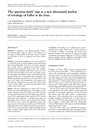
Figure 1 (a) Normal three vessels and trachea view, with both pulmonary artery and aorta showing similar size and a confluent shape
(‘V-shape’). (b) The ‘question-mark’ sign in a case of classical tetralogy of Fallot. The aorta is significantly larger than the pulmonary trunk,
and resembles a question mark in shape. A, aorta; P, pulmonary artery.
Copyright 2010 ISUOG. Published by John Wiley & Sons, Ltd. Ultrasound Obstet Gynecol 2010; 36: 556–560.
3. 558 Mart´ınez et al.
Figure 2 The ‘question-mark’ sign in a case of tetralogy of Fallot
with pulmonary atresia. Both the trachea (arrow) and the superior
vena cava (SVC) are positioned normally to the right of the aorta.
Color Doppler echocardiography demonstrates retrograde flow
(red) in the whole pulmonary artery, from the outflow tract to the
ductus arteriosus, while antegrade flow (red then blue) is shown in
the whole aortic arch.
gestational age at ultrasound examination was 21 (range,
12–40) weeks.
Complete follow-up was obtained in 98.2% of the preg-
nancies, among which 447 fetuses with CHD were identi-
fied, with an incidence of 11.2%. In this group a total of
42 (9.4%) cases were diagnosed with TOF. Among those
were 29 cases (69.0%) with classical TOF (pulmonary
stenosis), nine (21.4%) with pulmonary atresia and four
(9.5%) cases of absent pulmonary valve syndrome. A right
aortic arch was diagnosed in eight of the 29 cases (27.6%)
with classical TOF and in one of the nine (11.1%) with
pulmonary atresia. Termination of pregnancy was per-
formed in 24 cases (57.1%) at parental request because
of the presence of associated structural or chromosomal
anomalies (including four cases of 22q11 microdeletion).
The prenatal diagnosis of TOF was confirmed in all 42
cases, either by postnatal evaluation or by necropsy.
The prevalence of the question-mark sign in the whole
population and with regard to the type of anomaly and
the side of the aortic arch is summarized in Table 1. Over-
all, the question-mark sign was found in 24/42 (57.1%)
fetuses diagnosed with TOF, of which 16/29 (55.2%)
were in fetuses with classical TOF and 8/9 (88.9%) were
in fetuses with TOF and pulmonary atresia. The sign was
not present in any of the nine (21.4%) cases in which the
aortic arch was right sided (Figure 3). Therefore, if only
cases with left-sided aorta are considered, the detection
rate was 76.2% (16/21) for classical TOF and 100% (8/8)
for pulmonary atresia.
The question-mark sign was observed in none of the
3551 fetuses with a normal cardiac scan and in 1/405
(0.2%) fetuses with cardiac anomalies other than TOF.
This case was a fetus with a complex cardiac malforma-
tion consisting of tricuspid and pulmonary atresia with a
large perimembranous ventricular septal defect. Thus, the
Table 1 Prevalence of the ‘question-mark’ (‘?’) sign in the studied
population with regard to the type of anomaly and the side of the
aortic arch
Fetal heart n
‘?’ absent
(n (%))
‘?’ present
(n (%))
Normal 3551 3551 (100) 0
CHD 447 422 (94.4) 25 (5.6)
TOF 42 18 (42.9) 24 (57.1)
Type of TOF
PS-TOF 29 13 (44.8) 16 (55.2)
PA-TOF 9 1 (11.1)* 8 (88.9)
AbsP-TOF 4 4 (100) 0
Side of aortic arch
TOF + RAA 9 9 (100) 0
TOF + LAA 33 9 (27.3) 24 (72.7)
Other CHD 405 404 (99.8) 1 (0.2)†
*Case with right aortic arch. †Complex malformation: pulmonary
and tricuspid atresia with a large perimembranous ventricular
septal defect. AbsP, absent pulmonary valve; CHD, congenital
heart disease; LAA, left aortic arch; PA, pulmonary atresia; PS,
pulmonary stenosis; RAA, right aortic arch; TOF, tetralogy of
Fallot.
Figure 3 Absence of the ‘question-mark’ sign in a case of tetralogy
of Fallot with pulmonary atresia and right aortic arch, showing the
malaligned ventricular septal defect with the overriding aorta ( ).
The trachea is located to the left of the aorta (arrow) while the
superior vena cava is normally placed to the right of the aorta. Ao,
aorta; SVC, superior vena cava.
positive predictive value of the question-mark sign for the
diagnosis TOF was 96.0% (24/25) in this series.
DISCUSSION
Our investigation has shown that the finding of an
enlarged aorta with a marked ‘?’ shape at the media-
stinum level can be considered as a valuable marker for
improving the detection of TOF in the fetus; the sign was
found to be particularly associated with the variant of
TOF with pulmonary atresia. The question-mark sign was
never observed in cases with right aortic arch, in which the
Copyright 2010 ISUOG. Published by John Wiley & Sons, Ltd. Ultrasound Obstet Gynecol 2010; 36: 556–560.
4. A new ultrasound marker of tetralogy of Fallot 559
aorta always follows a straight anteroposterior trajectory
due to its right disposition with respect to the trachea.
TOF is a cardiac malformation characterized by an
anterior aorta overriding a subaortic ventricular septal
defect, caused by malalignment between the infundibular
and the trabecular septum, and obstruction of the right
outflow tract with varying severity. Consequently, there
is hypertrophy of the right ventricle, but it is not usually
patent after birth. The prenatal diagnosis of TOF remains
a challenge, with detection rates as low as 15–40%7,9,14
.
A main reason for this low detection rate is that the
four-chamber view in TOF is usually unremarkable in
the majority of cases4–7
, apart from a leftward devia-
tion of the cardiac apex15,16
. Likewise, in cases of TOF
with absent pulmonary valve the four-chamber view may
demonstrate some degree of cardiomegaly, although this
is not a constant feature in the second trimester17,18
.
The diagnosis of TOF cannot be made unless the out-
flow tracts are evaluated, thus the aorta overriding a
ventricular septal defect has to be demonstrated4–6,19–21.
Once a suspicious diagnosis has been arrived at, color
Doppler sonography can be used to demonstrate either
antegrade or reversed flow through the pulmonary out-
flow tract and arterial duct. Importantly, the severity of
the pulmonary obstruction may evolve during pregnancy.
Thus, pulmonary stenosis, which can be absent at the time
of diagnosis in the second trimester, may become evident
later on in gestation. In addition, in some cases a narrow
pulmonary outflow may progress to atresia, which entails
a significantly worse prognosis for the newborn22
. Serial
ultrasound monitoring is therefore warranted in order to
detect retrograde flow through the pulmonary artery late
in gestation, thus warning of the need for prostaglandin
therapy immediately after birth4–6,15–17
.
Although comprehensive fetal echocardiography may
detect most cases of TOF, such an examination can-
not be routinely offered in most settings because it is
time-consuming and requires advanced knowledge of car-
diac anatomy and substantial sonographic skills. That
is the reason why any effort to improve the prenatal
detection of TOF should be encouraged, and several
authors have suggested the inclusion of other sonographic
planes or measurements of aortic and pulmonary artery
diameters11,23. The addition of the direction of flow by
color Doppler examination provides further support for
the establishment of normal outflow tract examination
and complements the four-chamber view24
. The findings
of this study suggest that the question-mark sign could
constitute an additional marker that might help to raise
the suspicion of, or confirm the diagnosis of, TOF during
the standard anomaly screening examination, given that
the sonographic demonstration of an overriding aorta
has proved to be difficult. However, it is important to
emphasize that sequential examination of the heart, pay-
ing particular attention to the origin of the aorta from the
left ventricle, cannot be replaced by observation or not of
the question-mark sign.
Since morphology of the vessels and functional hemo-
dynamic abnormalities are closely related, we hypothesize
that a possible explanation for the ‘?’ shape might be
the presence of increased flow through the overriding
aorta together with reduced flow through the pulmonary
valve. This hypothesis is consistent with the fact that the
question-mark sign was not observed in any case of TOF
with absent pulmonary valve, in which there is high to-
and-fro flow through the right outflow tract. However,
we acknowledge that we do not have a clear explanation
for the fact that the sign was not present in any other
conotruncal anomaly – such as truncus arteriosus or dou-
ble outlet right ventricle – in which similar patterns of
outflow tract hemodynamics may be found, so other fac-
tors may be related to the anatomical morphology of the
vessels. On the other hand, in cases of TOF with a right
aortic arch the straight course of the aorta seems logical
and is most probably forced by the anatomical position
of the aorta to the right of the trachea, thus precluding
observation of the ‘?’ shape.
The major drawback of our study is that it was per-
formed in a high-risk population by experts in fetal
echocardiography. It remains an open question whether
observation of the question-mark sign is reproducible
enough in other settings. In our opinion, the fact that the
sign was found to be obvious in most of the cases with
TOF supports the potential contribution of the question-
mark sign during non-specialized examination at universal
screening.
To conclude, when exploring the fetal heart the finding
of the question-mark sign at the three vessels and tra-
chea view strongly suggests the presence of TOF in the
fetus, particularly the form with pulmonary atresia, but
not in the form with either pulmonary stenosis or absent
pulmonary valve, nor in cases with right aortic arch. We
suggest that the potential value of this sign for improv-
ing the prenatal diagnosis of TOF deserves to be further
explored in the general screening population.
ACKNOWLEDGMENTS
F. C. is supported by a Rio Hortega grant from the Spanish
Fondo de Investigaciones Cient´ıficas.
REFERENCES
1. Mitchell SC, Korones SB, Berendes HW. Congenital heart
disease in 56,109 births. Incidence and natural history.
Circulation 1971; 43: 323–332.
2. Allan L. Prenatal diagnosis of structural cardiac defects. Am J
Med Genet 2007; 145: 73–76.
3. Allan LD, Sharland GK, Milburn A, Lockhart SM, Groves AM,
Anderson RH, Cook AC, Fagg NL. Prospective diagnosis of
1,006 consecutive cases of congenital heart disease in the fetus.
J Am Coll Cardiol 1994; 23: 1452–1458.
4. Paladini D, Rustico M, Todros T, Palmieri S, Gaglioti P, Benet-
toni A, Russo MG, Chiappa E, D’Ottavio G. Conotruncal
anomalies in prenatal life. Ultrasound Obstet Gynecol 1996; 8:
241–246.
5. Tometzki AP, Suda K, Kohl T, Kovalchin JP, Silverman NH.
Accuracy of prenatal echocardiographic diagnosis and prognosis
of fetuses with conotruncal anomalies. J Am Coll Cardiol 1999;
33: 1696–1701.
Copyright 2010 ISUOG. Published by John Wiley & Sons, Ltd. Ultrasound Obstet Gynecol 2010; 36: 556–560.
5. 560 Mart´ınez et al.
6. Sivanandam S, Gilckstein JS, Printz BF, Allan LD, Altmann K,
Solowiejczyk DE, Simpson L, Perez-Delboy A, Kleinman CS.
Prenatal diagnosis of conotruncal malformations: diagnostic
accuracy, outcome, chromosomal abnormalities, and extracar-
diac anomalies. Am J Perinatol 2006; 23: 241–245.
7. Chew C, Halliday JL, Riley MM, Penny DJ. Population-based
study of antenatal detection of congenital heart disease by
ultrasound examination. Ultrasound Obstet Gynecol 2007; 29:
619–624.
8. Yates RS. The influence of prenatal diagnosis on postnatal
outcome in patients with structural congenital heart disease.
Prenat Diagn 2004; 24: 1143–1149.
9. Tegnander E, Williams W, Johansen OJ, Blaas HGK, Eik-
Nes SH. Prenatal detection of heart defects in a nonselected
population of 30 149 fetuses: detection rates and outcome.
Ultrasound Obstet Gynecol 2006; 27: 252–265.
10. Sharland G. Routine fetal cardiac screening: what are we doing
and what should we do? Prenat Diagn 2004; 24: 1123–1129.
11. Yagel S, Cohen SM, Achiron R. Examination of the fetal heart
by five short-axis views: a proposed screening method for
comprehensive cardiac evaluation. Ultrasound Obstet Gynecol
2001; 17: 367–369.
12. Allan L. Technique of fetal echocardiography. Pediatr Cardiol
2004; 25: 223–233.
13. International Society of Ultrasound in Obstetrics and Gyne-
cology. Cardiac screening examination of the fetus: guidelines
for performing the ‘basic’ and ‘extended basic’ cardiac scan.
Ultrasound Obstet Gynecol 2006; 27: 107–113.
14. Garne E, Stoll C, Clementi M; Euroscan Group. Evaluation of
prenatal diagnosis of congenital heart diseases by ultrasound:
experience from 20 European registries. Ultrasound Obstet
Gynecol 2001; 17: 386–391.
15. Shipp TD, Bromley B, Hornberger LS, Nadel A, Benacerraf BR.
Levorotation of the fetal cardiac axis: a clue for the presence of
congenital heart disease. Obstet Gynecol 1995; 85: 97–102.
16. Smith RS, Comstock CH, Kirk JS, Lee W. Ultrasonographic left
cardiac axis deviation: a marker for fetal anomalies. Obstet
Gynecol 1995; 85: 187–191.
17. Volpe P, Paladini D, Marasini M, Buonadonna AL, Russo MG,
Caruso G, Marzullo A, Arciprete P, Martinelli P, Gentile M.
Characteristics, associations and outcome of absent pulmonary
valve syndrome in the fetus. Ultrasound Obstet Gynecol 2004;
24: 623–628.
18. Galindo A, Guti´errez-Larraya F, Mart´ınez JM, del Rio M,
Gra˜neras A, Velasco JM, Puerto B, Gratacos E. Prenatal diag-
nosis and outcome for fetuses with congenital absence of the
pulmonary valve. Ultrasound Obstet Gynecol 2006; 28: 32–39.
19. Poon LGY, Huggon IC, Zidere V, Allan LD. Tetralogy of Fallot
in the fetus in the current era. Ultrasound Obstet Gynecol 2007;
29: 625–627.
20. Vesel S, Rollings S, Jones A, Callaghan N, Simpson J, Shar-
land GK. Prenatally diagnosed pulmonary atresia with ven-
tricular septal defect: echocardiography, genetics, associated
anomalies and outcome. Heart 2006; 92: 1501–1505.
21. Galindo A, Mendoza A, Arbues J, Gra˜neras A, Escribano D,
Nieto O. Conotruncal anomalies in fetal life: accuracy of
diagnosis, associated defects and outcome. Eur J Obstet Gynecol
Reprod Biol 2009; 146: 55–60.
22. Hornberger LK, Sanders SP, Sahn DJ, Rice MJ, Spevak PJ,
Benacerraf BR, McDonald RW, Colan SD. In utero pulmonary
artery and aortic growth and potential for progression of
pulmonary outflow tract obstruction in tetralogy of Fallot.
J Am Coll Cardiol 1995; 25: 739–745.
23. Wong SF, Ward C, Lee-Tannock A, Le S, Chan FY. Pulmonary
artery/aorta ratio in simple screening for fetal outflow tract
abnormalities during the second trimester. Ultrasound Obstet
Gynecol 2007; 30: 275–280.
24. Vi˜nals F, Tapia J, Giuliano A. Prenatal detection of ductal-
dependent congenital heart disease: how can things be made
easier? Ultrasound Obstet Gynecol 2002; 19: 246–249.
Copyright 2010 ISUOG. Published by John Wiley & Sons, Ltd. Ultrasound Obstet Gynecol 2010; 36: 556–560.