SlideShare a Scribd company logo
1 of 8
Ultrasound Obstet Gynecol 2018; 51: 323–330
Published online in Wiley Online Library (wileyonlinelibrary.com). DOI: 10.1002/uog.17546
Outcome of prenatally diagnosed fetal heterotaxy:
systematic review and meta-analysis
D. I. P. BUCA1
, A. KHALIL2
, G. RIZZO3
, A. FAMILIARI4
, S. DI GIOVANNI1
, M. LIBERATI1
,
D. MURGANO1
, A. RICCIARDULLI1
, F. FANFANI1
, G. SCAMBIA4
and F. D’ANTONIO5,6
1Department of Obstetrics and Gynaecology, SS. Annunziata Hospital, G. D’Annunzio University of Chieti-Pescara, Chieti, Italy;
2St George’s, University of London & St George’s University Hospitals NHS Foundation Trust Molecular and Clinical Sciences Research
Institute, London, UK; 3Department of Obstetrics and Gynaecology, Universit`a di Roma Tor Vergata, Rome, Italy; 4Department of
Obstetrics and Gynaecology, Catholic University of the Sacred Heart, Rome, Italy; 5Department of Clinical Medicine, Faculty of Health
Sciences, UiT - The Arctic University of Norway, Tromsø, Norway; 6Department of Obstetrics and Gynaecology, University Hospital of
Northern Norway, Tromsø, Norway
KEYWORDS: atrial isomerism; cardiac defects; cardiosplenic syndromes; echocardiography; heart block; prenatal diagnosis
ABSTRACT
Objectives The main aim of this systematic review
was to evaluate the prevalence and type of associated
anomalies in fetuses with heterotaxy diagnosed prenatally
on ultrasound; the perinatal outcome of these fetuses was
also studied.
Methods An electronic search of MEDLINE, EMBASE
and CINAHL databases was performed. Only studies
reporting the prenatal diagnosis of isomerism were
included. Outcomes observed included associated cardiac
and extracardiac anomalies, fetal arrhythmia, abnormal
karyotype, type of surgical repair and perinatal mortality.
The analysis was stratified according to the type of
heterotaxy syndrome (left (LAI) or right (RAI) atrial
isomerism). Meta-analyses of proportions were used to
combine data. Quality assessment of the included studies
was performed using the Newcastle–Ottawa Scale for
cohort studies.
Results Sixteen studies (647 fetuses) were included in
the analysis. Atrioventricular septal defect was the most
common associated major cardiac anomaly found both
in fetuses with LAI (pooled proportion (PP), 59.3%
(95% CI, 44.0–73.7%)), with obstructive lesions of
the right outflow tract occurring in 35.5% of these
cases, and in fetuses with RAI (PP, 72.9% (95% CI,
60.4–83.7%)). Fetal arrhythmias occurred in 36.7%
(95% CI, 26.9–47.2%) of cases with LAI and were
mainly represented by complete atrioventricular block,
while this finding was uncommon in cases with RAI (PP,
1.3% (95% CI, 0.2–3.2%)). Abnormal stomach and
liver position were found, respectively, in 59.4% (95%
Correspondence to: Dr F. D’Antonio, Department of Obstetrics and Gynecology, University Hospital of Northern Norway, Department of
Clinical Medicine, Faculty of Health Sciences, UiT - The Arctic University of Norway, Hansine Hansens veg 18, 9019 Tromsø, Norway
(e-mail: francesco.dantonio@uit.no)
Accepted: 2 June 2017
CI, 38.1–79.0%) and 32.5% (95% CI, 11.9–57.6%) of
cases with LAI, and in 54.5% (95% CI, 38.5–70.1%) and
45.9% (95% CI, 11.3–83.0%) of cases with RAI, while
intestinal malrotation was detected in 14.2% (95% CI,
2.5–33.1%) of LAI and 27.1% (95% CI, 7.9–52.0%)
of RAI cases. Hydrops developed in 11.8% (95% CI,
2.9–25.6%) of fetuses diagnosed prenatally with LAI.
Biventricular repair was accomplished in 78.2% (95%
CI, 64.3–89.4%) of cases with LAI, while univentricular
repair or palliation was needed in 17.0% (95% CI,
9.7–25.9%); death during or after surgery occurred in
26.8% (95% CI, 4.6–58.7%) of LAI cases. Most children
with RAI had univentricular repair and 27.8% (95% CI,
15.5–42.1%) died during or after surgery.
Conclusions Fetal heterotaxy is associated with a high pre-
valence of cardiac and extracardiac anomalies. Approxi-
mately one quarter of fetuses with heterotaxy died during
or after surgery. Abnormal heart rhythm, especially heart
block, is common in fetuses with LAI, while this finding
is uncommon in RAI. Biventricular repair was common
in LAI while univentricular repair was required in the
majority of children affected by RAI. Copyright © 2017
ISUOG. Published by John Wiley & Sons Ltd.
INTRODUCTION
Heterotaxy encompasses a wide spectrum of conditions
characterized by an abnormal arrangement of thoracoab-
dominal organs across the left–right axis of the heart,
differing from complete situs solitus and inversus1–4
.
Although its precise nomenclature and classification are
still under debate, heterotaxy is commonly referred to as
Copyright © 2017 ISUOG. Published by John Wiley & Sons Ltd. SYSTEMATIC REVIEW
324 Buca et al.
‘isomerism’, which describes a situation in which right
or left morphological structures are found on both sides
of the body, and is the currently accepted term used to
describe a heart with isomeric atria and atrial appendages.
Right atrial isomerism (RAI) is typically, but not invari-
ably, associated with asplenia while left atrial isomerism
(LAI) is linked to polysplenia (55% of cases)2,5
.
Associated major cardiac anomalies are the main
determinant in anticipating the outcome for children
affected by heterotaxy; however, even those presenting
with isolated defects can experience short- and long-term
morbidity such as intestinal malrotation, biliary atresia
and respiratory and immune disorders6–9
. Assessment
of the thoracoabdominal situs is an integral part of
the sonographic screening examination of the fetal
heart and has led to an increase in the prenatal
diagnosis of heterotaxy10
. Despite this, assessment of
atrial morphology and the anatomy of the spleen on
prenatal ultrasound is challenging and is still to be
validated in the general population, while lung lobulation
cannot be described reliably in utero11,12. In this context,
the prenatal diagnosis of fetal heterotaxy relies mainly
on the identification of cardiac and extracardiac defects
reported to be commonly associated with these anomalies
postnatally. Interruption of the inferior vena cava (IVC)
with azygos continuation is usually considered a proxy
for LAI, while juxtaposition of the aorta and the IVC
in fetuses presenting with major congenital heart disease
(CHD) is highly suggestive of RAI.
The medical pediatric literature reports high rates of
associated cardiac and extracardiac anomalies in children
affected by heterotaxy1,13,14
. However, these reports
might be biased by the fact that only symptomatic children
or those requiring surgery were included, and it is not
entirely certain whether the information contained in
the literature could be used to counsel parents with a
pregnancy affected by these defects.
The main aim of this systematic review was to evaluate
the prevalence and type of associated anomaly in fetuses
with heterotaxy diagnosed prenatally on ultrasound; the
perinatal outcome of these fetuses was also assessed.
METHODS
This review was performed according to an a-priori
designed protocol recommended for systematic reviews
and meta-analyses15–18. The electronic databases MED-
LINE, EMBASE and CINAHL were searched on 20
December 2016, utilizing combinations of the rele-
vant medical subject heading terms, keywords and
word variants for ‘isomerism’, ‘heterotaxy’, ‘cardios-
plenic syndromes’ and ‘outcome’ (Table S1). The search
and selection criteria were limited to the last two
decades (1996–2016) and restricted to the English lan-
guage. Reference lists of relevant articles and reviews
were hand searched for additional reports. PRISMA
guidelines were followed19
. The review was registered
with the PROSPERO database (registration number: CRD
42016053972).
The observed outcomes were associated cardiac anoma-
lies, associated fetal arrhythmias, associated extracardiac
anomalies, abnormal karyotype, termination of preg-
nancy (TOP), intrauterine death (IUD), neonatal death
(NND), late death (LD) and need for uni- or biventricular
repair and associated intra- and postsurgical mortality.
All outcomes were observed separately in fetuses
with LAI and RAI. To assess the associated cardiac
anomalies, the prevalence of the following defects
was explored: atrioventricular septal defect (AVSD);
atrial septal defect or common atrium; ventricular
septal defect; single ventricle; left ventricular outflow
tract obstruction (LVOTO), including aortic stenosis
and atresia; coarctation of the aorta (CoA); right
ventricular outflow tract obstruction (RVOTO), including
pulmonary stenosis and atresia; conotruncal anomalies,
including tetralogy of Fallot, double outlet right or
left ventricle and common arterial trunk; transposition
of the great arteries (TGA), including complete or
congenitally corrected TGA and malposition; persistent
left superior vena cava (PLSVC); interruption of the
IVC with azygos continuation; abnormal pulmonary
venous return, including partial and total types of the
anomaly; dextrocardia, defined as an abnormal cardiac
axis pointing to the right; and arrhythmias.
To evaluate associated extracardiac anomalies, the
prevalence of the following defects was explored:
polysplenia, asplenia, abnormal stomach and liver posi-
tion, intestinal obstruction and malrotation, abnormali-
ties of the hepatobiliary tract, hydrops, abdominal wall
defects and central nervous system, thoracic, facial, renal,
limb and spinal anomalies. Abnormal karyotype was
observed only in fetuses for which full karyotype test-
ing was performed, either pre- or postnatally. Finally,
the need for uni- or biventricular repair and the asso-
ciated intra- and postsurgical mortality were assessed
only in liveborn infants. IUD was defined as fetal loss at
or after 20 weeks’ gestation, while NND and LD were
defined as death occurring within and after 28 days of
age, respectively.
Only studies reporting the prenatal diagnosis of LAI
or RAI were considered suitable for inclusion in the
systematic review. Postnatal studies and studies from
which cases diagnosed prenatally could not be extracted
were excluded. Studies reporting fetal isomerism in the
setting of a specific cardiac or extracardiac anomaly
were not considered suitable for inclusion. Autopsy-based
studies were also excluded on the basis that fetuses
undergoing TOP are more likely to show associated
major structural and chromosomal anomalies. Finally,
studies not providing a clear classification of the anomaly
and studies not differentiating between LAI and RAI
were not considered suitable for inclusion in the review.
The wide heterogeneity in nomenclature among published
studies resulted in heterogeneity in risk stratification of
these fetuses, therefore we included only studies providing
definitions of the anomalies in accordance with those
reported above. Only full-text articles were considered
eligible for inclusion. Case reports, conference abstracts
Copyright © 2017 ISUOG. Published by John Wiley & Sons Ltd. Ultrasound Obstet Gynecol 2018; 51: 323–330.
Outcome of prenatally diagnosed fetal heterotaxy 325
and case series with fewer than three cases of isomerism
were also excluded in order to avoid publication bias.
The studies were assessed according to the following
criteria: population, outcome, type of heterotaxy syn-
drome and time of follow-up. Two authors (D.B., F.D.)
reviewed all abstracts independently, and agreement
regarding potential relevance was reached by consensus.
Full-text copies of those papers identified as relevant from
the abstracts were obtained and the same two reviewers
independently extracted relevant data related to study
characteristics and pregnancy outcomes. Inconsistencies
were discussed by the reviewers and consensus reached
by discussion with a third author. If more than one study
had been published for the same cohort with identical
endpoints, the report containing the most comprehensive
information on the population was included to avoid
overlapping populations. For articles in which informa-
tion of interest was not reported but the methodology
was such that this information was expected to have been
recorded initially, the authors were contacted requesting
the data.
Quality assessment of the included studies was
performed using the Newcastle–Ottawa Scale (NOS) for
cohort studies. According to the NOS, each study is
judged on three broad perspectives: selection of the study
groups; comparability of the groups; and ascertainment
of the outcome of interest20. Assessment of the selection
of a study includes evaluation of the representativeness of
the exposed cohort, selection of the non-exposed cohort,
ascertainment of exposure and demonstration that the
outcome of interest was not present at the beginning of
the study. Assessment of the comparability of the study
includes evaluation of the comparability of cohorts on the
basis of the design or analysis. Finally, ascertainment of
the outcome of interest includes evaluation of the type
of assessment of the outcome of interest and length and
adequacy of follow-up20
. According to the NOS, a study
can be awarded a maximum of one star for each numbered
item within the selection and outcome categories, and a
maximum of two stars can be given for comparability.
Statistical analysis
Meta-analyses of proportions were used to combine
the data21
. Funnel plots displaying the outcome rate
from individual studies vs their precision (1/standard
error) were produced with an exploratory aim. Tests
for funnel-plot asymmetry were not used when the total
number of publications included for each outcome was
less than 10. In such cases, the power of the tests to
distinguish chance from real asymmetry is too low22–24
.
Between-study heterogeneity was explored using the I2
statistic, which represents the percentage of between-study
variation that is due to heterogeneity rather than chance.
A value of 0% indicates no observed heterogeneity,
while I2
values of ≥ 50% indicate a substantial level
of heterogeneity. A random-effects model was used for
all analyses. All proportion meta-analysis was carried out
using StatsDirect 2.7.9 (StatsDirect Ltd, Altrincham, UK).
RESULTS
A total of 2037 articles were identified from the electronic
search, of which 134 were assessed with respect to their
eligibility for inclusion (Table S2) and a total of 16
studies were eventually included in the systematic review
(Figure 1, Table 1)12,14,25–38
. These 16 studies included
647 fetuses with a prenatal diagnosis of isomerism, of
which LAI was confirmed at birth in 61.7% (95% CI,
57.8–65.4%) and RAI in 38.3% (95% CI, 34.6–42.2%).
All studies included a relatively small number of cases
and different periods of follow-up. Most of the included
studies showed an overall good rate with regard to
selection and comparability of the study groups and
ascertainment of the outcome of interest (Table 2). The
main weaknesses of the studies were their retrospective
design, small sample size and the inclusion of high-risk
populations. Furthermore, the relatively short period
of follow-up after birth did not allow for a precise
estimate of the long-term outcome for these children,
especially those not undergoing surgery immediately
after birth.
Records screened
(n = 2037)
Full-text articles assessed
for eligibility
(n = 133)
Records excluded
(n = 1904)
Records identified
through database
search
(n = 2025)
Additional records
identified through
other sources
(n = 12)
Records after duplicates
removed
(n = 2037)
Full-text articles
excluded, with reasons
(n = 117)
Studies included in
qualitative synthesis
(n = 16)
Studies included in
quantitative synthesis
(meta-analysis)
(n = 16)
ScreeningIdentificationEligibilityIncluded
Figure 1 Flowchart summarizing inclusion of studies in systematic
review.
Copyright © 2017 ISUOG. Published by John Wiley & Sons Ltd. Ultrasound Obstet Gynecol 2018; 51: 323–330.
326 Buca et al.
Table 1 Summary of characteristics of studies included in systematic review evaluating cardiac and extracardiac anomalies and perinatal
outcome of fetuses diagnosed prenatally with left (LAI) or right (RAI) atrial isomerism
Study Country Study design
Period
analyzed (years)
GA at
diagnosis (weeks)* Heterotaxy Fetuses (n)
Duration of
follow-up†
Gottschalk (2016)25 Germany Retrospective 1999–2013 24.0 ± 6.7 LAI, RAI 165 29 months
Gaur (2016)26 USA Retrospective 2008–2013 23.0 ± 4.2 LAI, RAI 15 NS
Escobar-Diaz (2014)27 USA Retrospective 1995–2011 22 (19–30) LAI, RAI 154 5 years
Lee (2014)28
Korea Retrospective 1999–2011 26.9 (16.5–38.6) LAI, RAI 71 26.5 (1–138) months
Nemec (2012)29
Austria Retrospective 2002–2009 NS LAI, RAI 5 NS
Ozkutlu (2011)30
Turkey Retrospective 1999–2006 29.2 (21–37) LAI, RAI 6 NS
Paladini (2011)31
Italy Retrospective 2004–2009 22 (13–34) LAI, RAI 22 NS
Pepes (2009)32
UK Retrospective 1998–2008 18 (12–29) LAI 41 NS
Yan (2008)33 Singapore Retrospective 1998–2005 21.3 (18–33) LAI, RAI 22 NS
Taketazu (2006)34 Canada Retrospective 1992–2002 24.3 ± 6.4 LAI, RAI 71 NS
Pasquini (2005)35 UK Prospective 1997–2003 25 (18–39) LAI, RAI 8 NS
Lin (2000)14 Taiwan Retrospective 1994–1998 NS LAI, RAI 29 0–2 years
Hoefstaetter (2000)36 Germany Retrospective 1995–2000 22.2 (21–25) LAI, RAI 7 NS
Abuhamad (1999)12
USA Prospective 1995–1998 20.1 (17.4–24.4) LAI, RAI 8 NS
Atkinson (1998)37
USA Retrospective 1991–1997 26 (18–36) LAI, RAI 13 NS
Phoon (1996)38
USA Retrospective 1990–1994 NS LAI 10 1–24 months
Only first author of each study is given. *Median (range) or mean ± SD. †Mean, median (range) or range. GA, gestational age, NS, not
specified.
Table 2 Quality assessment of included studies according to
Newcastle–Ottawa Scale
Author Selection Comparability Outcome
Gottschalk (2016)25
Gaur (2016)26
Escobar-Diaz (2014)27
Lee (2014)28
Nemec (2012)29
Ozkutlu (2011)30
Paladini (2011)31
Pepes (2009)32
Yan (2008)33
Taketazu (2006)34
Pasquini (2005)35
Lin (2000)14
Hoefstaetter (2000)36
Abuhamad (1999)12
Atkinson (1998)37
Phoon (1996)38
Only first author of each study is given. A study can be awarded a
maximum of one star for each numbered item within the selection
and outcome categories and a maximum of two stars for
comparability.
Left atrial isomerism
Associated major cardiac anomalies occurred in 83.4%
(95% CI, 74.4–90.7%; I2
, 69.7%) of cases (Figure S1).
AVSD was the most common associated cardiac defect,
with a prevalence (pooled proportion (PP)) of 59.3%
(95% CI, 44.0–73.7%) in the overall population of
fetuses with a prenatal diagnosis of LAI, while RVOTO
and transposition or malposition of the great arteries
occurred in 35.5% (95% CI, 21.4–51.0%) and 11.0%
(95% CI, 5.0–18.8%) of the cases, respectively (Table 3).
Conotruncal anomalies, especially double outlet right
ventricle (DORV), were found in 21.2% (95% CI,
16.0–27.0%) of LAI cases while LVOTO and CoA were
less common (8.2% (95% CI, 3.7–14.2%) and 4.6%
(95% CI, 1.4–9.4%), respectively). Anomalies of systemic
veins such as PLSVC 28.5% (95% CI, 17.0–41.6%))
and interruption of the IVC (PP, 89.2% (95% CI,
80.5–95.5%)) occurred in the majority of cases, while
those of pulmonary venous return, either partial or total,
were less common (PP, 9.9% (95% CI, 5.4–15.5%)).
Fetal arrhythmia occurred in 36.7% (95% CI,
26.9–47.2%) of all cases with LAI and was mainly rep-
resented by complete atrioventricular block (CAVB) (PP,
26.5% (95% CI, (15.0–40.0%)) (Table 4 and Figure S2).
Extracardiac anomalies occurred in 55.4% (95%
CI, 35.1–74.8%) of fetuses with a prenatal diagnosis
of LAI (Figure S3). When excluding anomalies of the
abdominal situs and gastrointestinal tract, the rate of
associated extracardiac anomalies was 16.0% (95% CI,
7.5–26.9%).
Abnormal stomach and liver position were found,
respectively, in 59.4% (95% CI, 38.1–79.0%) and
32.5% (95% CI, 11.9–57.6%) of cases with LAI, while
malformations of the biliary tract such as biliary atresia
were found in 8.0% (95% CI, 3.5–14.3%) (Table 5).
Obstructive lesions of the bowel occurred in 4.9% (95%
CI, 2.7–7.7%) of cases while malrotation was found in
14.2% (95% CI, 2.5–33.1%). Hydrops developed in
11.8% (95% CI, 2.9–25.6%) of fetuses with LAI mainly
owing to the presence of CAVB (Table 5). Polysplenia
was detected after birth in 56.6% (95% CI, 48.1–65.0%)
of cases, while only 7.1% (95% CI, 2.1–14.9%) had
asplenia (Table 5). Associated extracardiac anomalies
involving other systems were less common. Finally, the
prevalence of chromosomal anomalies was 3.0% (95%
CI, 1.2–5.6%).
TOP was performed in 24.8% (95% CI, 14.9–36.2%)
of cases with LAI, while 6.7% (95% CI, 3.9–10.2%) of
fetuses died in utero (Table 6 and Figure S4). The majority
of fetuses with a prenatal diagnosis of LAI (60.7% (95%
CI, 44.3–76.0%)) were born alive, while NND or LD
occurred in 11.1% (95% CI, 6.1–17.3%) and 6.2%
(95% CI, 4.0–8.9%) of cases, respectively (Table 6 and
Copyright © 2017 ISUOG. Published by John Wiley & Sons Ltd. Ultrasound Obstet Gynecol 2018; 51: 323–330.
Outcome of prenatally diagnosed fetal heterotaxy 327
Table 3 Pooled proportions (PP) for prevalence of associated intracardiac anomalies in fetuses diagnosed prenatally with left (LAI) or right
(RAI) atrial isomerism
LAI RAI
Intracardiac
anomaly
Studies
(n)
Fetuses
(n/N)
PP (95% CI)
(%)
I2
(%)
Studies
(n)
Fetuses
(n/N)
PP (95% CI)
(%)
I2
(%)
AVSD 15 205/386 59.28 (44.0–73.7) 85.5 13 180/246 72.86 (60.4–83.7) 70.3
VSD 13 27/233 10.99 (3.1–22.9) 77.2 12 4/193 2.65 (0.9–5.3) 0
ASD 13 22/233 19.61 (6.8–37.0) 85.2 12 36/193 21.49 (2.8–50.9) 94.0
Single ventricle 13 56/347 13.76 (6.5–23.3) 71.8 12 104/238 37.84 (18.1–60.0) 89.9
LVOTO 15 35/386 8.17 (3.7–14.2) 59.4 13 18/246 6.91 (3.5–11.3) 21.8
CoA 15 18/386 4.57 (1.4–9.4) 58.8 13 5/246 3.01 (1.3–5.5) 0
RVOTO 15 108/386 35.49 (21.4–51.0) 86.3 13 173/246 67.43 (56.2–77.8) 61.3
Conotruncal anomaly 15 75/386 21.23 (16.0–27.0) 26.0 13 109/246 40.06 (28.0–52.8) 68.2
TGA 15 36/386 10.98 (5.0–18.8) 70.1 13 42/246 21.33 (10.9–34.1) 74.8
PLSVC 13 109/341 28.49 (17.0–41.6) 77.8 12 100/221 41.80 (28.9–55.3) 67.2
Interrupted IVC 15 336/386 89.15 (80.5–95.5) 75.4 13 11/246 4.86 (2.6–7.8) 0
Juxtaposition Ao/IVC 15 2/386 1.19 (0.3–2.5) 0 9 127/173 81.50 (59.2–96.2) 89.1
TAPVR 15 30/381 9.85 (5.4–15.5) 48.4 13 102/234 41.87 (28.4–56.0) 72.9
Dextrocardia 11 65/262 25.87 (13.3–40.9) 77.6 12 57/239 24.25 (10.4–41.6) 85.1
Arrhythmia 13 123/378 36.73 (26.9–47.2) 66.9 10 2/218 1.30 (0.2–3.2) 0
Ao, aorta; ASD, atrial septal defect; AVSD, atrioventricular septal defect; CoA, coarctation of aorta; IVC, inferior vena cava; LVOTO, left
ventricular outflow tract obstruction; PLSVC, persistent left superior vena cava; RVOTO, right ventricular outflow tract obstruction;
TAPVR, total anomalous pulmonary venous return; TGA, transposition of the great arteries; VSD, ventricular septal defect.
Table 4 Pooled proportions (PP) for prevalence of associated cardiac arrhythmia in fetuses diagnosed prenatally with left (LAI) or right
(RAI) atrial isomerism
LAI RAI
Cardiac
arrhythmia
Studies
(n)
Fetuses
(n/N)
PP (95% CI)
(%)
I2
(%)
Studies
(n)
Fetuses
(n/N)
PP (95% CI)
(%)
I2
(%)
Overall 13 123/378 36.73 (26.9–47.2) 66.9 10 2/218 1.30 (0.2–3.2) 0
AVB (overall) 11 96/367 28.82 (16.9–42.5) 82.3 9 1/216 1.14 (0.1–3.0) 0
First-degree AVB 11 0/367 0 (0–1.5) 0 9 0/216 0 (0–2.4) 0
Second-degree AVB 11 2/367 0.72 (0.1–1.9) 0 9 0/216 0 (0–2.4) 0
Third-degree AVB (CAVB) 11 93/367 26.51 (15.0–40.0) 82.5 9 1/216 1.14 (0.1–3.0) 0
Tachycardia 11 5/367 1.64 (0.3–4.2) 41.0 9 0/216 0 (0–2.4) 0
AVB, atrioventricular block; CAVB, complete AVB.
Table 5 Pooled proportions (PP) for prevalence of associated extracardiac anomalies in fetuses diagnosed prenatally with left (LAI) or right
(RAI) atrial isomerism
LAI RAI
Extracardiac
anomaly
Studies
(n)
Fetuses
(n/N)
PP (95% CI)
(%)
I2
(%)
Studies
(n)
Fetuses
(n/N)
PP (95% CI)
(%)
I2
(%)
Polysplenia 10 134/236 56.63 (48.1–65.0) 65.3 9 2/159 2.03 (0.4–4.8) 0
Asplenia 9 14/226 7.14 (2.1–14.9) 60 9 138/159 87.59 (75.5–96.0) 69.4
Abnormal stomach position 8 37/65 59.39 (38.1–79.0) 58.7 7 44/76 54.53 (38.5–70.1) 36.8
Abnormal liver position 6 24/53 32.54 (11.9–57.6) 68.5 7 50/76 45.89 (11.3–83.0) 90.1
Abnormal hepatobiliary tract 9 17/278 8.02 (3.5–14.3) 46.7 9 5/179 3.75 (0.5–9.8) 54.6
Intestinal malrotation 9 46/278 14.21 (2.5–33.1) 90.2 9 47/179 27.12 (7.9–52.0) 90.5
Gastrointestinal obstruction 9 12/278 4.91 (2.7–7.7) 0 9 6/179 3.97 (1.6–7.3) 0
Hydrops 11 45/258 11.76 (2.9–25.6) 83.4 10 6/180 3.67 (1.4–6.8) 0
Central nervous system anomaly 9 4/249 2.43 (0.9–4.7) 0 9 3/143 2.77 (0.7–6.0) 0
Facial anomaly 9 3/249 1.57 (0.3–3.9) 10 8 1/120 1.45 (0.1–4.3) 0
Thoracic anomaly 9 2/249 1.10 (0.2–2.7) 0 8 1/120 1.92 (0.3–5.1) 0
Abdominal wall defect 9 1/249 0.97 (0.1–2.5) 0 8 2/120 2.38 (0.4–5.8) 0
Limb anomaly 9 0/249 0 (0–1.8) 0 8 2/120 2.44 (0.2–7.2) 0
Kidney anomaly 9 7/249 3.24 (0.6–7.8) 37.8 8 3/120 3.43 (1.0–7.3) 0
Spine anomaly 9 2/249 1.10 (0.2–2.7) 0 8 0/120 0 (0–3.7) 0
Copyright © 2017 ISUOG. Published by John Wiley & Sons Ltd. Ultrasound Obstet Gynecol 2018; 51: 323–330.
328 Buca et al.
Table 6 Pooled proportions (PP) for prevalence of abnormal perinatal outcome in fetuses diagnosed prenatally with left (LAI) or right (RAI)
atrial isomerism
LAI RAI
Perinatal
outcome
Studies
(n)
Fetuses
(n/N)
PP (95% CI)
(%)
I2
(%)
Studies
(n)
Fetuses
(n/N)
PP (95% CI)
(%)
I2
(%)
Termination of pregnancy 15 99/380 24.79 (14.9–36.2) 76.2 13 77/238 33.46 (22.6–45.3) 63.3
Intrauterine death 15 25/380 6.73 (3.9–10.2) 19.4 13 9/238 4.32 (2.1–7.3) 1.7
Neonatal death 14 34/668 11.12 (6.1–17.3) 50.1 11 36/232 17.55 (8.7–28.7) 69.4
Late death 13 21/365 6.24 (4.0–8.9) 0 11 37/232 14.69 (7.9–23.1) 54.3
Table 7 Pooled proportions (PP) for prevalence of abnormal surgical outcome in fetuses diagnosed prenatally with left (LAI) or right (RAI)
atrial isomerism
LAI RAI
Surgical
outcome
Studies
(n)
Fetuses
(n/N)
PP (95% CI)
(%)
I2
(%)
Studies
(n)
Fetuses
(n/N)
PP (95% CI)
(%)
I2
(%)
Need for surgery 3 82/109 73.43 (44.4–94.3) 82.9 3 41/62 70.06 (20.7–99.6) 93.1
Biventricular repair 2 64/80 78.16 (64.3–89.4) 34.6 2 3/39 7.42 (0.5–33.7) 79.3
Univentricular repair 2 13/80 17.03 (9.7–25.9) 0 2 36/39 92.60 (66.3–99.5) 79.3
Deaths during or after surgery 3 11/82 26.80 (4.6–58.7) 78.3 3 11/41 27.81 (15.5–42.1) 27.0
Figure S5). Surgical intervention for CHD was performed
in 73.4% (95% CI, 44.4–94.3%) of fetuses with LAI that
were born alive. Biventricular repair was accomplished
in 78.2% (95% CI, 64.3–89.4%) of the cases, while
univentricular repair or palliation was required in 17.0%
(95% CI, 9.7–25.9%) of children. Overall, death during
or after surgery occurred in 26.8% (95% CI, 4.6–58.7%)
of cases with LAI (Table 7).
Right atrial isomerism
Associated cardiac anomalies were present in the majority
of fetuses with a prenatal diagnosis of RAI (PP, 97.8%
(95% CI, 95.3–99.4%); I2, 11.9%) (Figure S1). AVSD
was the commonest heart defect, with a prevalence
of 72.9% (95% CI, 60.4–83.7%) in the overall pop-
ulation of fetuses with RAI, while RVOTO and
conotruncal anomalies (mainly DORV) occurred in
67.4% (95% CI, 56.2–77.8%) and 40.1% (95% CI,
28.0–52.8%) of cases, respectively (Table 3). PLSVC and
total anomalous pulmonary venous return were common,
with a prevalence of 41.8% (95% CI, 28.9–55.3%) and
41.9% (95% CI, 28.4–56.0%), respectively. Juxtaposi-
tion of the aorta and IVC, which constitutes one of the
main prenatal diagnostic features of RAI in utero, was
found in 81.5% (95% CI, 59.2–96.2%) of cases, while
interruption of the IVC was less common (PP, 4.9%
(95% CI, 2.6–7.8%)) (Table 3). Abnormal heart rhythm
was not common, with a prevalence of 1.3% (95% CI,
0.2–3.2%) (Table 4 and Figure S2).
Associated extracardiac anomalies occurred in 62.9%
(95% CI, 32.5–88.4%; I2
, 93.6%) of cases with a prena-
tal diagnosis of RAI (Figure S3). Among other structural
abnormalities, those involving the gastrointestinal
tract were the most common. Abnormal position of
the stomach and liver were found, respectively, in
54.5% (95% CI, 38.5–70.1%) and 45.9% (95% CI,
11.3–83.0%) of fetuses with RAI, while malformations
of the biliary tract, such as atresia, were less common
(Table 5). Intestinal malrotation occurred in 27.1%
(95% CI, 7.9–52.0%). Asplenia was present in the
majority of cases (PP, 87.6% (95% CI, 75.5–96.0%))
while polysplenia, a common feature of LAI, was rare
(PP, 2.0% (95% CI, 0.4–4.8%)) (Table 5). Only a small
proportion (PP, 3.7% (95% CI, 1.4–6.8%)) of fetuses
with RAI presented with hydrops, which is likely to be
associated with the low incidence of CAVB. Finally, 4.0%
(95% CI, 0.8–9.6%) of cases had abnormal karyotype.
About one third of the pregnancies affected by RAI were
terminated, while 4.3% (95% CI, 2.1–7.3%) experienced
IUD (Table 6, Figure S4). Of the fetuses with a prenatal
diagnosis of RAI born alive, 17.6% (95% CI, 8.7–28.7%)
and 14.7% (95% CI, 7.9–23.1%) died during or after
the neonatal period, respectively (Table 6, Figure S5).
Surgical intervention for CHD was performed in 70.1%
(95% CI, 20.7–99.6%) of children (Table 7). A large
majority of those children required univentricular repair,
while in a small proportion of cases (PP, 7.4% (95%
CI, 0.5–33.7%)) biventricular repair was accomplished
(Table 7). Overall, 27.8% (95% CI, 15.5–42.1%) died
during or after surgery.
DISCUSSION
Main findings
This systematic review shows that fetuses with heterotaxy
are affected by a high rate of associated cardiac
and extracardiac anomalies, with abnormalities of the
atrioventricular valves being among the most common.
Abnormal heart rhythm, especially CAVB, occurs in about
a third of cases of LAI and is responsible for the high rate
of cases presenting with hydrops at an early gestational
age, while this finding is rare in cases with RAI. In cases
undergoing surgery after birth, biventricular repair was
common in LAI while univentricular repair was required
in a large majority of children affected by RAI. Intra- or
post-surgical mortality was high in both LAI and RAI and
occurred in about a quarter of these cases.
Copyright © 2017 ISUOG. Published by John Wiley & Sons Ltd. Ultrasound Obstet Gynecol 2018; 51: 323–330.
Outcome of prenatally diagnosed fetal heterotaxy 329
Strengths and limitations of the study
The small number of cases in each study, their
retrospective design, different periods of follow-up and
inclusion criteria, with most of the series involving
high-risk populations, represent the major limitations of
this systematic review. Unfortunately, the low number of
studies did not permit meaningful stratified meta-analyses
to explore the test performance in subgroups of studies
that may be less or more susceptible to bias. Assessment of
potential publication bias was also problematic, because
of both the outcome nature (rates with the left side limited
to the value zero), which limits the reliability of funnel
plots, and the low number of individual studies, which
strongly limits the reliability of formal tests.
Most of the studies included in this review involved
high-risk populations, which might have led to an
overestimation of the associated cardiac and extracardiac
anomalies. Finally, the different times of follow-up and
postnatal imaging protocols among the different studies
might have masked the real occurrence of associated
cardiac and extracardiac anomalies, since some anomalies
may manifest only later in childhood, while others can be
easily overlooked at a standard clinical examination.
Despite these limitations, however, the present review
represents the best published estimate of a number of
outcomes in fetuses diagnosed with isomerism.
Interpretation of findings
Assessment of the fetal situs is an integral part of the
second-trimester routine anomaly scan and has led to an
increase in the detection rate of heterotaxy syndromes10
.
Despite this, precise characterization of the type of
heterotaxy syndrome is not always feasible in utero.
Autopsy studies have reported that morphology of
the atrial appendages can differentiate reliably between
LAI and RAI39
. Characterization of the morphology
of the atrial appendages in utero has been reported
to be feasible and can improve the detection of these
anomalies, especially when new ultrasound techniques,
such as four-dimensional ultrasound, are employed, but
it may be challenging11,31
.
Prenatal diagnosis of heterotaxy on ultrasound mainly
relies on the presence of associated cardiac and extracar-
diac anomalies, which are typically reported to co-exist
with isomerism. RAI is almost invariably associated
with CHD, especially conotruncal anomalies, RVOTO,
transposition and hypoplastic left heart syndrome, and
it is generally affected by a worse perinatal outcome
compared with LAI in view of the higher incidence of
associated CHD. Furthermore, total abnormal pulmonary
venous return occurs in almost half the cases with RAI
and represents one of the major determinants of postnatal
outcome in these children, especially when obstructed.
Conversely, interruption of the IVC can be the only sign
of LAI, thus making prenatal diagnosis of this anomaly
challenging, although it has also been also reported in
fetuses not affected by isomerism40
.
Hydrops developed in approximately 10% of fetuses
affected by LAI, mainly because of the presence of a
CAVB, which occurs in a quarter of cases. Fetal hydrops
is one of the major determinants of poor perinatal
outcome in fetuses with LAI and can be evident from
the first trimester of pregnancy41
. CAVB was the most
common fetal arrhythmia seen in fetuses with LAI and was
associated with a high burden of perinatal mortality and
morbidity. Furthermore, CAVB in the setting of LAI can
be associated with myocardial non-compaction, which
is characterized by regional ventricular wall thickening
and deep trabecular recesses and is almost invariably
associated with a poor prognosis42.
Chromosomal anomalies are not common in hetero-
taxy; in the present review, abnormal karyotype was
present in 3.0% (95% CI, 1.2–5.6%) of cases with LAI
and 4.0% (95% CI, 0.8–9.6%) of cases with RAI. These
data might influence the parental decision as to whether
or not to proceed with an invasive prenatal diagnosis.
Abnormalities of the biliary tract, such as biliary atresia,
are typically associated with heterotaxy. Newborns with
biliary atresia develop progressive cholestasis, which may
evolve towards irreversible cirrhosis and liver failure if
untreated. Prompt recognition and treatment are essential
to improve the outcome for these children43,44
; however,
the prenatal diagnosis of biliary atresia is challenging.
Non-visualization of the fetal gallbladder, combined
with assessment of amniotic fluid digestive enzymes, has
been reported to have an overall good detection rate
for biliary atresia, though validation in larger studies
is required45–48. Fetuses suspected of being affected by
heterotaxy should be referred for a detailed assessment
of the hepatobiliary tract in order to rule out biliary
atresia. Nevertheless, urgent postnatal assessment should
be performed in order to rule out this anomaly43,44.
Intestinal malrotation has been variably associated
with heterotaxy, with a reported incidence ranging from
30% to 90% in the pediatric literature49–51
. Intestinal
malrotation increases the risk of midgut volvulus, which
can potentially result in significant bowel loss, intestinal
insufficiency and death52,53
. A recent systematic review54
exploring the association between intestinal malrotation
and heterotaxy reported a prevalence of 58% (95%
CI, 36–78%) in children with heterotaxy undergoing
screening for bowel anomalies, although there was wide
heterogeneity in screening regimens among the included
studies. In the present review, intestinal malrotation
occurred in 14.2% and 27.1% of cases with LAI and RAI,
respectively. However, there was significant heterogeneity
in the reported incidence owing to different screening poli-
cies for intestinal anomalies among the included studies.
Fetal magnetic resonance imaging (MRI) is an impor-
tant adjunct imaging modality to ultrasound and has been
shown to provide optimal anatomical characterization of
the bronchial, hepatic, intestinal and splenic anatomy. In
view of these findings, it might seem reasonable to refer
cases of heterotaxy suspected on ultrasound to undergo
fetal MRI, in order to determine the status of the spleen
and rule out associated abnormalities, such as biliary
atresia and intestinal malrotation, which might influence
the prognosis for children with heterotaxy29,55,56.
Copyright © 2017 ISUOG. Published by John Wiley & Sons Ltd. Ultrasound Obstet Gynecol 2018; 51: 323–330.
330 Buca et al.
REFERENCES
1. Degenhardt K, Rychik J. Fetal Situs, Isomerism, Heterotaxy Syndrome: Diagnostic
Evaluation and Implication for Postnatal Management. Curr Treat Options
Cardiovasc Med 2016; 18: 77.
2. Jacobs JP, Anderson RH, Weinberg PM, Walters HL 3rd
, Tchervenkov CI, Del Duca
D, Franklin RC, Aiello VD, B´eland MJ, Colan SD, Gaynor JW, Krogmann ON,
Kurosawa H, Maruszewski B, Stellin G, Elliott MJ. The nomenclature, definition and
classification of cardiac structures in the setting of heterotaxy. Cardiol Young 2007;
17 (Suppl): 1–28.
3. Loomba RS, Hlavacek AM, Spicer DE, Anderson RH. Isomerism or heterotaxy:
which term leads to better understanding? Cardiol Young 2015; 25: 1037–1043.
4. Icardo JM, Sanchez de Vega MJ. Spectrum of heart malformations in mice with
situs solitus, situs inversus, and associated visceral heterotaxy. Circulation 1991; 84:
2547–2558.
5. Peoples WM, Moller JH, Edwards JE. Polysplenia: a review of 146 cases. Pediatr
Cardiol 1983; 4: 129–137.
6. Kothari SS. Non-cardiac issues in patients with heterotaxy syndrome. Ann Pediatr
Cardiol 2014; 7: 187–192.
7. Tan YW, Khalil A, Kakade M, Carvalho JS, Bradley S, Cleeve S, Giuliani S. Screening
and Treatment of Intestinal Rotational Abnormalities in Heterotaxy: A Systematic
Review and Meta-Analysis. J Pediatr 2016; 171: 153–162.e1–3.
8. Brueckner M. Heterotaxia, congenital heart disease, and primary ciliary dyskinesia.
Circulation 2007; 115: 2793–2795.
9. Kennedy MP, Omran H, Leigh MW, Dell S, Morgan L, Molina PL, Robinson BV,
Minnix SL, Olbrich H, Severin T, Ahrens P, Lange L, Morillas HN, Noone PG,
Zariwala MA, Knowles MR. Congenital heart disease and other heterotaxic defects
in a large cohort of patients with primary ciliary dyskinesia. Circulation 2007; 115:
2814–2821.
10. International Society of Ultrasound in Obstetrics and Gynecology, Carvalho JS, Allan
LD, Chaoui R, Copel JA, DeVore GR, Hecher K, Lee W, Munoz H, Paladini D,
Tutschek B, Yagel S. ISUOG Practice Guidelines (updated): sonographic screening
examination of the fetal heart. Ultrasound Obstet Gynecol 2013; 41: 348–359.
11. Berg C, Geipel A, Kohl T, Smrcek J, Germer U, Baschat AA, Hansmann M, Gembruch
U. Fetal echocardiographic evaluation of atrial morphology and the prediction of
laterality in cases of heterotaxy syndromes. Ultrasound Obstet Gynecol 2005; 26:
538–545.
12. Abuhamad AZ, Robinson JN, Bogdan D, Tannous RJ. Color Doppler of the splenic
artery in the prenatal diagnosis of heterotaxic syndromes. Am J Perinatol 1999; 16:
469–473.
13. Lim JS, McCrindle BW, Smallhorn JF, Golding F, Caldarone CA, Taketazu M, Jaeggi
ET. Clinical features, management, and outcome of children with fetal and postnatal
diagnoses of isomerism syndromes. Circulation 2005; 112: 2454–2461.
14. Lin AE, Ticho BS, Houde K, Westgate MN, Holmes LB. Heterotaxy: associated
conditions and hospital-based prevalence in newborns. Genet Med 2000; 2:
157–172.
15. Henderson LK, Craig JC, Willis NS, Tovey D, Webster AC. How to write a Cochrane
systematic review. Nephrology 2010; 15: 617–624.
16. NHS Centre for Reviews and Dissemination. Systematic reviews: CRD’s guidance
for undertaking reviews in health care. University of York: York, UK, 2009.
17. Leeflang MM, Deeks JJ, Gatsonis C, Bossuyt PM; Cochrane Diagnostic Test Accuracy
Working Group. Systematic reviews of diagnostic test accuracy. Ann Intern Med
2008; 149: 889–897.
18. Cochrane Handbook for Systematic Reviews of Diagnostic Test Accuracy. http://
srdta.cochrane.org/handbook-dta-reviews.
19. Prisma statement. http://www.prisma-statement.org/ [accessed 10 July 2016].
20. Wells GA, Shea B, O’Connell D, Peterson J, Welch V, Losos M, Tugwell
P. Newcastle–Ottawa Scale for assessing the quality of nonrandomised stud-
ies in meta-analyses. http://www.ohri.ca/programs/clinical&uscore;epidemiology/
oxford.asp [accessed 20 November 2015].
21. Hunter JP, Saratzis A, Sutton AJ, Boucher RH, Sayers RD, Bown MJ. In meta-analyses
of proportion studies, funnel plots were found to be an inaccurate method of assessing
publication bias. J Clin Epidemiol 2014; 67: 897–903.
22. Egger M, Davey Smith G, Schneider M, Minder C. Bias in meta-analysis detected by
a simple, graphical test. BMJ 1997; 315: 629–634.
23. Manzoli L, De Vito C, Salanti G, D’Addario M, Villari P, Ioannidis JP. Meta-analysis
of the immunogenicity and tolerability of pandemic influenza A 2009 (H1N1)
vaccines. PLoS One 2011; 6: e24384.
24. Higgins JPT, Green S. Cochrane Handbook for Systematic Reviews of Interventions
Version 5.0.2 [updated September 2009]. The Cochrane Collaboration 2009. www
.cochrane-handbook.org. [Accessed 10 December 2016].
25. Gottschalk I, Stressig R, Ritgen J, Herberg U, Breuer J, Vorndamme A, Strizek
B, Willruth A, Geipel A, Gembruch U, Berg C. Extracardiac anomalies in
prenatally diagnosed heterotaxy syndrome. Ultrasound Obstet Gynecol 2016; 47:
443–449.
26. Gaur L, Talemal L, Bulas D, Donofrio MT. Utility of fetal magnetic resonance
imaging in assessing the fetus with cardiac malposition. Prenat Diagn 2016; 36:
752–759.
27. Escobar-Diaz MC, Friedman K, Salem Y, Marx GR, Kalish BT, Lafranchi T, Rathod
RH, Emani S, Geva T, Tworetzky W. Perinatal and infant outcomes of prenatal
diagnosis of heterotaxy syndrome (asplenia and polysplenia). Am J Cardiol 2014;
114: 612–617.
28. Lee MY, Won HS, Shim JY, Lee PR, Lee BS, Kim EA, Kim YH, Park JJ, Yun TJ, Kim
A. Prenatal diagnosis of atrial isomerism in the Korean population. Obstet Gynecol
Sci 2014; 57: 193–200.
29. Nemec SF, Brugger PC, Nemec U, Bettelheim D, Kasprian G, Amann G, Rimoin DL,
Graham JM Jr, Prayer D. Situs anomalies on pre-natal MRI. Eur J Radiol 2012; 81:
e495–501.
30. Ozkutlu S, Bostan OM, Deren O, Ondero˘glu L, Kale G, G¨uc¸er S, Orhan D. Prenatal
echocardiographic diagnosis of cardiac right/left axis and malpositions according to
standardized Cordes technique. Anadolu Kardiyol Derg 2011; 11: 131–136.
31. Paladini D, Sglavo G, Masucci A, Pastore G, Nappi C. Role of four-dimensional
ultrasound (spatiotemporal image correlation and sonography-based automated
volume count) in prenatal assessment of atrial morphology in cardiosplenic
syndromes. Ultrasound Obstet Gynecol 2011; 38: 337–343.
32. Pepes S, Zidere V, Allan LD. Prenatal diagnosis of left atrial isomerism. Heart 2009;
95: 1974–1977.
33. Yan YL, Tan KB, Yeo GS. Right atrial isomerism: preponderance in Asian fetuses.
Using the stomach–distance ratio as a possible diagnostic tool for prediction of right
atrial isomerism. Ann Acad Med Singapore 2008; 37: 906–912.
34. Taketazu M, Lougheed J, Yoo SJ, Lim JS, Hornberger LK. Spectrum of cardiovascular
disease, accuracy of diagnosis, and outcome in fetal heterotaxy syndrome. Am J
Cardiol 2006; 97: 720–724.
35. Pasquini L, Tan T, Yen Ho S, Gardiner H. The implications for fetal outcome of an
abnormal arrangement of the abdominal vessels. Cardiol Young 2005; 15: 35–42.
36. Hoefstaetter C, Plath H, Hansmann M. Prenatal diagnosis of abnormalities of the
fetal venous system. Ultrasound Obstet Gynecol 2000; 15: 231–241.
37. Atkinson DE, Drant S. Diagnosis of heterotaxy syndrome by fetal echocardiography.
Am J Cardiol 1998; 82: 1147–1149.
38. Phoon CK, Villegas MD, Ursell PC, Silverman NH. Left atrial isomerism detected in
fetal life. Am J Cardiol 1996; 77: 1083–1088.
39. Ho SY, Cook A, Anderson RH, Allan LD, Fagg N. Isomerism of the atrial appendages
in the fetus. Pediatr Pathol 1991; 11: 589–608.
40. Bronshtein M, Khatib N, Blumenfeld Z. Prenatal diagnosis and outcome of isolated
interrupted inferior vena cava. Am J Obstet Gynecol 2010; 202: 398.e1–4.
41. Baschat AA, Gembruch U, Kn¨opfle G, Hansmann M. First-trimester fetal heart block:
a marker for cardiac anomaly. Ultrasound Obstet Gynecol 1999; 14: 311–314.
42. Friedberg M, Ursell P, Silverman N. Isomerism of the left atrial appendage associated
with ventricular noncompaction. Am J Cardiol 2006; 96: 985–990.
43. McKiernan PJ, Baker AJ, Kelly DA. The frequency and outcome of biliary atresia in
the UK and Ireland. Lancet 2000; 355: 25–29.
44. Varela-Fascinetto G, Castaldo P, Fox IJ, Sudan D, Heffron TG, Shaw BW, Langnas
AN. Biliary atresia–polysplenia syndrome: surgical and clinical relevance in liver
transplantation. Ann Surg 1998; 227: 583–589.
45. Shen O, Rabinowitz R, Yagel S, Gal M. Absent gallbladder on fetal ultrasound:
prenatal findings and postnatal outcome. Ultrasound Obstet Gynecol 2011; 37:
673–677.
46. Dreux S, Boughanim M, Lepinard C, Guichet A, Rival JM, de Becdelievre A,
Dugueperoux I, Muller F. Relationship of non-visualization of the fetal gallbladder
and amniotic fluid digestive enzymes analysis to outcome. Prenat Diagn 2012; 32:
423–426.
47. Bardin R, Ashwal E, Davidov B, Danon D, Shohat M, Meizner I. Nonvisualization of
the Fetal Gallbladder: Can Levels of Gamma-Glutamyl Transpeptidase in Amniotic
Fluid Predict Fetal Prognosis? Fetal Diagn Ther 2016; 39: 50–55.
48. Shen O, Rabinowitz R, Yagel S, Gal M. Comment on ‘‘Relationship of
nonvisualization of the fetal gallbladder and amniotic fluid digestive enzymes analysis
to outcome’’. Prenat Diagn 2012; 32: 1119–1120; author reply 1121.
49. Nakada K, Kawaguchi F, Wakisaka M, Nakada M, Enami T, Yamate N. Digestive
tract disorders associated with asplenia/polysplenia syndrome. J Pediatr Surg 1997;
32: 91–94.
50. Ditchfield MR, Hutson JM. Intestinal rotational abnormalities in polysplenia and
asplenia syndromes. Pediatr Radiol 1998; 28: 303–306.
51. Collins DC. 71 000 human appendix specimens. A final report, summarizing forty
years’ study. Am J Proctol 1963; 14: 265–281.
52. Ladd WE. Congenital obstruction of the duodenum in children. N Engl J Med 1932;
206: 277–280.
53. Pockett CR, Dicken BJ, Rebeyka IM, Ross DB, Ryerson LM. Heterotaxy syndrome
and intestinal rotation abnormalities: a survey of institutional practice. J Pediatr Surg
2013; 48: 2078–2083.
54. Tan YW, Khalil A, Kakade M, Carvalho JS, Bradley S, Cleeve S, Giuliani S. Screening
and treatment of intestinal rotational abnormalities in heterotaxy: a systematic review
and meta-analysis. J Pediatr 2016; 171: 153–162.e1–3.
55. Loomba R, Shah PH, Anderson RH. Fetal magnetic resonance imaging of
malformations associated with heterotaxy. Cureus 2015; 7: e269.
56. Kheiri M, Lesieur E, Dabadie A, Colombani M, Capelle M, Sigaudy S, Guidicelli
B, Heckenroth H, Delagausie P, Pico H, Philip N, Bretelle F, Gorincour G. Prenatal
diagnosis of bowel malposition using T2-weighted fetal MRI sequences. Diagn Interv
Imaging 2016; 97: 857–861.
SUPPORTING INFORMATION ON THE INTERNET
Tables S1 and S2 and Figures S1–S5 may be found in the online version of this article.
Copyright © 2017 ISUOG. Published by John Wiley & Sons Ltd. Ultrasound Obstet Gynecol 2018; 51: 323–330.

More Related Content

What's hot

Fetal echocardiographic measures to improve the prenatal diagnosis of coarcta...
Fetal echocardiographic measures to improve the prenatal diagnosis of coarcta...Fetal echocardiographic measures to improve the prenatal diagnosis of coarcta...
Fetal echocardiographic measures to improve the prenatal diagnosis of coarcta...gisa_legal
 
Prevalência e MI por CC na Europa de 2000 a 2005
Prevalência e MI por CC na Europa de 2000 a 2005Prevalência e MI por CC na Europa de 2000 a 2005
Prevalência e MI por CC na Europa de 2000 a 2005gisa_legal
 
Thrombosis in children with bt shunts, glenns and fontans
Thrombosis in children with bt shunts, glenns and fontansThrombosis in children with bt shunts, glenns and fontans
Thrombosis in children with bt shunts, glenns and fontansgisa_legal
 
Indometacina em pca a termo
Indometacina em pca a termoIndometacina em pca a termo
Indometacina em pca a termogisa_legal
 
Fetal interventions for congenital heart disease in brazil
Fetal interventions for congenital heart disease in brazilFetal interventions for congenital heart disease in brazil
Fetal interventions for congenital heart disease in brazilgisa_legal
 
Pulmonary outflow tract obstruction in fetuses with complex
Pulmonary outflow tract obstruction in fetuses with complexPulmonary outflow tract obstruction in fetuses with complex
Pulmonary outflow tract obstruction in fetuses with complexgisa_legal
 
Fluorinated steroids do not improve
Fluorinated steroids do not improveFluorinated steroids do not improve
Fluorinated steroids do not improvegisa_legal
 
00001503 201306000-00011
00001503 201306000-0001100001503 201306000-00011
00001503 201306000-00011Kiria Bárcenas
 
Prediction for rashkind procedure in fetuses with d tgvb
Prediction for rashkind procedure in fetuses with d tgvbPrediction for rashkind procedure in fetuses with d tgvb
Prediction for rashkind procedure in fetuses with d tgvbgisa_legal
 
Novo metodo prediz rashkind em tgvb
Novo metodo prediz rashkind em tgvbNovo metodo prediz rashkind em tgvb
Novo metodo prediz rashkind em tgvbgisa_legal
 
Diagnosis, characterization and outcome of congenitally
Diagnosis, characterization and outcome of congenitallyDiagnosis, characterization and outcome of congenitally
Diagnosis, characterization and outcome of congenitallygisa_legal
 
Lindsay impacto
Lindsay impactoLindsay impacto
Lindsay impactogisa_legal
 
Inside the issues
Inside the issuesInside the issues
Inside the issuesdrucsamal
 
Cmwh 8-2015-039
Cmwh 8-2015-039Cmwh 8-2015-039
Cmwh 8-2015-039gisa_legal
 
Prenatal detection of critical cardiac outflow tract anomalies
Prenatal detection of critical cardiac outflow tract anomaliesPrenatal detection of critical cardiac outflow tract anomalies
Prenatal detection of critical cardiac outflow tract anomaliesgisa_legal
 
92 Moorehead Bovine Arch
92 Moorehead Bovine Arch92 Moorehead Bovine Arch
92 Moorehead Bovine ArchTejas Kashyap
 
Reversal of warfarin associated coagulopathy prothrombin complex concentrates
Reversal of warfarin associated coagulopathy prothrombin complex concentratesReversal of warfarin associated coagulopathy prothrombin complex concentrates
Reversal of warfarin associated coagulopathy prothrombin complex concentratesTÀI LIỆU NGÀNH MAY
 
ECMO in peripartum cardiomyopathy - an ELSO review
ECMO in peripartum cardiomyopathy - an ELSO reviewECMO in peripartum cardiomyopathy - an ELSO review
ECMO in peripartum cardiomyopathy - an ELSO reviewTexas Children's Hospital
 

What's hot (20)

Fetal echocardiographic measures to improve the prenatal diagnosis of coarcta...
Fetal echocardiographic measures to improve the prenatal diagnosis of coarcta...Fetal echocardiographic measures to improve the prenatal diagnosis of coarcta...
Fetal echocardiographic measures to improve the prenatal diagnosis of coarcta...
 
Prevalência e MI por CC na Europa de 2000 a 2005
Prevalência e MI por CC na Europa de 2000 a 2005Prevalência e MI por CC na Europa de 2000 a 2005
Prevalência e MI por CC na Europa de 2000 a 2005
 
Thrombosis in children with bt shunts, glenns and fontans
Thrombosis in children with bt shunts, glenns and fontansThrombosis in children with bt shunts, glenns and fontans
Thrombosis in children with bt shunts, glenns and fontans
 
Indometacina em pca a termo
Indometacina em pca a termoIndometacina em pca a termo
Indometacina em pca a termo
 
Fetal interventions for congenital heart disease in brazil
Fetal interventions for congenital heart disease in brazilFetal interventions for congenital heart disease in brazil
Fetal interventions for congenital heart disease in brazil
 
Pulmonary outflow tract obstruction in fetuses with complex
Pulmonary outflow tract obstruction in fetuses with complexPulmonary outflow tract obstruction in fetuses with complex
Pulmonary outflow tract obstruction in fetuses with complex
 
Fluorinated steroids do not improve
Fluorinated steroids do not improveFluorinated steroids do not improve
Fluorinated steroids do not improve
 
00001503 201306000-00011
00001503 201306000-0001100001503 201306000-00011
00001503 201306000-00011
 
Prediction for rashkind procedure in fetuses with d tgvb
Prediction for rashkind procedure in fetuses with d tgvbPrediction for rashkind procedure in fetuses with d tgvb
Prediction for rashkind procedure in fetuses with d tgvb
 
Novo metodo prediz rashkind em tgvb
Novo metodo prediz rashkind em tgvbNovo metodo prediz rashkind em tgvb
Novo metodo prediz rashkind em tgvb
 
Diagnosis, characterization and outcome of congenitally
Diagnosis, characterization and outcome of congenitallyDiagnosis, characterization and outcome of congenitally
Diagnosis, characterization and outcome of congenitally
 
Lindsay geral
Lindsay geralLindsay geral
Lindsay geral
 
Lindsay impacto
Lindsay impactoLindsay impacto
Lindsay impacto
 
Inside the issues
Inside the issuesInside the issues
Inside the issues
 
Cmwh 8-2015-039
Cmwh 8-2015-039Cmwh 8-2015-039
Cmwh 8-2015-039
 
Prenatal detection of critical cardiac outflow tract anomalies
Prenatal detection of critical cardiac outflow tract anomaliesPrenatal detection of critical cardiac outflow tract anomalies
Prenatal detection of critical cardiac outflow tract anomalies
 
92 Moorehead Bovine Arch
92 Moorehead Bovine Arch92 Moorehead Bovine Arch
92 Moorehead Bovine Arch
 
ECLS – where are we now? Young ones by Dr Adrian Mattke
ECLS – where are we now? Young ones by Dr Adrian MattkeECLS – where are we now? Young ones by Dr Adrian Mattke
ECLS – where are we now? Young ones by Dr Adrian Mattke
 
Reversal of warfarin associated coagulopathy prothrombin complex concentrates
Reversal of warfarin associated coagulopathy prothrombin complex concentratesReversal of warfarin associated coagulopathy prothrombin complex concentrates
Reversal of warfarin associated coagulopathy prothrombin complex concentrates
 
ECMO in peripartum cardiomyopathy - an ELSO review
ECMO in peripartum cardiomyopathy - an ELSO reviewECMO in peripartum cardiomyopathy - an ELSO review
ECMO in peripartum cardiomyopathy - an ELSO review
 

Similar to Outcome of prenatally diagnosed fetal heterotaxy: systematic review and meta-analysis

Outcome cc lindsay
Outcome cc lindsayOutcome cc lindsay
Outcome cc lindsaygisa_legal
 
Functional echocardiography in the fetus with non cardiac disease
Functional echocardiography in the fetus with non cardiac diseaseFunctional echocardiography in the fetus with non cardiac disease
Functional echocardiography in the fetus with non cardiac diseaseLuis Carlos Murillo Valencia
 
A new ultrasound marker of tetralogy of fallot
A new ultrasound marker of tetralogy of fallotA new ultrasound marker of tetralogy of fallot
A new ultrasound marker of tetralogy of fallotTony Terrones
 
Functional echocardiography in the fetus with non cardiac disease
Functional echocardiography in the  fetus with non cardiac diseaseFunctional echocardiography in the  fetus with non cardiac disease
Functional echocardiography in the fetus with non cardiac diseasegisa_legal
 
Functional echocardiography in the fetus with non cardiac disease
Functional echocardiography in the fetus with non cardiac diseaseFunctional echocardiography in the fetus with non cardiac disease
Functional echocardiography in the fetus with non cardiac diseasegisa_legal
 
Single umbilical artery and chd
Single umbilical artery and chdSingle umbilical artery and chd
Single umbilical artery and chdTony Terrones
 
The fetal venous system part ii
The fetal venous system part iiThe fetal venous system part ii
The fetal venous system part iigisa_legal
 
ISUOG consensus statement: what constitutes a fetal echocardiogram?
ISUOG consensus statement: what constitutes a fetal echocardiogram?ISUOG consensus statement: what constitutes a fetal echocardiogram?
ISUOG consensus statement: what constitutes a fetal echocardiogram?Tony Terrones
 
Gardiner et al-2016-ultrasound_in_obstetrics_&_gynecology (1)
Gardiner et al-2016-ultrasound_in_obstetrics_&_gynecology (1)Gardiner et al-2016-ultrasound_in_obstetrics_&_gynecology (1)
Gardiner et al-2016-ultrasound_in_obstetrics_&_gynecology (1)gisa_legal
 
Stratified Management of Cardiac Surgery for Structural Heart Disease during ...
Stratified Management of Cardiac Surgery for Structural Heart Disease during ...Stratified Management of Cardiac Surgery for Structural Heart Disease during ...
Stratified Management of Cardiac Surgery for Structural Heart Disease during ...semualkaira
 
Diagnosisoftetralogyoffallotanditsvariantsinthe 130818165058-phpapp01
Diagnosisoftetralogyoffallotanditsvariantsinthe 130818165058-phpapp01Diagnosisoftetralogyoffallotanditsvariantsinthe 130818165058-phpapp01
Diagnosisoftetralogyoffallotanditsvariantsinthe 130818165058-phpapp01accoll
 
Diagnosis of tetralogy of fallot and its variants in the
Diagnosis of tetralogy of fallot and its variants in theDiagnosis of tetralogy of fallot and its variants in the
Diagnosis of tetralogy of fallot and its variants in thegisa_legal
 
Short Guide For the Management of Transfusion-Dependent Thalassaemia (2nd Edi...
Short Guide For the Management of Transfusion-Dependent Thalassaemia (2nd Edi...Short Guide For the Management of Transfusion-Dependent Thalassaemia (2nd Edi...
Short Guide For the Management of Transfusion-Dependent Thalassaemia (2nd Edi...Thalassaemia International Federation
 
Clinical profile of paediatric patients with rheumatic heart disease at moi t...
Clinical profile of paediatric patients with rheumatic heart disease at moi t...Clinical profile of paediatric patients with rheumatic heart disease at moi t...
Clinical profile of paediatric patients with rheumatic heart disease at moi t...Alexander Decker
 

Similar to Outcome of prenatally diagnosed fetal heterotaxy: systematic review and meta-analysis (19)

Jurnal pediatric
Jurnal pediatricJurnal pediatric
Jurnal pediatric
 
Outcome cc lindsay
Outcome cc lindsayOutcome cc lindsay
Outcome cc lindsay
 
Functional echocardiography in the fetus with non cardiac disease
Functional echocardiography in the fetus with non cardiac diseaseFunctional echocardiography in the fetus with non cardiac disease
Functional echocardiography in the fetus with non cardiac disease
 
A new ultrasound marker of tetralogy of fallot
A new ultrasound marker of tetralogy of fallotA new ultrasound marker of tetralogy of fallot
A new ultrasound marker of tetralogy of fallot
 
Functional echocardiography in the fetus with non cardiac disease
Functional echocardiography in the  fetus with non cardiac diseaseFunctional echocardiography in the  fetus with non cardiac disease
Functional echocardiography in the fetus with non cardiac disease
 
Functional echocardiography in the fetus with non cardiac disease
Functional echocardiography in the fetus with non cardiac diseaseFunctional echocardiography in the fetus with non cardiac disease
Functional echocardiography in the fetus with non cardiac disease
 
Single umbilical artery and chd
Single umbilical artery and chdSingle umbilical artery and chd
Single umbilical artery and chd
 
The fetal venous system part ii
The fetal venous system part iiThe fetal venous system part ii
The fetal venous system part ii
 
ISUOG consensus statement: what constitutes a fetal echocardiogram?
ISUOG consensus statement: what constitutes a fetal echocardiogram?ISUOG consensus statement: what constitutes a fetal echocardiogram?
ISUOG consensus statement: what constitutes a fetal echocardiogram?
 
Gardiner et al-2016-ultrasound_in_obstetrics_&_gynecology (1)
Gardiner et al-2016-ultrasound_in_obstetrics_&_gynecology (1)Gardiner et al-2016-ultrasound_in_obstetrics_&_gynecology (1)
Gardiner et al-2016-ultrasound_in_obstetrics_&_gynecology (1)
 
Stratified Management of Cardiac Surgery for Structural Heart Disease during ...
Stratified Management of Cardiac Surgery for Structural Heart Disease during ...Stratified Management of Cardiac Surgery for Structural Heart Disease during ...
Stratified Management of Cardiac Surgery for Structural Heart Disease during ...
 
Us e fetal
Us e fetalUs e fetal
Us e fetal
 
Diagnosisoftetralogyoffallotanditsvariantsinthe 130818165058-phpapp01
Diagnosisoftetralogyoffallotanditsvariantsinthe 130818165058-phpapp01Diagnosisoftetralogyoffallotanditsvariantsinthe 130818165058-phpapp01
Diagnosisoftetralogyoffallotanditsvariantsinthe 130818165058-phpapp01
 
Diagnosis of tetralogy of fallot and its variants in the
Diagnosis of tetralogy of fallot and its variants in theDiagnosis of tetralogy of fallot and its variants in the
Diagnosis of tetralogy of fallot and its variants in the
 
Pediatric radiology
Pediatric radiologyPediatric radiology
Pediatric radiology
 
Corazón Fetal Dos Vistas Radiología
Corazón Fetal Dos Vistas RadiologíaCorazón Fetal Dos Vistas Radiología
Corazón Fetal Dos Vistas Radiología
 
Short Guide For the Management of Transfusion-Dependent Thalassaemia (2nd Edi...
Short Guide For the Management of Transfusion-Dependent Thalassaemia (2nd Edi...Short Guide For the Management of Transfusion-Dependent Thalassaemia (2nd Edi...
Short Guide For the Management of Transfusion-Dependent Thalassaemia (2nd Edi...
 
Clinical profile of paediatric patients with rheumatic heart disease at moi t...
Clinical profile of paediatric patients with rheumatic heart disease at moi t...Clinical profile of paediatric patients with rheumatic heart disease at moi t...
Clinical profile of paediatric patients with rheumatic heart disease at moi t...
 
Clinical profile of paediatric patients with rheumatic heart disease at moi t...
Clinical profile of paediatric patients with rheumatic heart disease at moi t...Clinical profile of paediatric patients with rheumatic heart disease at moi t...
Clinical profile of paediatric patients with rheumatic heart disease at moi t...
 

More from Võ Tá Sơn

Thai bam vet mo cu RMT - VOTASON 2023.pdf
Thai bam vet mo cu RMT - VOTASON 2023.pdfThai bam vet mo cu RMT - VOTASON 2023.pdf
Thai bam vet mo cu RMT - VOTASON 2023.pdfVõ Tá Sơn
 
YHSS 55 (13_10 S)-3 NGUYEN HAI DANG.pdf
YHSS 55 (13_10 S)-3 NGUYEN HAI DANG.pdfYHSS 55 (13_10 S)-3 NGUYEN HAI DANG.pdf
YHSS 55 (13_10 S)-3 NGUYEN HAI DANG.pdfVõ Tá Sơn
 
Sinh thiết gai rau CVS những điều mẹ bầu nên biết
Sinh thiết gai rau CVS những điều mẹ bầu nên biếtSinh thiết gai rau CVS những điều mẹ bầu nên biết
Sinh thiết gai rau CVS những điều mẹ bầu nên biếtVõ Tá Sơn
 
Chọc ối amniocentesis những điều mẹ bầu cần biết
Chọc ối amniocentesis những điều mẹ bầu cần biếtChọc ối amniocentesis những điều mẹ bầu cần biết
Chọc ối amniocentesis những điều mẹ bầu cần biếtVõ Tá Sơn
 
Mang thai ở từ tuổi 35 - Pregnancy at 35 years or older - ACOG SMFM 2022.pdf
Mang thai ở từ tuổi 35 - Pregnancy at 35 years or older - ACOG SMFM 2022.pdfMang thai ở từ tuổi 35 - Pregnancy at 35 years or older - ACOG SMFM 2022.pdf
Mang thai ở từ tuổi 35 - Pregnancy at 35 years or older - ACOG SMFM 2022.pdfVõ Tá Sơn
 
wigby2016 Expanding the phenotype of Triple X syndrome- A comparison of prena...
wigby2016 Expanding the phenotype of Triple X syndrome- A comparison of prena...wigby2016 Expanding the phenotype of Triple X syndrome- A comparison of prena...
wigby2016 Expanding the phenotype of Triple X syndrome- A comparison of prena...Võ Tá Sơn
 
Prenatal Diagnosis - 2022 - Wu - Prenatal diagnosis of Cornelia de Lange synd...
Prenatal Diagnosis - 2022 - Wu - Prenatal diagnosis of Cornelia de Lange synd...Prenatal Diagnosis - 2022 - Wu - Prenatal diagnosis of Cornelia de Lange synd...
Prenatal Diagnosis - 2022 - Wu - Prenatal diagnosis of Cornelia de Lange synd...Võ Tá Sơn
 
Day ron bam mang - mach mau tien dao - 2021 bv tu du
Day ron bam mang -  mach mau tien dao - 2021 bv tu duDay ron bam mang -  mach mau tien dao - 2021 bv tu du
Day ron bam mang - mach mau tien dao - 2021 bv tu duVõ Tá Sơn
 
Prediction and prevention of spontaneous preterm birth 2021 [votason.net]
Prediction and prevention of spontaneous preterm birth 2021 [votason.net]Prediction and prevention of spontaneous preterm birth 2021 [votason.net]
Prediction and prevention of spontaneous preterm birth 2021 [votason.net]Võ Tá Sơn
 
VISUOG fetal goitre images- HÌNH ẢNH BƯỚU GIÁP THAI NHI
VISUOG fetal goitre images- HÌNH ẢNH BƯỚU GIÁP THAI NHIVISUOG fetal goitre images- HÌNH ẢNH BƯỚU GIÁP THAI NHI
VISUOG fetal goitre images- HÌNH ẢNH BƯỚU GIÁP THAI NHIVõ Tá Sơn
 
Oligohydramnios - etiology, diagnosis, and management - uptodate 7/2021
Oligohydramnios - etiology, diagnosis, and management  - uptodate 7/2021Oligohydramnios - etiology, diagnosis, and management  - uptodate 7/2021
Oligohydramnios - etiology, diagnosis, and management - uptodate 7/2021Võ Tá Sơn
 
Amnioinfusion - uptodate 7 2021
Amnioinfusion  - uptodate 7 2021Amnioinfusion  - uptodate 7 2021
Amnioinfusion - uptodate 7 2021Võ Tá Sơn
 
2020 HƯỚNG DẪN VỀ CHUYÊN MÔN KỸ THUẬT TRONG SÀNG LỌC, CHẨN ĐOÁN, ĐIỀU TRỊ TRƯ...
2020 HƯỚNG DẪN VỀ CHUYÊN MÔN KỸ THUẬT TRONG SÀNG LỌC, CHẨN ĐOÁN, ĐIỀU TRỊ TRƯ...2020 HƯỚNG DẪN VỀ CHUYÊN MÔN KỸ THUẬT TRONG SÀNG LỌC, CHẨN ĐOÁN, ĐIỀU TRỊ TRƯ...
2020 HƯỚNG DẪN VỀ CHUYÊN MÔN KỸ THUẬT TRONG SÀNG LỌC, CHẨN ĐOÁN, ĐIỀU TRỊ TRƯ...Võ Tá Sơn
 
Toxoplasma và thai kỳ, Toxoplasmosis and pregnancy, uptodate 6 2021
Toxoplasma và thai kỳ, Toxoplasmosis and pregnancy, uptodate 6 2021Toxoplasma và thai kỳ, Toxoplasmosis and pregnancy, uptodate 6 2021
Toxoplasma và thai kỳ, Toxoplasmosis and pregnancy, uptodate 6 2021Võ Tá Sơn
 
ACOG screening for fetal chromosomal abnormalities 2020
ACOG screening for fetal chromosomal abnormalities 2020ACOG screening for fetal chromosomal abnormalities 2020
ACOG screening for fetal chromosomal abnormalities 2020Võ Tá Sơn
 
Hướng dẫn sàng lọc và dự phòng tiền sản giật 2021 Bộ Y Tế
Hướng dẫn sàng lọc và dự phòng tiền sản giật 2021 Bộ Y TếHướng dẫn sàng lọc và dự phòng tiền sản giật 2021 Bộ Y Tế
Hướng dẫn sàng lọc và dự phòng tiền sản giật 2021 Bộ Y TếVõ Tá Sơn
 
Posttest isuog mid trimester ultrasound course 2021
Posttest isuog mid trimester ultrasound course 2021Posttest isuog mid trimester ultrasound course 2021
Posttest isuog mid trimester ultrasound course 2021Võ Tá Sơn
 
Visuog miscarriage puv say thai bs vo ta son 2020
Visuog miscarriage puv say thai bs vo ta son 2020Visuog miscarriage puv say thai bs vo ta son 2020
Visuog miscarriage puv say thai bs vo ta son 2020Võ Tá Sơn
 
Siêu âm chẩn đoán sẩy thai, miscarriage - isuog - bs vo ta son 2020
Siêu âm chẩn đoán sẩy thai, miscarriage - isuog - bs vo ta son 2020Siêu âm chẩn đoán sẩy thai, miscarriage - isuog - bs vo ta son 2020
Siêu âm chẩn đoán sẩy thai, miscarriage - isuog - bs vo ta son 2020Võ Tá Sơn
 
Thai đoạn kẽ vòi tử cung, interstitial ectopic pregnancy, siêu âm, võ tá sơn
Thai đoạn kẽ vòi tử cung, interstitial ectopic pregnancy, siêu âm, võ tá sơnThai đoạn kẽ vòi tử cung, interstitial ectopic pregnancy, siêu âm, võ tá sơn
Thai đoạn kẽ vòi tử cung, interstitial ectopic pregnancy, siêu âm, võ tá sơnVõ Tá Sơn
 

More from Võ Tá Sơn (20)

Thai bam vet mo cu RMT - VOTASON 2023.pdf
Thai bam vet mo cu RMT - VOTASON 2023.pdfThai bam vet mo cu RMT - VOTASON 2023.pdf
Thai bam vet mo cu RMT - VOTASON 2023.pdf
 
YHSS 55 (13_10 S)-3 NGUYEN HAI DANG.pdf
YHSS 55 (13_10 S)-3 NGUYEN HAI DANG.pdfYHSS 55 (13_10 S)-3 NGUYEN HAI DANG.pdf
YHSS 55 (13_10 S)-3 NGUYEN HAI DANG.pdf
 
Sinh thiết gai rau CVS những điều mẹ bầu nên biết
Sinh thiết gai rau CVS những điều mẹ bầu nên biếtSinh thiết gai rau CVS những điều mẹ bầu nên biết
Sinh thiết gai rau CVS những điều mẹ bầu nên biết
 
Chọc ối amniocentesis những điều mẹ bầu cần biết
Chọc ối amniocentesis những điều mẹ bầu cần biếtChọc ối amniocentesis những điều mẹ bầu cần biết
Chọc ối amniocentesis những điều mẹ bầu cần biết
 
Mang thai ở từ tuổi 35 - Pregnancy at 35 years or older - ACOG SMFM 2022.pdf
Mang thai ở từ tuổi 35 - Pregnancy at 35 years or older - ACOG SMFM 2022.pdfMang thai ở từ tuổi 35 - Pregnancy at 35 years or older - ACOG SMFM 2022.pdf
Mang thai ở từ tuổi 35 - Pregnancy at 35 years or older - ACOG SMFM 2022.pdf
 
wigby2016 Expanding the phenotype of Triple X syndrome- A comparison of prena...
wigby2016 Expanding the phenotype of Triple X syndrome- A comparison of prena...wigby2016 Expanding the phenotype of Triple X syndrome- A comparison of prena...
wigby2016 Expanding the phenotype of Triple X syndrome- A comparison of prena...
 
Prenatal Diagnosis - 2022 - Wu - Prenatal diagnosis of Cornelia de Lange synd...
Prenatal Diagnosis - 2022 - Wu - Prenatal diagnosis of Cornelia de Lange synd...Prenatal Diagnosis - 2022 - Wu - Prenatal diagnosis of Cornelia de Lange synd...
Prenatal Diagnosis - 2022 - Wu - Prenatal diagnosis of Cornelia de Lange synd...
 
Day ron bam mang - mach mau tien dao - 2021 bv tu du
Day ron bam mang -  mach mau tien dao - 2021 bv tu duDay ron bam mang -  mach mau tien dao - 2021 bv tu du
Day ron bam mang - mach mau tien dao - 2021 bv tu du
 
Prediction and prevention of spontaneous preterm birth 2021 [votason.net]
Prediction and prevention of spontaneous preterm birth 2021 [votason.net]Prediction and prevention of spontaneous preterm birth 2021 [votason.net]
Prediction and prevention of spontaneous preterm birth 2021 [votason.net]
 
VISUOG fetal goitre images- HÌNH ẢNH BƯỚU GIÁP THAI NHI
VISUOG fetal goitre images- HÌNH ẢNH BƯỚU GIÁP THAI NHIVISUOG fetal goitre images- HÌNH ẢNH BƯỚU GIÁP THAI NHI
VISUOG fetal goitre images- HÌNH ẢNH BƯỚU GIÁP THAI NHI
 
Oligohydramnios - etiology, diagnosis, and management - uptodate 7/2021
Oligohydramnios - etiology, diagnosis, and management  - uptodate 7/2021Oligohydramnios - etiology, diagnosis, and management  - uptodate 7/2021
Oligohydramnios - etiology, diagnosis, and management - uptodate 7/2021
 
Amnioinfusion - uptodate 7 2021
Amnioinfusion  - uptodate 7 2021Amnioinfusion  - uptodate 7 2021
Amnioinfusion - uptodate 7 2021
 
2020 HƯỚNG DẪN VỀ CHUYÊN MÔN KỸ THUẬT TRONG SÀNG LỌC, CHẨN ĐOÁN, ĐIỀU TRỊ TRƯ...
2020 HƯỚNG DẪN VỀ CHUYÊN MÔN KỸ THUẬT TRONG SÀNG LỌC, CHẨN ĐOÁN, ĐIỀU TRỊ TRƯ...2020 HƯỚNG DẪN VỀ CHUYÊN MÔN KỸ THUẬT TRONG SÀNG LỌC, CHẨN ĐOÁN, ĐIỀU TRỊ TRƯ...
2020 HƯỚNG DẪN VỀ CHUYÊN MÔN KỸ THUẬT TRONG SÀNG LỌC, CHẨN ĐOÁN, ĐIỀU TRỊ TRƯ...
 
Toxoplasma và thai kỳ, Toxoplasmosis and pregnancy, uptodate 6 2021
Toxoplasma và thai kỳ, Toxoplasmosis and pregnancy, uptodate 6 2021Toxoplasma và thai kỳ, Toxoplasmosis and pregnancy, uptodate 6 2021
Toxoplasma và thai kỳ, Toxoplasmosis and pregnancy, uptodate 6 2021
 
ACOG screening for fetal chromosomal abnormalities 2020
ACOG screening for fetal chromosomal abnormalities 2020ACOG screening for fetal chromosomal abnormalities 2020
ACOG screening for fetal chromosomal abnormalities 2020
 
Hướng dẫn sàng lọc và dự phòng tiền sản giật 2021 Bộ Y Tế
Hướng dẫn sàng lọc và dự phòng tiền sản giật 2021 Bộ Y TếHướng dẫn sàng lọc và dự phòng tiền sản giật 2021 Bộ Y Tế
Hướng dẫn sàng lọc và dự phòng tiền sản giật 2021 Bộ Y Tế
 
Posttest isuog mid trimester ultrasound course 2021
Posttest isuog mid trimester ultrasound course 2021Posttest isuog mid trimester ultrasound course 2021
Posttest isuog mid trimester ultrasound course 2021
 
Visuog miscarriage puv say thai bs vo ta son 2020
Visuog miscarriage puv say thai bs vo ta son 2020Visuog miscarriage puv say thai bs vo ta son 2020
Visuog miscarriage puv say thai bs vo ta son 2020
 
Siêu âm chẩn đoán sẩy thai, miscarriage - isuog - bs vo ta son 2020
Siêu âm chẩn đoán sẩy thai, miscarriage - isuog - bs vo ta son 2020Siêu âm chẩn đoán sẩy thai, miscarriage - isuog - bs vo ta son 2020
Siêu âm chẩn đoán sẩy thai, miscarriage - isuog - bs vo ta son 2020
 
Thai đoạn kẽ vòi tử cung, interstitial ectopic pregnancy, siêu âm, võ tá sơn
Thai đoạn kẽ vòi tử cung, interstitial ectopic pregnancy, siêu âm, võ tá sơnThai đoạn kẽ vòi tử cung, interstitial ectopic pregnancy, siêu âm, võ tá sơn
Thai đoạn kẽ vòi tử cung, interstitial ectopic pregnancy, siêu âm, võ tá sơn
 

Recently uploaded

Krishnagiri call girls Tamil aunty 7877702510
Krishnagiri call girls Tamil aunty 7877702510Krishnagiri call girls Tamil aunty 7877702510
Krishnagiri call girls Tamil aunty 7877702510Vipesco
 
Ozhukarai Call Girls 👙 6297143586 👙 Genuine WhatsApp Number for Real Meet
Ozhukarai Call Girls 👙 6297143586 👙 Genuine WhatsApp Number for Real MeetOzhukarai Call Girls 👙 6297143586 👙 Genuine WhatsApp Number for Real Meet
Ozhukarai Call Girls 👙 6297143586 👙 Genuine WhatsApp Number for Real MeetCall Girls Service
 
VIP Call Girls Sector 67 Gurgaon Just Call Me 9711199012
VIP Call Girls Sector 67 Gurgaon Just Call Me 9711199012VIP Call Girls Sector 67 Gurgaon Just Call Me 9711199012
VIP Call Girls Sector 67 Gurgaon Just Call Me 9711199012Call Girls Service Gurgaon
 
💚😋Mumbai Escort Service Call Girls, ₹5000 To 25K With AC💚😋
💚😋Mumbai Escort Service Call Girls, ₹5000 To 25K With AC💚😋💚😋Mumbai Escort Service Call Girls, ₹5000 To 25K With AC💚😋
💚😋Mumbai Escort Service Call Girls, ₹5000 To 25K With AC💚😋Sheetaleventcompany
 
Enjoyment ★ 8854095900 Indian Call Girls In Dehradun 🍆🍌 By Dehradun Call Girl ★
Enjoyment ★ 8854095900 Indian Call Girls In Dehradun 🍆🍌 By Dehradun Call Girl ★Enjoyment ★ 8854095900 Indian Call Girls In Dehradun 🍆🍌 By Dehradun Call Girl ★
Enjoyment ★ 8854095900 Indian Call Girls In Dehradun 🍆🍌 By Dehradun Call Girl ★indiancallgirl4rent
 
VIP Call Girls Noida Jhanvi 9711199171 Best VIP Call Girls Near Me
VIP Call Girls Noida Jhanvi 9711199171 Best VIP Call Girls Near MeVIP Call Girls Noida Jhanvi 9711199171 Best VIP Call Girls Near Me
VIP Call Girls Noida Jhanvi 9711199171 Best VIP Call Girls Near Memriyagarg453
 
Nepali Escort Girl * 9999965857 Naughty Call Girls Service in Faridabad
Nepali Escort Girl * 9999965857 Naughty Call Girls Service in FaridabadNepali Escort Girl * 9999965857 Naughty Call Girls Service in Faridabad
Nepali Escort Girl * 9999965857 Naughty Call Girls Service in Faridabadgragteena
 
Call Girl In Zirakpur ❤️♀️@ 9988299661 Zirakpur Call Girls Near Me ❤️♀️@ Sexy...
Call Girl In Zirakpur ❤️♀️@ 9988299661 Zirakpur Call Girls Near Me ❤️♀️@ Sexy...Call Girl In Zirakpur ❤️♀️@ 9988299661 Zirakpur Call Girls Near Me ❤️♀️@ Sexy...
Call Girl In Zirakpur ❤️♀️@ 9988299661 Zirakpur Call Girls Near Me ❤️♀️@ Sexy...Sheetaleventcompany
 
Jalandhar Female Call Girls Contact Number 9053900678 💚Jalandhar Female Call...
Jalandhar  Female Call Girls Contact Number 9053900678 💚Jalandhar Female Call...Jalandhar  Female Call Girls Contact Number 9053900678 💚Jalandhar Female Call...
Jalandhar Female Call Girls Contact Number 9053900678 💚Jalandhar Female Call...Call Girls Service Chandigarh Ayushi
 
Hot Call Girl In Chandigarh 👅🥵 9053'900678 Call Girls Service In Chandigarh
Hot  Call Girl In Chandigarh 👅🥵 9053'900678 Call Girls Service In ChandigarhHot  Call Girl In Chandigarh 👅🥵 9053'900678 Call Girls Service In Chandigarh
Hot Call Girl In Chandigarh 👅🥵 9053'900678 Call Girls Service In ChandigarhVip call girls In Chandigarh
 
Vip Kolkata Call Girls Cossipore 👉 8250192130 ❣️💯 Available With Room 24×7
Vip Kolkata Call Girls Cossipore 👉 8250192130 ❣️💯 Available With Room 24×7Vip Kolkata Call Girls Cossipore 👉 8250192130 ❣️💯 Available With Room 24×7
Vip Kolkata Call Girls Cossipore 👉 8250192130 ❣️💯 Available With Room 24×7Miss joya
 
❤️♀️@ Jaipur Call Girl Agency ❤️♀️@ Manjeet Russian Call Girls Service in Jai...
❤️♀️@ Jaipur Call Girl Agency ❤️♀️@ Manjeet Russian Call Girls Service in Jai...❤️♀️@ Jaipur Call Girl Agency ❤️♀️@ Manjeet Russian Call Girls Service in Jai...
❤️♀️@ Jaipur Call Girl Agency ❤️♀️@ Manjeet Russian Call Girls Service in Jai...Gfnyt.com
 
Vip sexy Call Girls Service In Sector 137,9999965857 Young Female Escorts Ser...
Vip sexy Call Girls Service In Sector 137,9999965857 Young Female Escorts Ser...Vip sexy Call Girls Service In Sector 137,9999965857 Young Female Escorts Ser...
Vip sexy Call Girls Service In Sector 137,9999965857 Young Female Escorts Ser...Call Girls Noida
 
Chandigarh Call Girls 👙 7001035870 👙 Genuine WhatsApp Number for Real Meet
Chandigarh Call Girls 👙 7001035870 👙 Genuine WhatsApp Number for Real MeetChandigarh Call Girls 👙 7001035870 👙 Genuine WhatsApp Number for Real Meet
Chandigarh Call Girls 👙 7001035870 👙 Genuine WhatsApp Number for Real Meetpriyashah722354
 
Call Girls Service Faridabad 📲 9999965857 ヅ10k NiGhT Call Girls In Faridabad
Call Girls Service Faridabad 📲 9999965857 ヅ10k NiGhT Call Girls In FaridabadCall Girls Service Faridabad 📲 9999965857 ヅ10k NiGhT Call Girls In Faridabad
Call Girls Service Faridabad 📲 9999965857 ヅ10k NiGhT Call Girls In Faridabadgragmanisha42
 
VIP Call Girl Sector 25 Gurgaon Just Call Me 9899900591
VIP Call Girl Sector 25 Gurgaon Just Call Me 9899900591VIP Call Girl Sector 25 Gurgaon Just Call Me 9899900591
VIP Call Girl Sector 25 Gurgaon Just Call Me 9899900591adityaroy0215
 
Call Girl Raipur 📲 9999965857 ヅ10k NiGhT Call Girls In Raipur
Call Girl Raipur 📲 9999965857 ヅ10k NiGhT Call Girls In RaipurCall Girl Raipur 📲 9999965857 ヅ10k NiGhT Call Girls In Raipur
Call Girl Raipur 📲 9999965857 ヅ10k NiGhT Call Girls In Raipurgragmanisha42
 
(Sonam Bajaj) Call Girl in Jaipur- 09257276172 Escorts Service 50% Off with C...
(Sonam Bajaj) Call Girl in Jaipur- 09257276172 Escorts Service 50% Off with C...(Sonam Bajaj) Call Girl in Jaipur- 09257276172 Escorts Service 50% Off with C...
(Sonam Bajaj) Call Girl in Jaipur- 09257276172 Escorts Service 50% Off with C...indiancallgirl4rent
 
Russian Escorts Aishbagh Road * 9548273370 Naughty Call Girls Service in Lucknow
Russian Escorts Aishbagh Road * 9548273370 Naughty Call Girls Service in LucknowRussian Escorts Aishbagh Road * 9548273370 Naughty Call Girls Service in Lucknow
Russian Escorts Aishbagh Road * 9548273370 Naughty Call Girls Service in Lucknowgragteena
 
Nanded Call Girls 👙 6297143586 👙 Genuine WhatsApp Number for Real Meet
Nanded Call Girls 👙 6297143586 👙 Genuine WhatsApp Number for Real MeetNanded Call Girls 👙 6297143586 👙 Genuine WhatsApp Number for Real Meet
Nanded Call Girls 👙 6297143586 👙 Genuine WhatsApp Number for Real MeetCall Girls Service
 

Recently uploaded (20)

Krishnagiri call girls Tamil aunty 7877702510
Krishnagiri call girls Tamil aunty 7877702510Krishnagiri call girls Tamil aunty 7877702510
Krishnagiri call girls Tamil aunty 7877702510
 
Ozhukarai Call Girls 👙 6297143586 👙 Genuine WhatsApp Number for Real Meet
Ozhukarai Call Girls 👙 6297143586 👙 Genuine WhatsApp Number for Real MeetOzhukarai Call Girls 👙 6297143586 👙 Genuine WhatsApp Number for Real Meet
Ozhukarai Call Girls 👙 6297143586 👙 Genuine WhatsApp Number for Real Meet
 
VIP Call Girls Sector 67 Gurgaon Just Call Me 9711199012
VIP Call Girls Sector 67 Gurgaon Just Call Me 9711199012VIP Call Girls Sector 67 Gurgaon Just Call Me 9711199012
VIP Call Girls Sector 67 Gurgaon Just Call Me 9711199012
 
💚😋Mumbai Escort Service Call Girls, ₹5000 To 25K With AC💚😋
💚😋Mumbai Escort Service Call Girls, ₹5000 To 25K With AC💚😋💚😋Mumbai Escort Service Call Girls, ₹5000 To 25K With AC💚😋
💚😋Mumbai Escort Service Call Girls, ₹5000 To 25K With AC💚😋
 
Enjoyment ★ 8854095900 Indian Call Girls In Dehradun 🍆🍌 By Dehradun Call Girl ★
Enjoyment ★ 8854095900 Indian Call Girls In Dehradun 🍆🍌 By Dehradun Call Girl ★Enjoyment ★ 8854095900 Indian Call Girls In Dehradun 🍆🍌 By Dehradun Call Girl ★
Enjoyment ★ 8854095900 Indian Call Girls In Dehradun 🍆🍌 By Dehradun Call Girl ★
 
VIP Call Girls Noida Jhanvi 9711199171 Best VIP Call Girls Near Me
VIP Call Girls Noida Jhanvi 9711199171 Best VIP Call Girls Near MeVIP Call Girls Noida Jhanvi 9711199171 Best VIP Call Girls Near Me
VIP Call Girls Noida Jhanvi 9711199171 Best VIP Call Girls Near Me
 
Nepali Escort Girl * 9999965857 Naughty Call Girls Service in Faridabad
Nepali Escort Girl * 9999965857 Naughty Call Girls Service in FaridabadNepali Escort Girl * 9999965857 Naughty Call Girls Service in Faridabad
Nepali Escort Girl * 9999965857 Naughty Call Girls Service in Faridabad
 
Call Girl In Zirakpur ❤️♀️@ 9988299661 Zirakpur Call Girls Near Me ❤️♀️@ Sexy...
Call Girl In Zirakpur ❤️♀️@ 9988299661 Zirakpur Call Girls Near Me ❤️♀️@ Sexy...Call Girl In Zirakpur ❤️♀️@ 9988299661 Zirakpur Call Girls Near Me ❤️♀️@ Sexy...
Call Girl In Zirakpur ❤️♀️@ 9988299661 Zirakpur Call Girls Near Me ❤️♀️@ Sexy...
 
Jalandhar Female Call Girls Contact Number 9053900678 💚Jalandhar Female Call...
Jalandhar  Female Call Girls Contact Number 9053900678 💚Jalandhar Female Call...Jalandhar  Female Call Girls Contact Number 9053900678 💚Jalandhar Female Call...
Jalandhar Female Call Girls Contact Number 9053900678 💚Jalandhar Female Call...
 
Hot Call Girl In Chandigarh 👅🥵 9053'900678 Call Girls Service In Chandigarh
Hot  Call Girl In Chandigarh 👅🥵 9053'900678 Call Girls Service In ChandigarhHot  Call Girl In Chandigarh 👅🥵 9053'900678 Call Girls Service In Chandigarh
Hot Call Girl In Chandigarh 👅🥵 9053'900678 Call Girls Service In Chandigarh
 
Vip Kolkata Call Girls Cossipore 👉 8250192130 ❣️💯 Available With Room 24×7
Vip Kolkata Call Girls Cossipore 👉 8250192130 ❣️💯 Available With Room 24×7Vip Kolkata Call Girls Cossipore 👉 8250192130 ❣️💯 Available With Room 24×7
Vip Kolkata Call Girls Cossipore 👉 8250192130 ❣️💯 Available With Room 24×7
 
❤️♀️@ Jaipur Call Girl Agency ❤️♀️@ Manjeet Russian Call Girls Service in Jai...
❤️♀️@ Jaipur Call Girl Agency ❤️♀️@ Manjeet Russian Call Girls Service in Jai...❤️♀️@ Jaipur Call Girl Agency ❤️♀️@ Manjeet Russian Call Girls Service in Jai...
❤️♀️@ Jaipur Call Girl Agency ❤️♀️@ Manjeet Russian Call Girls Service in Jai...
 
Vip sexy Call Girls Service In Sector 137,9999965857 Young Female Escorts Ser...
Vip sexy Call Girls Service In Sector 137,9999965857 Young Female Escorts Ser...Vip sexy Call Girls Service In Sector 137,9999965857 Young Female Escorts Ser...
Vip sexy Call Girls Service In Sector 137,9999965857 Young Female Escorts Ser...
 
Chandigarh Call Girls 👙 7001035870 👙 Genuine WhatsApp Number for Real Meet
Chandigarh Call Girls 👙 7001035870 👙 Genuine WhatsApp Number for Real MeetChandigarh Call Girls 👙 7001035870 👙 Genuine WhatsApp Number for Real Meet
Chandigarh Call Girls 👙 7001035870 👙 Genuine WhatsApp Number for Real Meet
 
Call Girls Service Faridabad 📲 9999965857 ヅ10k NiGhT Call Girls In Faridabad
Call Girls Service Faridabad 📲 9999965857 ヅ10k NiGhT Call Girls In FaridabadCall Girls Service Faridabad 📲 9999965857 ヅ10k NiGhT Call Girls In Faridabad
Call Girls Service Faridabad 📲 9999965857 ヅ10k NiGhT Call Girls In Faridabad
 
VIP Call Girl Sector 25 Gurgaon Just Call Me 9899900591
VIP Call Girl Sector 25 Gurgaon Just Call Me 9899900591VIP Call Girl Sector 25 Gurgaon Just Call Me 9899900591
VIP Call Girl Sector 25 Gurgaon Just Call Me 9899900591
 
Call Girl Raipur 📲 9999965857 ヅ10k NiGhT Call Girls In Raipur
Call Girl Raipur 📲 9999965857 ヅ10k NiGhT Call Girls In RaipurCall Girl Raipur 📲 9999965857 ヅ10k NiGhT Call Girls In Raipur
Call Girl Raipur 📲 9999965857 ヅ10k NiGhT Call Girls In Raipur
 
(Sonam Bajaj) Call Girl in Jaipur- 09257276172 Escorts Service 50% Off with C...
(Sonam Bajaj) Call Girl in Jaipur- 09257276172 Escorts Service 50% Off with C...(Sonam Bajaj) Call Girl in Jaipur- 09257276172 Escorts Service 50% Off with C...
(Sonam Bajaj) Call Girl in Jaipur- 09257276172 Escorts Service 50% Off with C...
 
Russian Escorts Aishbagh Road * 9548273370 Naughty Call Girls Service in Lucknow
Russian Escorts Aishbagh Road * 9548273370 Naughty Call Girls Service in LucknowRussian Escorts Aishbagh Road * 9548273370 Naughty Call Girls Service in Lucknow
Russian Escorts Aishbagh Road * 9548273370 Naughty Call Girls Service in Lucknow
 
Nanded Call Girls 👙 6297143586 👙 Genuine WhatsApp Number for Real Meet
Nanded Call Girls 👙 6297143586 👙 Genuine WhatsApp Number for Real MeetNanded Call Girls 👙 6297143586 👙 Genuine WhatsApp Number for Real Meet
Nanded Call Girls 👙 6297143586 👙 Genuine WhatsApp Number for Real Meet
 

Outcome of prenatally diagnosed fetal heterotaxy: systematic review and meta-analysis

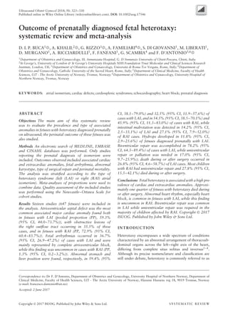
  • 1. Ultrasound Obstet Gynecol 2018; 51: 323–330 Published online in Wiley Online Library (wileyonlinelibrary.com). DOI: 10.1002/uog.17546 Outcome of prenatally diagnosed fetal heterotaxy: systematic review and meta-analysis D. I. P. BUCA1 , A. KHALIL2 , G. RIZZO3 , A. FAMILIARI4 , S. DI GIOVANNI1 , M. LIBERATI1 , D. MURGANO1 , A. RICCIARDULLI1 , F. FANFANI1 , G. SCAMBIA4 and F. D’ANTONIO5,6 1Department of Obstetrics and Gynaecology, SS. Annunziata Hospital, G. D’Annunzio University of Chieti-Pescara, Chieti, Italy; 2St George’s, University of London & St George’s University Hospitals NHS Foundation Trust Molecular and Clinical Sciences Research Institute, London, UK; 3Department of Obstetrics and Gynaecology, Universit`a di Roma Tor Vergata, Rome, Italy; 4Department of Obstetrics and Gynaecology, Catholic University of the Sacred Heart, Rome, Italy; 5Department of Clinical Medicine, Faculty of Health Sciences, UiT - The Arctic University of Norway, Tromsø, Norway; 6Department of Obstetrics and Gynaecology, University Hospital of Northern Norway, Tromsø, Norway KEYWORDS: atrial isomerism; cardiac defects; cardiosplenic syndromes; echocardiography; heart block; prenatal diagnosis ABSTRACT Objectives The main aim of this systematic review was to evaluate the prevalence and type of associated anomalies in fetuses with heterotaxy diagnosed prenatally on ultrasound; the perinatal outcome of these fetuses was also studied. Methods An electronic search of MEDLINE, EMBASE and CINAHL databases was performed. Only studies reporting the prenatal diagnosis of isomerism were included. Outcomes observed included associated cardiac and extracardiac anomalies, fetal arrhythmia, abnormal karyotype, type of surgical repair and perinatal mortality. The analysis was stratified according to the type of heterotaxy syndrome (left (LAI) or right (RAI) atrial isomerism). Meta-analyses of proportions were used to combine data. Quality assessment of the included studies was performed using the Newcastle–Ottawa Scale for cohort studies. Results Sixteen studies (647 fetuses) were included in the analysis. Atrioventricular septal defect was the most common associated major cardiac anomaly found both in fetuses with LAI (pooled proportion (PP), 59.3% (95% CI, 44.0–73.7%)), with obstructive lesions of the right outflow tract occurring in 35.5% of these cases, and in fetuses with RAI (PP, 72.9% (95% CI, 60.4–83.7%)). Fetal arrhythmias occurred in 36.7% (95% CI, 26.9–47.2%) of cases with LAI and were mainly represented by complete atrioventricular block, while this finding was uncommon in cases with RAI (PP, 1.3% (95% CI, 0.2–3.2%)). Abnormal stomach and liver position were found, respectively, in 59.4% (95% Correspondence to: Dr F. D’Antonio, Department of Obstetrics and Gynecology, University Hospital of Northern Norway, Department of Clinical Medicine, Faculty of Health Sciences, UiT - The Arctic University of Norway, Hansine Hansens veg 18, 9019 Tromsø, Norway (e-mail: francesco.dantonio@uit.no) Accepted: 2 June 2017 CI, 38.1–79.0%) and 32.5% (95% CI, 11.9–57.6%) of cases with LAI, and in 54.5% (95% CI, 38.5–70.1%) and 45.9% (95% CI, 11.3–83.0%) of cases with RAI, while intestinal malrotation was detected in 14.2% (95% CI, 2.5–33.1%) of LAI and 27.1% (95% CI, 7.9–52.0%) of RAI cases. Hydrops developed in 11.8% (95% CI, 2.9–25.6%) of fetuses diagnosed prenatally with LAI. Biventricular repair was accomplished in 78.2% (95% CI, 64.3–89.4%) of cases with LAI, while univentricular repair or palliation was needed in 17.0% (95% CI, 9.7–25.9%); death during or after surgery occurred in 26.8% (95% CI, 4.6–58.7%) of LAI cases. Most children with RAI had univentricular repair and 27.8% (95% CI, 15.5–42.1%) died during or after surgery. Conclusions Fetal heterotaxy is associated with a high pre- valence of cardiac and extracardiac anomalies. Approxi- mately one quarter of fetuses with heterotaxy died during or after surgery. Abnormal heart rhythm, especially heart block, is common in fetuses with LAI, while this finding is uncommon in RAI. Biventricular repair was common in LAI while univentricular repair was required in the majority of children affected by RAI. Copyright © 2017 ISUOG. Published by John Wiley & Sons Ltd. INTRODUCTION Heterotaxy encompasses a wide spectrum of conditions characterized by an abnormal arrangement of thoracoab- dominal organs across the left–right axis of the heart, differing from complete situs solitus and inversus1–4 . Although its precise nomenclature and classification are still under debate, heterotaxy is commonly referred to as Copyright © 2017 ISUOG. Published by John Wiley & Sons Ltd. SYSTEMATIC REVIEW
  • 2. 324 Buca et al. ‘isomerism’, which describes a situation in which right or left morphological structures are found on both sides of the body, and is the currently accepted term used to describe a heart with isomeric atria and atrial appendages. Right atrial isomerism (RAI) is typically, but not invari- ably, associated with asplenia while left atrial isomerism (LAI) is linked to polysplenia (55% of cases)2,5 . Associated major cardiac anomalies are the main determinant in anticipating the outcome for children affected by heterotaxy; however, even those presenting with isolated defects can experience short- and long-term morbidity such as intestinal malrotation, biliary atresia and respiratory and immune disorders6–9 . Assessment of the thoracoabdominal situs is an integral part of the sonographic screening examination of the fetal heart and has led to an increase in the prenatal diagnosis of heterotaxy10 . Despite this, assessment of atrial morphology and the anatomy of the spleen on prenatal ultrasound is challenging and is still to be validated in the general population, while lung lobulation cannot be described reliably in utero11,12. In this context, the prenatal diagnosis of fetal heterotaxy relies mainly on the identification of cardiac and extracardiac defects reported to be commonly associated with these anomalies postnatally. Interruption of the inferior vena cava (IVC) with azygos continuation is usually considered a proxy for LAI, while juxtaposition of the aorta and the IVC in fetuses presenting with major congenital heart disease (CHD) is highly suggestive of RAI. The medical pediatric literature reports high rates of associated cardiac and extracardiac anomalies in children affected by heterotaxy1,13,14 . However, these reports might be biased by the fact that only symptomatic children or those requiring surgery were included, and it is not entirely certain whether the information contained in the literature could be used to counsel parents with a pregnancy affected by these defects. The main aim of this systematic review was to evaluate the prevalence and type of associated anomaly in fetuses with heterotaxy diagnosed prenatally on ultrasound; the perinatal outcome of these fetuses was also assessed. METHODS This review was performed according to an a-priori designed protocol recommended for systematic reviews and meta-analyses15–18. The electronic databases MED- LINE, EMBASE and CINAHL were searched on 20 December 2016, utilizing combinations of the rele- vant medical subject heading terms, keywords and word variants for ‘isomerism’, ‘heterotaxy’, ‘cardios- plenic syndromes’ and ‘outcome’ (Table S1). The search and selection criteria were limited to the last two decades (1996–2016) and restricted to the English lan- guage. Reference lists of relevant articles and reviews were hand searched for additional reports. PRISMA guidelines were followed19 . The review was registered with the PROSPERO database (registration number: CRD 42016053972). The observed outcomes were associated cardiac anoma- lies, associated fetal arrhythmias, associated extracardiac anomalies, abnormal karyotype, termination of preg- nancy (TOP), intrauterine death (IUD), neonatal death (NND), late death (LD) and need for uni- or biventricular repair and associated intra- and postsurgical mortality. All outcomes were observed separately in fetuses with LAI and RAI. To assess the associated cardiac anomalies, the prevalence of the following defects was explored: atrioventricular septal defect (AVSD); atrial septal defect or common atrium; ventricular septal defect; single ventricle; left ventricular outflow tract obstruction (LVOTO), including aortic stenosis and atresia; coarctation of the aorta (CoA); right ventricular outflow tract obstruction (RVOTO), including pulmonary stenosis and atresia; conotruncal anomalies, including tetralogy of Fallot, double outlet right or left ventricle and common arterial trunk; transposition of the great arteries (TGA), including complete or congenitally corrected TGA and malposition; persistent left superior vena cava (PLSVC); interruption of the IVC with azygos continuation; abnormal pulmonary venous return, including partial and total types of the anomaly; dextrocardia, defined as an abnormal cardiac axis pointing to the right; and arrhythmias. To evaluate associated extracardiac anomalies, the prevalence of the following defects was explored: polysplenia, asplenia, abnormal stomach and liver posi- tion, intestinal obstruction and malrotation, abnormali- ties of the hepatobiliary tract, hydrops, abdominal wall defects and central nervous system, thoracic, facial, renal, limb and spinal anomalies. Abnormal karyotype was observed only in fetuses for which full karyotype test- ing was performed, either pre- or postnatally. Finally, the need for uni- or biventricular repair and the asso- ciated intra- and postsurgical mortality were assessed only in liveborn infants. IUD was defined as fetal loss at or after 20 weeks’ gestation, while NND and LD were defined as death occurring within and after 28 days of age, respectively. Only studies reporting the prenatal diagnosis of LAI or RAI were considered suitable for inclusion in the systematic review. Postnatal studies and studies from which cases diagnosed prenatally could not be extracted were excluded. Studies reporting fetal isomerism in the setting of a specific cardiac or extracardiac anomaly were not considered suitable for inclusion. Autopsy-based studies were also excluded on the basis that fetuses undergoing TOP are more likely to show associated major structural and chromosomal anomalies. Finally, studies not providing a clear classification of the anomaly and studies not differentiating between LAI and RAI were not considered suitable for inclusion in the review. The wide heterogeneity in nomenclature among published studies resulted in heterogeneity in risk stratification of these fetuses, therefore we included only studies providing definitions of the anomalies in accordance with those reported above. Only full-text articles were considered eligible for inclusion. Case reports, conference abstracts Copyright © 2017 ISUOG. Published by John Wiley & Sons Ltd. Ultrasound Obstet Gynecol 2018; 51: 323–330.
  • 3. Outcome of prenatally diagnosed fetal heterotaxy 325 and case series with fewer than three cases of isomerism were also excluded in order to avoid publication bias. The studies were assessed according to the following criteria: population, outcome, type of heterotaxy syn- drome and time of follow-up. Two authors (D.B., F.D.) reviewed all abstracts independently, and agreement regarding potential relevance was reached by consensus. Full-text copies of those papers identified as relevant from the abstracts were obtained and the same two reviewers independently extracted relevant data related to study characteristics and pregnancy outcomes. Inconsistencies were discussed by the reviewers and consensus reached by discussion with a third author. If more than one study had been published for the same cohort with identical endpoints, the report containing the most comprehensive information on the population was included to avoid overlapping populations. For articles in which informa- tion of interest was not reported but the methodology was such that this information was expected to have been recorded initially, the authors were contacted requesting the data. Quality assessment of the included studies was performed using the Newcastle–Ottawa Scale (NOS) for cohort studies. According to the NOS, each study is judged on three broad perspectives: selection of the study groups; comparability of the groups; and ascertainment of the outcome of interest20. Assessment of the selection of a study includes evaluation of the representativeness of the exposed cohort, selection of the non-exposed cohort, ascertainment of exposure and demonstration that the outcome of interest was not present at the beginning of the study. Assessment of the comparability of the study includes evaluation of the comparability of cohorts on the basis of the design or analysis. Finally, ascertainment of the outcome of interest includes evaluation of the type of assessment of the outcome of interest and length and adequacy of follow-up20 . According to the NOS, a study can be awarded a maximum of one star for each numbered item within the selection and outcome categories, and a maximum of two stars can be given for comparability. Statistical analysis Meta-analyses of proportions were used to combine the data21 . Funnel plots displaying the outcome rate from individual studies vs their precision (1/standard error) were produced with an exploratory aim. Tests for funnel-plot asymmetry were not used when the total number of publications included for each outcome was less than 10. In such cases, the power of the tests to distinguish chance from real asymmetry is too low22–24 . Between-study heterogeneity was explored using the I2 statistic, which represents the percentage of between-study variation that is due to heterogeneity rather than chance. A value of 0% indicates no observed heterogeneity, while I2 values of ≥ 50% indicate a substantial level of heterogeneity. A random-effects model was used for all analyses. All proportion meta-analysis was carried out using StatsDirect 2.7.9 (StatsDirect Ltd, Altrincham, UK). RESULTS A total of 2037 articles were identified from the electronic search, of which 134 were assessed with respect to their eligibility for inclusion (Table S2) and a total of 16 studies were eventually included in the systematic review (Figure 1, Table 1)12,14,25–38 . These 16 studies included 647 fetuses with a prenatal diagnosis of isomerism, of which LAI was confirmed at birth in 61.7% (95% CI, 57.8–65.4%) and RAI in 38.3% (95% CI, 34.6–42.2%). All studies included a relatively small number of cases and different periods of follow-up. Most of the included studies showed an overall good rate with regard to selection and comparability of the study groups and ascertainment of the outcome of interest (Table 2). The main weaknesses of the studies were their retrospective design, small sample size and the inclusion of high-risk populations. Furthermore, the relatively short period of follow-up after birth did not allow for a precise estimate of the long-term outcome for these children, especially those not undergoing surgery immediately after birth. Records screened (n = 2037) Full-text articles assessed for eligibility (n = 133) Records excluded (n = 1904) Records identified through database search (n = 2025) Additional records identified through other sources (n = 12) Records after duplicates removed (n = 2037) Full-text articles excluded, with reasons (n = 117) Studies included in qualitative synthesis (n = 16) Studies included in quantitative synthesis (meta-analysis) (n = 16) ScreeningIdentificationEligibilityIncluded Figure 1 Flowchart summarizing inclusion of studies in systematic review. Copyright © 2017 ISUOG. Published by John Wiley & Sons Ltd. Ultrasound Obstet Gynecol 2018; 51: 323–330.
  • 4. 326 Buca et al. Table 1 Summary of characteristics of studies included in systematic review evaluating cardiac and extracardiac anomalies and perinatal outcome of fetuses diagnosed prenatally with left (LAI) or right (RAI) atrial isomerism Study Country Study design Period analyzed (years) GA at diagnosis (weeks)* Heterotaxy Fetuses (n) Duration of follow-up† Gottschalk (2016)25 Germany Retrospective 1999–2013 24.0 ± 6.7 LAI, RAI 165 29 months Gaur (2016)26 USA Retrospective 2008–2013 23.0 ± 4.2 LAI, RAI 15 NS Escobar-Diaz (2014)27 USA Retrospective 1995–2011 22 (19–30) LAI, RAI 154 5 years Lee (2014)28 Korea Retrospective 1999–2011 26.9 (16.5–38.6) LAI, RAI 71 26.5 (1–138) months Nemec (2012)29 Austria Retrospective 2002–2009 NS LAI, RAI 5 NS Ozkutlu (2011)30 Turkey Retrospective 1999–2006 29.2 (21–37) LAI, RAI 6 NS Paladini (2011)31 Italy Retrospective 2004–2009 22 (13–34) LAI, RAI 22 NS Pepes (2009)32 UK Retrospective 1998–2008 18 (12–29) LAI 41 NS Yan (2008)33 Singapore Retrospective 1998–2005 21.3 (18–33) LAI, RAI 22 NS Taketazu (2006)34 Canada Retrospective 1992–2002 24.3 ± 6.4 LAI, RAI 71 NS Pasquini (2005)35 UK Prospective 1997–2003 25 (18–39) LAI, RAI 8 NS Lin (2000)14 Taiwan Retrospective 1994–1998 NS LAI, RAI 29 0–2 years Hoefstaetter (2000)36 Germany Retrospective 1995–2000 22.2 (21–25) LAI, RAI 7 NS Abuhamad (1999)12 USA Prospective 1995–1998 20.1 (17.4–24.4) LAI, RAI 8 NS Atkinson (1998)37 USA Retrospective 1991–1997 26 (18–36) LAI, RAI 13 NS Phoon (1996)38 USA Retrospective 1990–1994 NS LAI 10 1–24 months Only first author of each study is given. *Median (range) or mean ± SD. †Mean, median (range) or range. GA, gestational age, NS, not specified. Table 2 Quality assessment of included studies according to Newcastle–Ottawa Scale Author Selection Comparability Outcome Gottschalk (2016)25 Gaur (2016)26 Escobar-Diaz (2014)27 Lee (2014)28 Nemec (2012)29 Ozkutlu (2011)30 Paladini (2011)31 Pepes (2009)32 Yan (2008)33 Taketazu (2006)34 Pasquini (2005)35 Lin (2000)14 Hoefstaetter (2000)36 Abuhamad (1999)12 Atkinson (1998)37 Phoon (1996)38 Only first author of each study is given. A study can be awarded a maximum of one star for each numbered item within the selection and outcome categories and a maximum of two stars for comparability. Left atrial isomerism Associated major cardiac anomalies occurred in 83.4% (95% CI, 74.4–90.7%; I2 , 69.7%) of cases (Figure S1). AVSD was the most common associated cardiac defect, with a prevalence (pooled proportion (PP)) of 59.3% (95% CI, 44.0–73.7%) in the overall population of fetuses with a prenatal diagnosis of LAI, while RVOTO and transposition or malposition of the great arteries occurred in 35.5% (95% CI, 21.4–51.0%) and 11.0% (95% CI, 5.0–18.8%) of the cases, respectively (Table 3). Conotruncal anomalies, especially double outlet right ventricle (DORV), were found in 21.2% (95% CI, 16.0–27.0%) of LAI cases while LVOTO and CoA were less common (8.2% (95% CI, 3.7–14.2%) and 4.6% (95% CI, 1.4–9.4%), respectively). Anomalies of systemic veins such as PLSVC 28.5% (95% CI, 17.0–41.6%)) and interruption of the IVC (PP, 89.2% (95% CI, 80.5–95.5%)) occurred in the majority of cases, while those of pulmonary venous return, either partial or total, were less common (PP, 9.9% (95% CI, 5.4–15.5%)). Fetal arrhythmia occurred in 36.7% (95% CI, 26.9–47.2%) of all cases with LAI and was mainly rep- resented by complete atrioventricular block (CAVB) (PP, 26.5% (95% CI, (15.0–40.0%)) (Table 4 and Figure S2). Extracardiac anomalies occurred in 55.4% (95% CI, 35.1–74.8%) of fetuses with a prenatal diagnosis of LAI (Figure S3). When excluding anomalies of the abdominal situs and gastrointestinal tract, the rate of associated extracardiac anomalies was 16.0% (95% CI, 7.5–26.9%). Abnormal stomach and liver position were found, respectively, in 59.4% (95% CI, 38.1–79.0%) and 32.5% (95% CI, 11.9–57.6%) of cases with LAI, while malformations of the biliary tract such as biliary atresia were found in 8.0% (95% CI, 3.5–14.3%) (Table 5). Obstructive lesions of the bowel occurred in 4.9% (95% CI, 2.7–7.7%) of cases while malrotation was found in 14.2% (95% CI, 2.5–33.1%). Hydrops developed in 11.8% (95% CI, 2.9–25.6%) of fetuses with LAI mainly owing to the presence of CAVB (Table 5). Polysplenia was detected after birth in 56.6% (95% CI, 48.1–65.0%) of cases, while only 7.1% (95% CI, 2.1–14.9%) had asplenia (Table 5). Associated extracardiac anomalies involving other systems were less common. Finally, the prevalence of chromosomal anomalies was 3.0% (95% CI, 1.2–5.6%). TOP was performed in 24.8% (95% CI, 14.9–36.2%) of cases with LAI, while 6.7% (95% CI, 3.9–10.2%) of fetuses died in utero (Table 6 and Figure S4). The majority of fetuses with a prenatal diagnosis of LAI (60.7% (95% CI, 44.3–76.0%)) were born alive, while NND or LD occurred in 11.1% (95% CI, 6.1–17.3%) and 6.2% (95% CI, 4.0–8.9%) of cases, respectively (Table 6 and Copyright © 2017 ISUOG. Published by John Wiley & Sons Ltd. Ultrasound Obstet Gynecol 2018; 51: 323–330.
  • 5. Outcome of prenatally diagnosed fetal heterotaxy 327 Table 3 Pooled proportions (PP) for prevalence of associated intracardiac anomalies in fetuses diagnosed prenatally with left (LAI) or right (RAI) atrial isomerism LAI RAI Intracardiac anomaly Studies (n) Fetuses (n/N) PP (95% CI) (%) I2 (%) Studies (n) Fetuses (n/N) PP (95% CI) (%) I2 (%) AVSD 15 205/386 59.28 (44.0–73.7) 85.5 13 180/246 72.86 (60.4–83.7) 70.3 VSD 13 27/233 10.99 (3.1–22.9) 77.2 12 4/193 2.65 (0.9–5.3) 0 ASD 13 22/233 19.61 (6.8–37.0) 85.2 12 36/193 21.49 (2.8–50.9) 94.0 Single ventricle 13 56/347 13.76 (6.5–23.3) 71.8 12 104/238 37.84 (18.1–60.0) 89.9 LVOTO 15 35/386 8.17 (3.7–14.2) 59.4 13 18/246 6.91 (3.5–11.3) 21.8 CoA 15 18/386 4.57 (1.4–9.4) 58.8 13 5/246 3.01 (1.3–5.5) 0 RVOTO 15 108/386 35.49 (21.4–51.0) 86.3 13 173/246 67.43 (56.2–77.8) 61.3 Conotruncal anomaly 15 75/386 21.23 (16.0–27.0) 26.0 13 109/246 40.06 (28.0–52.8) 68.2 TGA 15 36/386 10.98 (5.0–18.8) 70.1 13 42/246 21.33 (10.9–34.1) 74.8 PLSVC 13 109/341 28.49 (17.0–41.6) 77.8 12 100/221 41.80 (28.9–55.3) 67.2 Interrupted IVC 15 336/386 89.15 (80.5–95.5) 75.4 13 11/246 4.86 (2.6–7.8) 0 Juxtaposition Ao/IVC 15 2/386 1.19 (0.3–2.5) 0 9 127/173 81.50 (59.2–96.2) 89.1 TAPVR 15 30/381 9.85 (5.4–15.5) 48.4 13 102/234 41.87 (28.4–56.0) 72.9 Dextrocardia 11 65/262 25.87 (13.3–40.9) 77.6 12 57/239 24.25 (10.4–41.6) 85.1 Arrhythmia 13 123/378 36.73 (26.9–47.2) 66.9 10 2/218 1.30 (0.2–3.2) 0 Ao, aorta; ASD, atrial septal defect; AVSD, atrioventricular septal defect; CoA, coarctation of aorta; IVC, inferior vena cava; LVOTO, left ventricular outflow tract obstruction; PLSVC, persistent left superior vena cava; RVOTO, right ventricular outflow tract obstruction; TAPVR, total anomalous pulmonary venous return; TGA, transposition of the great arteries; VSD, ventricular septal defect. Table 4 Pooled proportions (PP) for prevalence of associated cardiac arrhythmia in fetuses diagnosed prenatally with left (LAI) or right (RAI) atrial isomerism LAI RAI Cardiac arrhythmia Studies (n) Fetuses (n/N) PP (95% CI) (%) I2 (%) Studies (n) Fetuses (n/N) PP (95% CI) (%) I2 (%) Overall 13 123/378 36.73 (26.9–47.2) 66.9 10 2/218 1.30 (0.2–3.2) 0 AVB (overall) 11 96/367 28.82 (16.9–42.5) 82.3 9 1/216 1.14 (0.1–3.0) 0 First-degree AVB 11 0/367 0 (0–1.5) 0 9 0/216 0 (0–2.4) 0 Second-degree AVB 11 2/367 0.72 (0.1–1.9) 0 9 0/216 0 (0–2.4) 0 Third-degree AVB (CAVB) 11 93/367 26.51 (15.0–40.0) 82.5 9 1/216 1.14 (0.1–3.0) 0 Tachycardia 11 5/367 1.64 (0.3–4.2) 41.0 9 0/216 0 (0–2.4) 0 AVB, atrioventricular block; CAVB, complete AVB. Table 5 Pooled proportions (PP) for prevalence of associated extracardiac anomalies in fetuses diagnosed prenatally with left (LAI) or right (RAI) atrial isomerism LAI RAI Extracardiac anomaly Studies (n) Fetuses (n/N) PP (95% CI) (%) I2 (%) Studies (n) Fetuses (n/N) PP (95% CI) (%) I2 (%) Polysplenia 10 134/236 56.63 (48.1–65.0) 65.3 9 2/159 2.03 (0.4–4.8) 0 Asplenia 9 14/226 7.14 (2.1–14.9) 60 9 138/159 87.59 (75.5–96.0) 69.4 Abnormal stomach position 8 37/65 59.39 (38.1–79.0) 58.7 7 44/76 54.53 (38.5–70.1) 36.8 Abnormal liver position 6 24/53 32.54 (11.9–57.6) 68.5 7 50/76 45.89 (11.3–83.0) 90.1 Abnormal hepatobiliary tract 9 17/278 8.02 (3.5–14.3) 46.7 9 5/179 3.75 (0.5–9.8) 54.6 Intestinal malrotation 9 46/278 14.21 (2.5–33.1) 90.2 9 47/179 27.12 (7.9–52.0) 90.5 Gastrointestinal obstruction 9 12/278 4.91 (2.7–7.7) 0 9 6/179 3.97 (1.6–7.3) 0 Hydrops 11 45/258 11.76 (2.9–25.6) 83.4 10 6/180 3.67 (1.4–6.8) 0 Central nervous system anomaly 9 4/249 2.43 (0.9–4.7) 0 9 3/143 2.77 (0.7–6.0) 0 Facial anomaly 9 3/249 1.57 (0.3–3.9) 10 8 1/120 1.45 (0.1–4.3) 0 Thoracic anomaly 9 2/249 1.10 (0.2–2.7) 0 8 1/120 1.92 (0.3–5.1) 0 Abdominal wall defect 9 1/249 0.97 (0.1–2.5) 0 8 2/120 2.38 (0.4–5.8) 0 Limb anomaly 9 0/249 0 (0–1.8) 0 8 2/120 2.44 (0.2–7.2) 0 Kidney anomaly 9 7/249 3.24 (0.6–7.8) 37.8 8 3/120 3.43 (1.0–7.3) 0 Spine anomaly 9 2/249 1.10 (0.2–2.7) 0 8 0/120 0 (0–3.7) 0 Copyright © 2017 ISUOG. Published by John Wiley & Sons Ltd. Ultrasound Obstet Gynecol 2018; 51: 323–330.
  • 6. 328 Buca et al. Table 6 Pooled proportions (PP) for prevalence of abnormal perinatal outcome in fetuses diagnosed prenatally with left (LAI) or right (RAI) atrial isomerism LAI RAI Perinatal outcome Studies (n) Fetuses (n/N) PP (95% CI) (%) I2 (%) Studies (n) Fetuses (n/N) PP (95% CI) (%) I2 (%) Termination of pregnancy 15 99/380 24.79 (14.9–36.2) 76.2 13 77/238 33.46 (22.6–45.3) 63.3 Intrauterine death 15 25/380 6.73 (3.9–10.2) 19.4 13 9/238 4.32 (2.1–7.3) 1.7 Neonatal death 14 34/668 11.12 (6.1–17.3) 50.1 11 36/232 17.55 (8.7–28.7) 69.4 Late death 13 21/365 6.24 (4.0–8.9) 0 11 37/232 14.69 (7.9–23.1) 54.3 Table 7 Pooled proportions (PP) for prevalence of abnormal surgical outcome in fetuses diagnosed prenatally with left (LAI) or right (RAI) atrial isomerism LAI RAI Surgical outcome Studies (n) Fetuses (n/N) PP (95% CI) (%) I2 (%) Studies (n) Fetuses (n/N) PP (95% CI) (%) I2 (%) Need for surgery 3 82/109 73.43 (44.4–94.3) 82.9 3 41/62 70.06 (20.7–99.6) 93.1 Biventricular repair 2 64/80 78.16 (64.3–89.4) 34.6 2 3/39 7.42 (0.5–33.7) 79.3 Univentricular repair 2 13/80 17.03 (9.7–25.9) 0 2 36/39 92.60 (66.3–99.5) 79.3 Deaths during or after surgery 3 11/82 26.80 (4.6–58.7) 78.3 3 11/41 27.81 (15.5–42.1) 27.0 Figure S5). Surgical intervention for CHD was performed in 73.4% (95% CI, 44.4–94.3%) of fetuses with LAI that were born alive. Biventricular repair was accomplished in 78.2% (95% CI, 64.3–89.4%) of the cases, while univentricular repair or palliation was required in 17.0% (95% CI, 9.7–25.9%) of children. Overall, death during or after surgery occurred in 26.8% (95% CI, 4.6–58.7%) of cases with LAI (Table 7). Right atrial isomerism Associated cardiac anomalies were present in the majority of fetuses with a prenatal diagnosis of RAI (PP, 97.8% (95% CI, 95.3–99.4%); I2, 11.9%) (Figure S1). AVSD was the commonest heart defect, with a prevalence of 72.9% (95% CI, 60.4–83.7%) in the overall pop- ulation of fetuses with RAI, while RVOTO and conotruncal anomalies (mainly DORV) occurred in 67.4% (95% CI, 56.2–77.8%) and 40.1% (95% CI, 28.0–52.8%) of cases, respectively (Table 3). PLSVC and total anomalous pulmonary venous return were common, with a prevalence of 41.8% (95% CI, 28.9–55.3%) and 41.9% (95% CI, 28.4–56.0%), respectively. Juxtaposi- tion of the aorta and IVC, which constitutes one of the main prenatal diagnostic features of RAI in utero, was found in 81.5% (95% CI, 59.2–96.2%) of cases, while interruption of the IVC was less common (PP, 4.9% (95% CI, 2.6–7.8%)) (Table 3). Abnormal heart rhythm was not common, with a prevalence of 1.3% (95% CI, 0.2–3.2%) (Table 4 and Figure S2). Associated extracardiac anomalies occurred in 62.9% (95% CI, 32.5–88.4%; I2 , 93.6%) of cases with a prena- tal diagnosis of RAI (Figure S3). Among other structural abnormalities, those involving the gastrointestinal tract were the most common. Abnormal position of the stomach and liver were found, respectively, in 54.5% (95% CI, 38.5–70.1%) and 45.9% (95% CI, 11.3–83.0%) of fetuses with RAI, while malformations of the biliary tract, such as atresia, were less common (Table 5). Intestinal malrotation occurred in 27.1% (95% CI, 7.9–52.0%). Asplenia was present in the majority of cases (PP, 87.6% (95% CI, 75.5–96.0%)) while polysplenia, a common feature of LAI, was rare (PP, 2.0% (95% CI, 0.4–4.8%)) (Table 5). Only a small proportion (PP, 3.7% (95% CI, 1.4–6.8%)) of fetuses with RAI presented with hydrops, which is likely to be associated with the low incidence of CAVB. Finally, 4.0% (95% CI, 0.8–9.6%) of cases had abnormal karyotype. About one third of the pregnancies affected by RAI were terminated, while 4.3% (95% CI, 2.1–7.3%) experienced IUD (Table 6, Figure S4). Of the fetuses with a prenatal diagnosis of RAI born alive, 17.6% (95% CI, 8.7–28.7%) and 14.7% (95% CI, 7.9–23.1%) died during or after the neonatal period, respectively (Table 6, Figure S5). Surgical intervention for CHD was performed in 70.1% (95% CI, 20.7–99.6%) of children (Table 7). A large majority of those children required univentricular repair, while in a small proportion of cases (PP, 7.4% (95% CI, 0.5–33.7%)) biventricular repair was accomplished (Table 7). Overall, 27.8% (95% CI, 15.5–42.1%) died during or after surgery. DISCUSSION Main findings This systematic review shows that fetuses with heterotaxy are affected by a high rate of associated cardiac and extracardiac anomalies, with abnormalities of the atrioventricular valves being among the most common. Abnormal heart rhythm, especially CAVB, occurs in about a third of cases of LAI and is responsible for the high rate of cases presenting with hydrops at an early gestational age, while this finding is rare in cases with RAI. In cases undergoing surgery after birth, biventricular repair was common in LAI while univentricular repair was required in a large majority of children affected by RAI. Intra- or post-surgical mortality was high in both LAI and RAI and occurred in about a quarter of these cases. Copyright © 2017 ISUOG. Published by John Wiley & Sons Ltd. Ultrasound Obstet Gynecol 2018; 51: 323–330.
  • 7. Outcome of prenatally diagnosed fetal heterotaxy 329 Strengths and limitations of the study The small number of cases in each study, their retrospective design, different periods of follow-up and inclusion criteria, with most of the series involving high-risk populations, represent the major limitations of this systematic review. Unfortunately, the low number of studies did not permit meaningful stratified meta-analyses to explore the test performance in subgroups of studies that may be less or more susceptible to bias. Assessment of potential publication bias was also problematic, because of both the outcome nature (rates with the left side limited to the value zero), which limits the reliability of funnel plots, and the low number of individual studies, which strongly limits the reliability of formal tests. Most of the studies included in this review involved high-risk populations, which might have led to an overestimation of the associated cardiac and extracardiac anomalies. Finally, the different times of follow-up and postnatal imaging protocols among the different studies might have masked the real occurrence of associated cardiac and extracardiac anomalies, since some anomalies may manifest only later in childhood, while others can be easily overlooked at a standard clinical examination. Despite these limitations, however, the present review represents the best published estimate of a number of outcomes in fetuses diagnosed with isomerism. Interpretation of findings Assessment of the fetal situs is an integral part of the second-trimester routine anomaly scan and has led to an increase in the detection rate of heterotaxy syndromes10 . Despite this, precise characterization of the type of heterotaxy syndrome is not always feasible in utero. Autopsy studies have reported that morphology of the atrial appendages can differentiate reliably between LAI and RAI39 . Characterization of the morphology of the atrial appendages in utero has been reported to be feasible and can improve the detection of these anomalies, especially when new ultrasound techniques, such as four-dimensional ultrasound, are employed, but it may be challenging11,31 . Prenatal diagnosis of heterotaxy on ultrasound mainly relies on the presence of associated cardiac and extracar- diac anomalies, which are typically reported to co-exist with isomerism. RAI is almost invariably associated with CHD, especially conotruncal anomalies, RVOTO, transposition and hypoplastic left heart syndrome, and it is generally affected by a worse perinatal outcome compared with LAI in view of the higher incidence of associated CHD. Furthermore, total abnormal pulmonary venous return occurs in almost half the cases with RAI and represents one of the major determinants of postnatal outcome in these children, especially when obstructed. Conversely, interruption of the IVC can be the only sign of LAI, thus making prenatal diagnosis of this anomaly challenging, although it has also been also reported in fetuses not affected by isomerism40 . Hydrops developed in approximately 10% of fetuses affected by LAI, mainly because of the presence of a CAVB, which occurs in a quarter of cases. Fetal hydrops is one of the major determinants of poor perinatal outcome in fetuses with LAI and can be evident from the first trimester of pregnancy41 . CAVB was the most common fetal arrhythmia seen in fetuses with LAI and was associated with a high burden of perinatal mortality and morbidity. Furthermore, CAVB in the setting of LAI can be associated with myocardial non-compaction, which is characterized by regional ventricular wall thickening and deep trabecular recesses and is almost invariably associated with a poor prognosis42. Chromosomal anomalies are not common in hetero- taxy; in the present review, abnormal karyotype was present in 3.0% (95% CI, 1.2–5.6%) of cases with LAI and 4.0% (95% CI, 0.8–9.6%) of cases with RAI. These data might influence the parental decision as to whether or not to proceed with an invasive prenatal diagnosis. Abnormalities of the biliary tract, such as biliary atresia, are typically associated with heterotaxy. Newborns with biliary atresia develop progressive cholestasis, which may evolve towards irreversible cirrhosis and liver failure if untreated. Prompt recognition and treatment are essential to improve the outcome for these children43,44 ; however, the prenatal diagnosis of biliary atresia is challenging. Non-visualization of the fetal gallbladder, combined with assessment of amniotic fluid digestive enzymes, has been reported to have an overall good detection rate for biliary atresia, though validation in larger studies is required45–48. Fetuses suspected of being affected by heterotaxy should be referred for a detailed assessment of the hepatobiliary tract in order to rule out biliary atresia. Nevertheless, urgent postnatal assessment should be performed in order to rule out this anomaly43,44. Intestinal malrotation has been variably associated with heterotaxy, with a reported incidence ranging from 30% to 90% in the pediatric literature49–51 . Intestinal malrotation increases the risk of midgut volvulus, which can potentially result in significant bowel loss, intestinal insufficiency and death52,53 . A recent systematic review54 exploring the association between intestinal malrotation and heterotaxy reported a prevalence of 58% (95% CI, 36–78%) in children with heterotaxy undergoing screening for bowel anomalies, although there was wide heterogeneity in screening regimens among the included studies. In the present review, intestinal malrotation occurred in 14.2% and 27.1% of cases with LAI and RAI, respectively. However, there was significant heterogeneity in the reported incidence owing to different screening poli- cies for intestinal anomalies among the included studies. Fetal magnetic resonance imaging (MRI) is an impor- tant adjunct imaging modality to ultrasound and has been shown to provide optimal anatomical characterization of the bronchial, hepatic, intestinal and splenic anatomy. In view of these findings, it might seem reasonable to refer cases of heterotaxy suspected on ultrasound to undergo fetal MRI, in order to determine the status of the spleen and rule out associated abnormalities, such as biliary atresia and intestinal malrotation, which might influence the prognosis for children with heterotaxy29,55,56. Copyright © 2017 ISUOG. Published by John Wiley & Sons Ltd. Ultrasound Obstet Gynecol 2018; 51: 323–330.
  • 8. 330 Buca et al. REFERENCES 1. Degenhardt K, Rychik J. Fetal Situs, Isomerism, Heterotaxy Syndrome: Diagnostic Evaluation and Implication for Postnatal Management. Curr Treat Options Cardiovasc Med 2016; 18: 77. 2. Jacobs JP, Anderson RH, Weinberg PM, Walters HL 3rd , Tchervenkov CI, Del Duca D, Franklin RC, Aiello VD, B´eland MJ, Colan SD, Gaynor JW, Krogmann ON, Kurosawa H, Maruszewski B, Stellin G, Elliott MJ. The nomenclature, definition and classification of cardiac structures in the setting of heterotaxy. Cardiol Young 2007; 17 (Suppl): 1–28. 3. Loomba RS, Hlavacek AM, Spicer DE, Anderson RH. Isomerism or heterotaxy: which term leads to better understanding? Cardiol Young 2015; 25: 1037–1043. 4. Icardo JM, Sanchez de Vega MJ. Spectrum of heart malformations in mice with situs solitus, situs inversus, and associated visceral heterotaxy. Circulation 1991; 84: 2547–2558. 5. Peoples WM, Moller JH, Edwards JE. Polysplenia: a review of 146 cases. Pediatr Cardiol 1983; 4: 129–137. 6. Kothari SS. Non-cardiac issues in patients with heterotaxy syndrome. Ann Pediatr Cardiol 2014; 7: 187–192. 7. Tan YW, Khalil A, Kakade M, Carvalho JS, Bradley S, Cleeve S, Giuliani S. Screening and Treatment of Intestinal Rotational Abnormalities in Heterotaxy: A Systematic Review and Meta-Analysis. J Pediatr 2016; 171: 153–162.e1–3. 8. Brueckner M. Heterotaxia, congenital heart disease, and primary ciliary dyskinesia. Circulation 2007; 115: 2793–2795. 9. Kennedy MP, Omran H, Leigh MW, Dell S, Morgan L, Molina PL, Robinson BV, Minnix SL, Olbrich H, Severin T, Ahrens P, Lange L, Morillas HN, Noone PG, Zariwala MA, Knowles MR. Congenital heart disease and other heterotaxic defects in a large cohort of patients with primary ciliary dyskinesia. Circulation 2007; 115: 2814–2821. 10. International Society of Ultrasound in Obstetrics and Gynecology, Carvalho JS, Allan LD, Chaoui R, Copel JA, DeVore GR, Hecher K, Lee W, Munoz H, Paladini D, Tutschek B, Yagel S. ISUOG Practice Guidelines (updated): sonographic screening examination of the fetal heart. Ultrasound Obstet Gynecol 2013; 41: 348–359. 11. Berg C, Geipel A, Kohl T, Smrcek J, Germer U, Baschat AA, Hansmann M, Gembruch U. Fetal echocardiographic evaluation of atrial morphology and the prediction of laterality in cases of heterotaxy syndromes. Ultrasound Obstet Gynecol 2005; 26: 538–545. 12. Abuhamad AZ, Robinson JN, Bogdan D, Tannous RJ. Color Doppler of the splenic artery in the prenatal diagnosis of heterotaxic syndromes. Am J Perinatol 1999; 16: 469–473. 13. Lim JS, McCrindle BW, Smallhorn JF, Golding F, Caldarone CA, Taketazu M, Jaeggi ET. Clinical features, management, and outcome of children with fetal and postnatal diagnoses of isomerism syndromes. Circulation 2005; 112: 2454–2461. 14. Lin AE, Ticho BS, Houde K, Westgate MN, Holmes LB. Heterotaxy: associated conditions and hospital-based prevalence in newborns. Genet Med 2000; 2: 157–172. 15. Henderson LK, Craig JC, Willis NS, Tovey D, Webster AC. How to write a Cochrane systematic review. Nephrology 2010; 15: 617–624. 16. NHS Centre for Reviews and Dissemination. Systematic reviews: CRD’s guidance for undertaking reviews in health care. University of York: York, UK, 2009. 17. Leeflang MM, Deeks JJ, Gatsonis C, Bossuyt PM; Cochrane Diagnostic Test Accuracy Working Group. Systematic reviews of diagnostic test accuracy. Ann Intern Med 2008; 149: 889–897. 18. Cochrane Handbook for Systematic Reviews of Diagnostic Test Accuracy. http:// srdta.cochrane.org/handbook-dta-reviews. 19. Prisma statement. http://www.prisma-statement.org/ [accessed 10 July 2016]. 20. Wells GA, Shea B, O’Connell D, Peterson J, Welch V, Losos M, Tugwell P. Newcastle–Ottawa Scale for assessing the quality of nonrandomised stud- ies in meta-analyses. http://www.ohri.ca/programs/clinical&uscore;epidemiology/ oxford.asp [accessed 20 November 2015]. 21. Hunter JP, Saratzis A, Sutton AJ, Boucher RH, Sayers RD, Bown MJ. In meta-analyses of proportion studies, funnel plots were found to be an inaccurate method of assessing publication bias. J Clin Epidemiol 2014; 67: 897–903. 22. Egger M, Davey Smith G, Schneider M, Minder C. Bias in meta-analysis detected by a simple, graphical test. BMJ 1997; 315: 629–634. 23. Manzoli L, De Vito C, Salanti G, D’Addario M, Villari P, Ioannidis JP. Meta-analysis of the immunogenicity and tolerability of pandemic influenza A 2009 (H1N1) vaccines. PLoS One 2011; 6: e24384. 24. Higgins JPT, Green S. Cochrane Handbook for Systematic Reviews of Interventions Version 5.0.2 [updated September 2009]. The Cochrane Collaboration 2009. www .cochrane-handbook.org. [Accessed 10 December 2016]. 25. Gottschalk I, Stressig R, Ritgen J, Herberg U, Breuer J, Vorndamme A, Strizek B, Willruth A, Geipel A, Gembruch U, Berg C. Extracardiac anomalies in prenatally diagnosed heterotaxy syndrome. Ultrasound Obstet Gynecol 2016; 47: 443–449. 26. Gaur L, Talemal L, Bulas D, Donofrio MT. Utility of fetal magnetic resonance imaging in assessing the fetus with cardiac malposition. Prenat Diagn 2016; 36: 752–759. 27. Escobar-Diaz MC, Friedman K, Salem Y, Marx GR, Kalish BT, Lafranchi T, Rathod RH, Emani S, Geva T, Tworetzky W. Perinatal and infant outcomes of prenatal diagnosis of heterotaxy syndrome (asplenia and polysplenia). Am J Cardiol 2014; 114: 612–617. 28. Lee MY, Won HS, Shim JY, Lee PR, Lee BS, Kim EA, Kim YH, Park JJ, Yun TJ, Kim A. Prenatal diagnosis of atrial isomerism in the Korean population. Obstet Gynecol Sci 2014; 57: 193–200. 29. Nemec SF, Brugger PC, Nemec U, Bettelheim D, Kasprian G, Amann G, Rimoin DL, Graham JM Jr, Prayer D. Situs anomalies on pre-natal MRI. Eur J Radiol 2012; 81: e495–501. 30. Ozkutlu S, Bostan OM, Deren O, Ondero˘glu L, Kale G, G¨uc¸er S, Orhan D. Prenatal echocardiographic diagnosis of cardiac right/left axis and malpositions according to standardized Cordes technique. Anadolu Kardiyol Derg 2011; 11: 131–136. 31. Paladini D, Sglavo G, Masucci A, Pastore G, Nappi C. Role of four-dimensional ultrasound (spatiotemporal image correlation and sonography-based automated volume count) in prenatal assessment of atrial morphology in cardiosplenic syndromes. Ultrasound Obstet Gynecol 2011; 38: 337–343. 32. Pepes S, Zidere V, Allan LD. Prenatal diagnosis of left atrial isomerism. Heart 2009; 95: 1974–1977. 33. Yan YL, Tan KB, Yeo GS. Right atrial isomerism: preponderance in Asian fetuses. Using the stomach–distance ratio as a possible diagnostic tool for prediction of right atrial isomerism. Ann Acad Med Singapore 2008; 37: 906–912. 34. Taketazu M, Lougheed J, Yoo SJ, Lim JS, Hornberger LK. Spectrum of cardiovascular disease, accuracy of diagnosis, and outcome in fetal heterotaxy syndrome. Am J Cardiol 2006; 97: 720–724. 35. Pasquini L, Tan T, Yen Ho S, Gardiner H. The implications for fetal outcome of an abnormal arrangement of the abdominal vessels. Cardiol Young 2005; 15: 35–42. 36. Hoefstaetter C, Plath H, Hansmann M. Prenatal diagnosis of abnormalities of the fetal venous system. Ultrasound Obstet Gynecol 2000; 15: 231–241. 37. Atkinson DE, Drant S. Diagnosis of heterotaxy syndrome by fetal echocardiography. Am J Cardiol 1998; 82: 1147–1149. 38. Phoon CK, Villegas MD, Ursell PC, Silverman NH. Left atrial isomerism detected in fetal life. Am J Cardiol 1996; 77: 1083–1088. 39. Ho SY, Cook A, Anderson RH, Allan LD, Fagg N. Isomerism of the atrial appendages in the fetus. Pediatr Pathol 1991; 11: 589–608. 40. Bronshtein M, Khatib N, Blumenfeld Z. Prenatal diagnosis and outcome of isolated interrupted inferior vena cava. Am J Obstet Gynecol 2010; 202: 398.e1–4. 41. Baschat AA, Gembruch U, Kn¨opfle G, Hansmann M. First-trimester fetal heart block: a marker for cardiac anomaly. Ultrasound Obstet Gynecol 1999; 14: 311–314. 42. Friedberg M, Ursell P, Silverman N. Isomerism of the left atrial appendage associated with ventricular noncompaction. Am J Cardiol 2006; 96: 985–990. 43. McKiernan PJ, Baker AJ, Kelly DA. The frequency and outcome of biliary atresia in the UK and Ireland. Lancet 2000; 355: 25–29. 44. Varela-Fascinetto G, Castaldo P, Fox IJ, Sudan D, Heffron TG, Shaw BW, Langnas AN. Biliary atresia–polysplenia syndrome: surgical and clinical relevance in liver transplantation. Ann Surg 1998; 227: 583–589. 45. Shen O, Rabinowitz R, Yagel S, Gal M. Absent gallbladder on fetal ultrasound: prenatal findings and postnatal outcome. Ultrasound Obstet Gynecol 2011; 37: 673–677. 46. Dreux S, Boughanim M, Lepinard C, Guichet A, Rival JM, de Becdelievre A, Dugueperoux I, Muller F. Relationship of non-visualization of the fetal gallbladder and amniotic fluid digestive enzymes analysis to outcome. Prenat Diagn 2012; 32: 423–426. 47. Bardin R, Ashwal E, Davidov B, Danon D, Shohat M, Meizner I. Nonvisualization of the Fetal Gallbladder: Can Levels of Gamma-Glutamyl Transpeptidase in Amniotic Fluid Predict Fetal Prognosis? Fetal Diagn Ther 2016; 39: 50–55. 48. Shen O, Rabinowitz R, Yagel S, Gal M. Comment on ‘‘Relationship of nonvisualization of the fetal gallbladder and amniotic fluid digestive enzymes analysis to outcome’’. Prenat Diagn 2012; 32: 1119–1120; author reply 1121. 49. Nakada K, Kawaguchi F, Wakisaka M, Nakada M, Enami T, Yamate N. Digestive tract disorders associated with asplenia/polysplenia syndrome. J Pediatr Surg 1997; 32: 91–94. 50. Ditchfield MR, Hutson JM. Intestinal rotational abnormalities in polysplenia and asplenia syndromes. Pediatr Radiol 1998; 28: 303–306. 51. Collins DC. 71 000 human appendix specimens. A final report, summarizing forty years’ study. Am J Proctol 1963; 14: 265–281. 52. Ladd WE. Congenital obstruction of the duodenum in children. N Engl J Med 1932; 206: 277–280. 53. Pockett CR, Dicken BJ, Rebeyka IM, Ross DB, Ryerson LM. Heterotaxy syndrome and intestinal rotation abnormalities: a survey of institutional practice. J Pediatr Surg 2013; 48: 2078–2083. 54. Tan YW, Khalil A, Kakade M, Carvalho JS, Bradley S, Cleeve S, Giuliani S. Screening and treatment of intestinal rotational abnormalities in heterotaxy: a systematic review and meta-analysis. J Pediatr 2016; 171: 153–162.e1–3. 55. Loomba R, Shah PH, Anderson RH. Fetal magnetic resonance imaging of malformations associated with heterotaxy. Cureus 2015; 7: e269. 56. Kheiri M, Lesieur E, Dabadie A, Colombani M, Capelle M, Sigaudy S, Guidicelli B, Heckenroth H, Delagausie P, Pico H, Philip N, Bretelle F, Gorincour G. Prenatal diagnosis of bowel malposition using T2-weighted fetal MRI sequences. Diagn Interv Imaging 2016; 97: 857–861. SUPPORTING INFORMATION ON THE INTERNET Tables S1 and S2 and Figures S1–S5 may be found in the online version of this article. Copyright © 2017 ISUOG. Published by John Wiley & Sons Ltd. Ultrasound Obstet Gynecol 2018; 51: 323–330.