
Recommended
More Related Content
What's hot
What's hot (20)
Similar to Anatomy of Uvea track
Similar to Anatomy of Uvea track (20)
Recently uploaded
Recently uploaded (20)
Anatomy of Uvea track
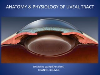
- 1. ANATOMY & PHYSIOLOGY OF UVEAL TRACT Dr.Lhacha Wangd(Resident) JDWNRH, KGUMSB
- 2. Outline • Anatomic classification of uveal track • Macroanatomy • Microanatomy • Physiology of uvea • Clinical correlates
- 3. Introduction The middle vascular layer of eyeball consists of three part- - iris, ciliary body, choroid Functions: -Regulates light entry & anchors the lens -Absorbs reflected light -Nourishes eye Walls of eye ball Middle vascular layer Outer Fibrous layer Inner nervous layer retina Uveal tract Sclera & cornea 2. Ciliary body 1.Iris Anterior uvea 3.choroid Posterior uvea
- 4. Embryology Iris- • Both the layers of epithelium are derived from the marginal region of optic cup(Neuroectoderm) • Sphincter and dilator derived from anterior epithelium • Stroma and vassels- mesoderm Ciliary body- • Proximal region of optic cup- neuroectoderm which undergoes convulation to form ciliary process(70-75) Choroid- • Vascular endothelium and the haemopoietic cells of choroid are derived from endoderm • Choroidal stroma ( vascular pericytes, smooth muscles, melanocytes and collagenous components) of choroid are derived from ectoderm Iris origin Ciliary body
- 5. Iris • Thin, pigmented contractile circular structure, analogous to the diaphragm of a camera • Anterior extension of uveal tract • Extends from iris root to iris margin that forms the pupil – Central aperture is called pupil (3-4mm) • Lies on anterior lens surface, surrounded by aqueous humour • Posteriorly, Central portion of iris is in contact with lens which give characteristic conical configuration • Functions; 1.Regulates light entry 2.Give distinctive eye colour
- 6. Iris- gross anatomy • Rough anterior surface & smooth posterior surface • Average diameter: 12mm • Collerate-thickened iris (0.6-1mm) wide located 2mm from the pupillary margin • Anteriorly iris divided into two parts by collarete 1.Inner pupillary zone (1-2mm wide) 2.Outer ciliary zone (3-4mm wide) Thickness: 0.5mm • Thickest at collarette • Thinnest at the root • Thin regions eg iris root & margin are more susceptible to tearing in injuries It is attached peripherally to the midline of the anterior surface of ciliary body
- 7. Iris- Macroscopic appearence Pupillary zone- 1.6mm wide • Begins at margin of pupil known as pupillary ruff( anterior termination of the iris pigmented epithelium) • it has lots of -connecting crest -deep radial slits/ridges known as (Fuchs crypts/somates)- due to radial arrangement of vessels and connective tissue Ciliary zone- divided into three area 1. Inner smooth area 2. Middle furrowed area (contraction) 3. Marginal cribriform area ( visible only in gonioscopy ) crypts Choroid
- 8. Iris microanatomy- 4 layers 2.Stroma & sphincter muscle • Loose, pigmented, highly vascular connective tissue • Composed of pigmented & non- pigmented cells, muscles, collagen fibrils & extensive ground substance – Pigmented: melanocytes & clump cells(Pigmented cell) – Non-pigmented: fibroblast, lymphocyte, macrophage, mast cells 3. Anterior epithelium 4. Posterior epithelium 1. Anterior limiting layer- 2. Iris stroma 1. Anterior border layer: • Thin, discontinuous (cryptic) condensed anterior stroma • Composed of collagen fibrils & fibroblasts • It contains contains number of melanocytes- regardless of iris color-in darker iris melanin granules are dense & numerous
- 9. Ctn… • Color of iris is determined by the character of melanin and amount of pigmentation not by the number of melanocytes • Blue iris is relatively free of pigmentation there for absorbs long waves of light but reflects shorter blue wave giving blue color • Brown iris has abundant stromal pigmentation. • Benign proliferation of uveal melanocytic cells- iris nevus • Malignant transformation of Iris nevus can lead into iris melenom
- 10. Iris muscle • Two types of iris muscle located in the stromal layer of iris. • Sphincter muscle in pupillary zone and dilator muscle in the ciliary zone Sphincter muscle • Oriented parallel to the pupillary margin • Measure 0.75mm in diameter and has thickness of 0.1-1.7mm, • Contractions causes constriction (Miosis) • Supplied by parasympathetic nerveDilator muscle • 4ųm in thickness • Located in ciliary zone • Oriented radially from iris root toward the pupil • Contraction cuase- dilatation of pupil • Supplied by sympathetic nerve
- 11. Ctn… 3.Anterior epithelium & dilator muscle • Composed of unique myoepithelial cells 4.Posterior epithelium • Single layer of heavily pigmented simple columnar cells • At posterior, continuous with inner non-pigmented epithelial layer of ciliary body • Curled to anterior surface at pupil margin – pupillary ruff
- 12. Iris nerve supply- sphincter muscle 1. Efferent presynaptic Parasympathetic nerve begins oculomoter nucleus -Edinger- Westphal nucleus(EWN) in midbrain 2. Axons of EWN extends into 111n (Oculomoter nerve) 3. Leaves at dorsomedial aspects of brain stem 4. Enters orbit via the inferior division of 111n 5. Terminates in ciliary ganlion 6. Axons from ciliary ganglion extends as post synaptic fiber via short ciliary nerve to innervate the sphincter pupillae.
- 13. Parasympathetic innervation of iris
- 14. Iris nerve- dilator muscle Sympathetic nerve to dilator muscle involves three neurons; 1. First(central) neurons- extends from posterior hypothalamus to terminates in ciliospinal center of Budge (C8-T2) 3. Third(post-ganglionic)- extends from cervical ganglion along the internal carotid artery, enter cavernous sinus and joins ophthalmic division of trigeminal nerve. From ciliary ganglion it innervates dilator muscle via nasociliary and long ciliary nerve. 2. Second( preganglionic)- axons extends from ciliospinal centre to superior cervical ganglion
- 15. Iris and pupil physiology • In bright light there is activation of parasympathetic nerve • In dim light there is activation of sympathetic nerve • Shining light to an eye cause causes pupillary constriction same- direct light reflex • Shining light to one eye cause pupillary constriction of other eye- consensual light reflux
- 16. Pupillary light reflux Light reflux is mediated by photoreceptors and subserved by four neurons 1. First(sensory) neurons-connects each retinal with pretectal nuclei.Axons originating from the nasal retina cross in the chiasm pass in opposite optic tract to terminate in contralateral pretectal nucleus, temporal axon pass uncrossed to terminate in ipstilateral pretectal nucleus 2. Second(interneuron)- connects each pretectal nucleus to both Edinger-Westphal nuclei 3. Third(preganglionic)- connects EDN with ciliary ganglion via oculomoter nerve along with parasympathetic nerve 4. Fourth( post ganglionic)- extends from ciliary ganglion as short ciliary nerve to sphincter pupillae
- 17. Relative affarent pupillary defect (RAPD) Normally when light is shine to one eye it cause constriction of eye- dual activation of parasympathetic innervation of both iris However when light is shone to disease eye both eye dilates instead of constriction (RAPD+)- due to deactivation of parasympathetic nerve as a result of reduced perception of light
- 18. CILIARY BODY • It extends from the posterior limit of limbus ( scleral spur & iris root) to Ora serrata • Its is triangular shape in cross section- base facing anterior chamber and apex at ora serrata • Dimension- 7mm wide temporally -6mm nasally (from limbus) It consists two parts 1. Pars plicata-2mm- Anterior vascular part 2. Pars plana- 4mm- Posterior flat avascular part Avascular pars plana is located 3-4mm from the corneal limbus which is the safest posterior surgical approach to the vitreous cavity Function 1. Aqueous humor production 2. Lens accomodation 3. Facilitates aqueuos drainage
- 19. Ciliary process o 70-80 finger-like projections with vascular core radiating from pars plicata – Occupy peripheral part of posterior chamber – Each process is about 2mm long and 0.5mm in diameter – Ciliary processes increases surface area for secretion • Grooves in between serve as attachment for lens zonules • Zonules are the suspensory ligament of lens produced by ciliary epithelium • Provide stability of lens and accommodation
- 20. Ciliary structure It consists of three structure; 1.Ciliary epithelium- a)Nonpigmented(Inner) b)Pigmented (outer) 2.Srtoma- vascular layer 3.Muscular layer
- 21. Epithelium 2. Non-pigmented epithelium(NPE)(inner – faces post chamber) - Columnar cells in pars plana, cuboidal cells in pars plicata - Anterior part continuous with posterior iris epithelium - Produces aquous humor & glycoprotein of vitreous - cells are interlinked by tight junction called zonulae occludentes - Diffusion barrier between blood & aquous ( blood aqueous barrier) Epithelium two layers ; 1.Pigmented epithelium(PE) (outer – next to stroma) – Anterior part continuous with anterior iris epithelium – Posteriorly it is continuous with retinal pigment epithelium
- 22. Structural anatomy- epithelium • Two layer of epithelial cells of ciliary body forms a unique combination • Bosolateral surface of NPE cells faces aqueuos humor and basolateral surface of PE cell faces towards the stroma • Apical surfaces of PE and NPE cells are justaposed with gap junction • Two Epithelial cell is considered functional unit. • This unique combination of two epithelial cells is important for secretion aqueous humor
- 23. Aqueuos humor formation It involves three essential steps; 1.Uptake of Fluid from the Stroma a. Na+/H+ counter-exchanger (the NHE-1 antiport)- intracellulare transport of NaCl is coupled H+ and HCO3- towards stroma( Bicarbonate is produced by carbonic anhydrase inhibitors) b. Na+−K+−2Cl− co-transporter (symport)-NaCl is transported into the cell via the concentration gradient. Water follows the solutes via water spore/aquaporin 2.Fluid Transfer Through Gap Junctions • Fluid transfer is parallely coupled between PE-NPE cells due numerous gap junction between them 3.Fluid Transfer into the Aqueous Humor • Final step is NaCle is actively trasported through the basolateral surface of NPE cell toward aqueuos via Na+/K+ ATPase • Water passes pasively via the aquaporin
- 24. Composition of aqueous humor • Water- 99.9% of nomal aqueous • Protein- 5-16/100ml, 1% less than plasma concetration • Glucose – 75 % of plasma concentration • Electrolytes - Na+- similar to plasma - Cl- ion concetration is high than plasma -Phosphate- low than plasma concentration • Ascorbic acid- very high in aqueous • Various proportion of coagulation and anti coagulation pathways are present
- 25. Aqueous flow 1. Produce by ciliary body 2. Passes from PC to AC via pupil 3. About 90% of aqueuos flows through Trabecular meshwork which drain through Schlemm canal to episcleral vein • 10% of aqueuos drain through Uveoscleral route across the face of ciliary body into the suprachoroidal spaces
- 26. Ciliary muscle • 3 groups of smooth muscle fibers which function as unit 1.Longitudinal muscle(Outer)- • Attached anteriorly to scleral spur, outer trabecular meshwork 2.Radial muscle/oblique- • Attached to the inner uveal mesh work 3.Circular muscle(inner muscle) • Primarily attached to the ciliary and iris stroma Posteriorly muscle are attached to- elastic structure of par planna vessels and Bruch’ s elastic
- 27. Ciliary muscle function • Ciliary muscle contraction also causes opening of intertrabecular spaces/spore- facilitates aqueous filtration • At the accommodation at rest zonules are in tension which stretch and flattened the lens Ciliary Action a) Contraction muscle causes anterior movement of ciliary body, b) Coordinated anterior-inward squeezing effects- displaces processes towards the lens equator. c) Relaxation of zonules on the lens capsule and lens assumes more spherical shape, d) Produces accommadation
- 28. 3. CHOROID • Highly pigmented, vascular loose connective tissue • Rich in melanocytes gives characteristic dark color • Situated between sclera & retina • Extends from optic nerve to ciliary body (at ora serrata) • Thickness decreases from post (0.22mm) to ant (0.1mm) • Functions: – Nourishment for adjacent retina – Block light entering through sclera, retain light entering through pupil
- 29. Choroid layer Choroid has three layers- 1. Inner Bruch’s membrane 2. Middle stroma/vascular layer 3.Suprachoroid- • Interface/transition zone between choroid and sclera • Composed of interconnected lamellar fibers that connected choroid and sclera • Potential space- may get filled up with blood/fluid in diseased eye
- 30. Ctn… Bruch’s membrane • Also called lamina vitrea • Extend from optic nerve head to ora serrata • Thin refractile connective tissue (membrane) between choriocapillaris (choroid) and RPE (retina) • Prevents choroid vessels from penetrating the retina but allows nutrients, proteins etc • Constitutes element of both retina and choroid Microscopically is composed of; 1.Basement membrane of RPE 2.Inner collagenous layer 3.Elastic tissue layer 4.Outer collagenous layer 5.Basement membrane of choriocapillaris
- 31. Ctn- With increasing age although there is atrophy of choriocapillaris, there is increase in Bruch’s membrane • Drusen- visible deposits between RPE basement membrane and inner layer of collagenous layer of Bruch’s membrane
- 32. Choroidal vasculature Choroidal circulatiob acounts for 85% of blood circulating through eye Choroid receives artery from the branches of ophthamic artery It has two arterial system; 1. Anterior arterial system 2. Posterior arterial system 1.Anterior arterial system • Comprised of muscular artery which follows there tendenous insertion at sclera as anterial cilairy artery (ACA) • There are two ACA in each rectus muscle except Lateral rectus muscle which form anterior uveal arterial arcades at iris
- 33. Posterior arterial Posterior ciliary artery branches several time in orbit giving rise into; 1.Short posterior ciliary artery(SPCA) • 10-20 cluster of short posterior ciliary artery(SPCA) perforate sclera (2-2.5)mm from the optic disc. • Nasal ciliary artery supplies nasal quadrant of choroid vise versa 2.long posterior ciliar artery(LPCA) • LPCA pierce the sclera (3-4) mm from the optic disc outside the ring of PSCAs • Supply choroid, give branches to choriocapillaries and terminate in main arterial circle of irs
- 34. Choriocapillaries • Mono layers of broad wide capillaries be the plane of artery and Bruch’s membrane • Arterioles and venules join from the external surface obliquely. • Choriocapillaries measure 20-50 um in diameter and appears as a continuous meshwork of reticulation Capillaries are flattened providing large surface are for metabolic exchanges. Function • It supplies oxygen and nutrients to Bruch’s membrane and outer third of retina except in macula.
- 35. Venous drainage • Choroid blood are drained by the four vortex vein; • 2 superior and 2 inferior vortex vein • Vortex vein lie (2.5-3.5)mm posterior to the equator • Vertex vein are closely related to IO and SO muscle • Superior and two inferior vortex vein drainins to superior and inferior orbital vein respectively
- 36. Choroidal stroma Stromal structures 1. Blood Vessels are the main constituent of choroid stroma 2. It has ramdomly oriented collagen fibers 3. Cells-Pigmented cells-Numerous melanocytes- produces pigmented melanin granules- absorbs scattered light Non pigmented cells • Fibroblas cells- produce collegen • Macrophages, • Plasma cells. Lymphocytes, mast cell- 4. Ground substances- water and mucinous substance of unknown origin
- 37. THANK YOU