
Recommended
More Related Content
Similar to 1. Anatomy of the eye.pptx
Similar to 1. Anatomy of the eye.pptx (20)
Recently uploaded
Recently uploaded (20)
1. Anatomy of the eye.pptx
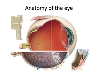
- 1. Anatomy of the eye
- 5. The roof: the lesser wing of the sphenoid, the orbital plate of the frontal. The lateral wall: the greater wing of the sphenoid, Zygomatic. The floor: Zygomatic, Maxillary, Palatine. The Medial wall: Maxillary, Lacrimal, Ethmoid, Sphenoid.
- 9. Lid anatomy Anterior lamella Posterior lamella • Skin • Orbicularis • Tarsus • Conjunctiva
- 10. Eyelid Structure 1. Orbicularis oculi (sphincter muscle); closes eyelids. 2. Levator muscle of the upper eyelid (striate muscle); opens eyelids. 3. Tarsal plate (connective tissue); contributes to eyelid form and support. 4. Locus of tarsal plate; set as a requirement for eyelid manipulation
- 12. Orbicularis oculi Two main parts: • Orbital part: plays a role when the eyelids need to be tightly shut. • Palpebral portion that plays a role in winking and blinking. Nerve supply: facial nerve
- 13. Levator Palpebrae Superioris Origin: Roof of the orbit, above the optic foramen Insertion: Anterior surface of superior tarsus and upper eyelid Innervation: Oculomotor nerve (CN III) Action: Retraction and elevation of eyelid.
- 14. Blood Supply of the Lid
- 17. Blood supply of Conjunctiva Arterial supply of conjunctiva derives from (1) Peripheral Tarsal arcades (2) Marginal Tarsal arcades (3) The anterior ciliary arteries
- 20. Lymphatics Lateral Side Preauricular Lymphnode Medial Side Submandibular Lymphnode
- 21. Lacrimal System Lacrimal System Secretory part Main lacrimal gland Accessory lacrimal glands Drainage part
- 22. Lacrimal gland • Located within the lacrimal fossa in the superior and outer edge of the orbital roof. • The tendon of the levator palpebrae muscle divides the lacrimal gland into a larger orbital part (2/3) and a smaller palpebral part (1/3). • Parasympathetic secretomotor nerve supply comes from the nervus intermedius. • Blood supply: lacrimal artery (a branch of the ophthalmic artery
- 26. Tear drainage The superior and inferior puncta lacrimales collect the tears, which then drain through the superior and inferior lacrimal canaliculi into the lacrimal sac. From there they pass through the nasolacrimal duct into the inferior concha
- 27. CoRNEA
- 28. The cornea is the transparent tissue that covers the front of the eye, forms anterior 1/6th of the outer coat of eyeball. • Diameter 11.5 mm. (11.7 mm * 11mm). It forms 3/4th of the total refractive power of eye. • Refractive power: 45D. • Refractive index: 1.376.
- 29. Microscopic Anatomy of cornea
- 30. FACTORS AFFECTING CORNEAL TRANSPERANCY Anatomical factors: • Uniform and regular arrangement of corneal epithelium • regular arrangement of corneal stromal lamellae. • Corneal avascularity. Physiological factors: • Relative state of corneal dehydration.
- 32. Iris • Iris is the anterior-most part of the uveal tract. • It is a thin, circular structure, forms a diaphragm like structure in front of the crystalline lens. • It has a central aperture known as the pupil. • The pupil determines the amount of light entering the eye.
- 33. Muscles in iris Sphincter pupillae: circular muscle, 1 mm wide, encircles the pupil. Innervated by the parasympathetic system. Contraction of the sphincter causes the pupil to constrict (miosis). Dilator pupillae: extends radially from the iris root to the sphincter. innervated by the sympathetic system Contraction of the dilator muscle causes the pupil to dilate (mydriasis).
- 34. Ciliary body The ciliary body is the middle part of the uveal tract forms a ring- shaped structure that projects posteriorly from the scleral spur. Anteriorly: it is confluent with the periphery of the iris (iris root). Posteriorly: it has a scalloped periphery, known as ora serrata, where it is continuous with the choroid and retina.
- 36. Ciliary muscle Nonstriated muscle primarily situated in the anterior 2/3 of the ciliary body stroma. The muscle has three parts: Outer longitudinal portion, Middle oblique portion, Inner circular portion. Action: Accommodation Nerve supply: The parasympathetic stimulation activates the muscle for contraction, whereas sympathetic innervation likely has an inhibitory effect.
- 37. Pars plicata: the portion of the ciliary body that contains finger-like projections (ciliary processes), extend into the posterior chamber. Pars plana: the smooth part of the ciliary body, terminates at the ora serrata, which is the transitional zone between the ciliary body and choroid.
- 38. Physiology of aqueous humor circulation
- 40. Choroid • The choroid is a thin but highly vascular layer, lining the inner surface of the sclera. • It extends from ora serrata anteriorly to the optic nerve posteriorly. • The outer surface is attached to the sclera at the optic nerve and at the exit of the vortex veins, The inner surface is attached to the retinal pigmented epithelium (RPE).
- 41. Microscopic structure of choroid
- 42. Blood supply of uveal tract: The blood supply of the uveal tract is mainly from three arteries namely • Short posterior ciliary arteries • Long posterior ciliary arteries • Anterior ciliary arteries. The posterior ciliary arteries are branches of the ophthalmic artery
- 43. Blood Supply of Uveal Tract
- 44. Retina
- 48. The optic disc lies a 3 mm medial to the center of the macula (fovea). There are no normal retinal layers in this zone (blind spot) as ganglion cell axons from the retina pierce the sclera to enter the optic nerve.
- 49. Vascular supply to the retina The inner layers of the retina (the internal limiting membrane through the inner nuclear layer) are supplied by the central artery of the retina. This originates at the ophthalmic artery, enters the eye with the optic nerve, and branches on the inner surface of the retina. It is a terminal artery without anastomoses and divides into four main branches Because the central artery is a terminal artery, occlusion will lead to retinal infarction. The outer layers (outer plexiform layer through the pigment epithelium) contain no capillaries. They are nourished by diffusion primarily from the richly supplied capillary layer of the choroid.
- 50. diagram of the human visual pathways and their neuronal components.