1) Dopamine transmission in the basal ganglia controls motor behavior through two pathways - the direct pathway which stimulates motion, and the indirect pathway which inhibits motion.
2) CK1δ over-expression disrupts this system, possibly through dopamine deficiency caused by downregulation of D1 and D2 receptors.
3) This study found an increase in calretinin-containing neurons in the striatum of CK1δ over-expressing mice, suggesting this imbalance contributes to their ADHD-like behaviors.
The role of vasopressin in light-induced c-Fos expression in the SCN
SandhuPoster2011
1. Dopamine transmission controls motor and perseverative behavior
and may be perturbed in disorders of attention and hyperactivity. The
striatum is host to many spiny projection neurons (SPN), neurons re-
sponsible for the striatal interactions in the basal ganglia. These SPNs
are sorted into two categories: SPNs involved in the direct pathway,
which stimulates motion, and SPNs involved in the indirect pathway,
which inhibits motion. Dopamine affects these pathways via the sub-
stantia nigra pars compacta (SNpc) – when the SNpc is activated, it
sends dopamine to the striatum, differentially activating both dopa-
mine 1 (D1) and dopamine 2 (D2) receptors (Kawaguchi, 1997). D1
receptors increase the excitability of SPNs the direct pathway, while D2
receptors decrease the excitability of SPNs in the indirect pathway. It
is not known whether it is a deficiency or a surplus of dopamine that
leads to ADHD (Swanson et al., 1998); the modus operandi of CK1δ
over-expression appears to be the former, that is, a deficiency of dopa-
mine as a result of the down-regulation of both D1 and D2 receptors.
By examining the densities of the four major subtypes of striatal inter-
neurons (calretinin-, calbindin-, parvalbumin-, and somatostatin-con-
taining) in CK1δ OE mice, we aimed to shed more light on the mech-
anism underlying the neurotransmitter imbalance for which CK1δ
over-expression is responsible.
Calretinin-containing neurons increase in the striata of CK1δ over-expressing mice
Burhan Sandhu, Mingming Zhou, Marc Flajolet, Dr. Paul Greengard
Laboratory of Cellular and Molecular Neuroscience, The Rockefeller University, New York, NY
A neural network including the orbitofrontal cortex, the dorsome-
dial striatum, and the subthalmic nucleus is responsible for inhibiting
many forms of behavior, including both impusivity and compulsivity;
casein kinase 1 isoform CK1δ over-expression (CK1δ OE) appears to
disrupt this network (Zhou et al., 2010). Through immunohistochemi-
cal staining, we found that calretinin-containing neurons increase in
the striata of CK1δ OE mice. These results suggest that an increase in
calretinin-containing neurons could be a part of the mechanism that
underlies the ADHD-like behavioral phenotype that CK1δ over-ex-
pressing mice express.
A transgenic mouse line overexpressing CK1δ was bred for 6
months. Adult mice were deeply anesthetized and then perfused with
saline buffer followed by 4% paraformaldehyde (PFA). Brains were re-
moved and post-fixed overnight in 4% PFA. After washing, brains were
cryoprotected in 25% sucrose, and free-floating cryostat sections (40
μm) were cut. The sections were then immunohistochemically stained
with DAB labeling (protocol from Zhuang et al., 2005). After the sec-
tions were mounted on slides, dried, and dehydrated with increasing
concentrations of ethanol as well as xylene, images were acquired with
a Zeiss wide-field brightfield microscope.
INTRODUCTION
ABSTRACT MATERIALS & METHODS
DISCUSSION
Calretinin is an integral protein in terms of calcium signalling be-
tween neurons. The observed imbalance - a ratio of nearly 3:1 - in
the distribution of calretinin-containing neurons in the striatum sug-
gests that the altered behavioral phenotype resulting from CK1δ over-
expression may be a result, at least in part, of a misconfigured neural
circuit that involves calretinin-containing neurons in the striatum. Al-
though a double-stain was not implemented in this experiment, the
widespread distribution of these calretinin-containing neurons sug-
gests that the striatal neurons are interneurons. If this is indeed the
case, then one can conclude that CK1δ over-expression targets the in-
terneuronal circuitry of the striatum to effect an imbalance between
the direct and indirect pathways of the basal ganglia. Further research
into protein distributions among striatal neurons could yield beneficial
information concerning possible targets for drug treatment of ADHD,
providing a more precise treatment than what is currently in use.
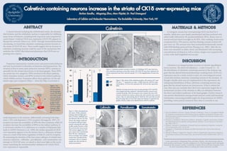
Figure 2. Calretinin positive neurons in striata. (A) Wildtype (WT) mice have low,
even calretinin expression across their striata. (B) CK1δ OE mice have clustered cal-
retinin expression near their external capsule. (C) A 10x magnification of some clus-
ters.
1. Zhou M, Rebholz H, Brocia C, Warner-Schmidt J, Fienberg A, Nairn A, Greengard P, Flajolet
M. Forebrain overexpression of CK1δ leads to down-regulation of dopamine receptors and
altered locomotor activity reminiscent of ADHD. Proceedings of the National Academy of Sci-
ence 2010; 107: 4401-6.
2. Swanson J, Castellanos F, Murias M, LaHoste G, Kennedy J. Cognitive neuroscience of atten-
tion deficit hyperactivity disorder and hyperkinetic disorder. Current Opinion in Neurobiol-
ogy 1998; 8: 263-71.
3. Gerfen C, Surmeier D. Modulation of Striatal Projection Systems by Dopamine. Annual Re-
view of Neuroscience 2011; 34: 441-66.
4. Kawaguchi Y. Neostriatal cell subtypes and their functional roles. Neuroscience Research
1997; 27: 1-8.
5. Zhuang X, Masson J, Gingrich JA, Rayport S, Hen R. Targeted gene expression in dopamine
and serotonin neurons of the mouse brain. Journal of Neuroscience Methods 2005; 143(1): 27-
32.
REFERENCES
I owe a great deal to Mingming Zhou and Marc Flajolet, who played huge roles as
both mentors and friends. I am also indebted to Dr. Paul Greengard, whose lab kindly
hosted me this summer. Lastly, I would like to thank Ted Scovell and Amaranta Viera
for their tremendous efforts in making this whole experience possible.
Figure 1. An
overview of the
various steps of
the basal ganglia
circuit. The direct
and indirect path-
ways provide op-
ponent regulation
of the the basal
ganglia output
interface. Green
arrows are gluta-
matergic, while
red are GABAer-
gic.
Gerfen et al., 2011
Calretinin
A B
C
0
10
20
30
40
50
60
Numberofcells
WT
OE
0
1
2
3
4
5
6
Percentagetototalcount(%)
*** ***
Figure 3. Bar charts of the calretinin positive cell counts in WT and
OE mice. *** represents a p value < 0.0001 from the ANOVA statis-
tical analysis. (A) Error bar represents SD. (B) Error bar represents
SEM.
Methods. Sections from four mice of each genotype (WT and OE)
were imaged and then analyzed. Calretinin positive neurons were
counted manually. After the neurons were counted, the sum of all
neurons counted was used as a divisor for each section’s own count to
create a data set representing chart B. Both the cell count data set and
the percentage data set were then input into Statview to generate the
graphs, whose p values were obtained from ANOVA analyses.
A B
Calbindin
Figure 4. Other attempted pro-
tein stains. While calretinin
staining acheived high speci-
ficity in striata, calbindin and
parvalbumin did not. For this
reason, the images could not
be properly analyzed. Soma-
tostatin staining did yield ana-
lyzable results; however, while
the sections that were success-
fully stained suggest that WT
mice express less somatostatin
than OE mice, staining sec-
tions of younger mice yielded
results that contradicted this
conclusion. Due to a limited
sample size, this trial also re-
mains inconclusive.
WT
OE
B
Parvalbumin Somatostatin