2. these preclinical studies, partial deletion of the EAAT1 gene has
been found in a subject with schizophrenia (Walsh et al., 2008),
while expression of a high-risk for schizophrenia allele of the GRM3
metabotropic glutamate receptor is associated with decreased EAAT2
mRNA expression in human prefrontal cortex (Egan et al., 2004;
Walsh et al., 2008). Taken together, these findings suggest that abnor-
mal expression of EAAT1 and EAAT2 may contribute to the pathophys-
iology of schizophrenia.
Converging evidence implicates temporal lobe areas in the patho-
physiology of schizophrenia, including data from neuroimaging, post-
mortem, and animal model studies (Bogerts et al., 1990; Benes et al.,
1991; Nelson et al., 1998; Young et al., 1998; Bruder et al., 1999;
Schroeder et al., 1999; Eastwood and Harrison, 2000; Le Corre et al.,
2000; Ohnuma et al., 2000; Rajarethinam et al., 2000; Sokolov et al.,
2000; Fatemi et al., 2001; Heckers, 2001; Heckers and Konradi,
2002; Harrison et al., 2003; Lipska, 2004; Rajarethinam et al., 2004;
Boyer et al., 2007; Tseng et al., 2009; Uezato et al., 2009). Volume
reduction is one of the most consistent structural abnormalities
found in the temporal lobe in schizophrenia (Nelson et al., 1998;
Rajarethinam et al., 2000, 2004), and volume change in these regions
correlates with the presence of severe thought disorder. Changes in
cellular density as well as expression of synaptic proteins have also
been reported in the temporal lobe in this illness. These findings
suggest that glutamate synapses may be remodeled due to a lifetime
of severe mental illness and antipsychotic treatment. Thus, we tested
the hypothesis that glutamate transmission may be abnormal by
examining expression of glutamate transporter proteins in the STG
and hippocampus in schizophrenia.
2. Materials and methods
2.1. Subjects and tissue preparation
Postmortem brain samples were provided by the Mount Sinai
Medical Center and Bronx Veterans Administration Medical Cen-
ter Brain Bank (Table 1 and Supplemental Table 1) and consisted
of twenty-three subjects with schizophrenia and twenty-seven
nonpsychiatrically ill comparison subjects. Subjects were excluded for
a history of alcoholism, death by suicide, or coma for more than 6 h be-
fore death. Next of kin consent was obtained for each subject.
Brain samples obtained upon autopsy were sliced mid-sagittally
and sectioned in 6–8 mm coronal slabs, immediately snap-frozen in
liquid nitrogen-cooled isopentane, and stored at −80 °C. The supe-
rior temporal gyrus was defined by sulcogyral position and dissected
from frozen slabs. Anterior hippocampus was dissected from the
0.8 mm thick coronal block at the level of the mammillary body.
The dissected tissue was pulverized into a fine powder at −190 °C,
aliquoted into individual Eppendorf tubes, and stored at −80 °C.
2.2. Animals
Twenty-two adult male Sprague–Dawley rats were ordered from
Harlan (Indianapolis, Indiana, USA) at approximately 60 days of age
and housed 2–3 to a cage with food and water ad libitum. Animals
received subcutaneous injections of haloperidol dissolved in sesame
oil (28.5 mg/kg) or vehicle (sesame oil) every 3 weeks for 9 months
(n=11 per group). 24 h following the last injection, the animals
were sacrificed, and brains were immediately removed, dissected,
and snap-frozen in isopentane cooled to −25 °C. For dissection, a
brain blocker (David Kopf Instruments, Tujunga, California, USA)
was used to block the brain into coronal planes containing the
rostrocaudal extension of the region of interest. Temporal associa-
tion cortex (TAC) and hippocampus were dissected using landmarks
from The Rat Brain in Stereotaxic Coordinates. Specifically, brains
were blocked from interaural 6.88 mm to 4.48 mm for HC and from
interaural 2.28 mm to 1.36 mm for TAC. Immediately after dissec-
tion, tissue was homogenized (10% w/v) in 50 mM Tris–HCl with a
Polytron homogenizer (Fisher Scientific International Inc., Pittsburg,
Pennsylvania, USA) for 30 s and stored at −80 °C in 0.5 ml aliquots.
2.3. Antibodies
Commercially available antisera for EAAT1 (1:1000, Santa Cruz
Biotechnology, Santa Cruz, California, USA), EAAT2 (1:1000, Millipore,
Billerica, Massachusetts, USA), EAAT3 (1:1000, Alpha Diagnostic Inter-
national, San Antonio, Texas, USA), VGLUT1 (1:2000, Alpha Diagnostic
International, San Antonia, Texas, USA) and VGLUT2 (1:1000, Synaptic
Systems, Göttingen, Germany) were used for Western blot analyses.
β-tubulin (1:10,000; Upstate, Lake Placid, New York, USA) was used
as a loading control.
2.4. Western blot analysis
Postmortem brain samples (50 mg) were homogenized in 1 ml of
5 mM Tris–HCl (pH 7.4), containing one complete, mini, ethylene
diaminetetraacetic acid-free protease inhibitor cocktail tablet (Roche
Applied Science, Indianapolis, Indiana, USA) per 10 ml, for 30 s with
a PowerGen 125 homogenizer (Fisher Scientific International, Inc.,
Hampton, New Hampshire, USA). Total protein concentration was deter-
mined with a bicinchoninic acid protein assay kit (Pierce Biotechnology,
Inc., Rockford, Illinouis, USA), and absorbance was measured on a
SpectraCount absorbance microplate reader (Packard/Perkin Elmer,
Wellesley, Massachusetts, USA) at 562 nm. Homogenates were stored
in 0.5 ml aliquots at −80 °C until assayed.
For electrophoresis, samples were prepared by combining ho-
mogenate with sample buffer and sterile water, and then denatured
at 95 °C for 4 min. For VGLUT1-2, samples were loaded in duplicate
on 7.5% or 12.5% precast Criterion polyacrylamide Tris–HCl gels
(Bio-Rad, Hercules, California, USA). For EAAT1-3, Nupage BisTris
gels were used (Invitrogen, Carlsbad, California, USA). Gels were
run at 120 V for 1.75 h. Following transblot onto nitrocellulose
(VGLUT1-2) or PVDF (EAAT1-3) membranes with a semidry transblotter
(Biorad, Hercules, California, USA), blots were rinsed several times in
distilled water and blocked in 3% dried milk phosphate buffered saline
(PBS) (VGLUT1), 1% BSA in PBS (VGLUT2), or LiCor/Oddyssey blocking
buffer (LiCor, Lincoln, Nebraska, USA) (EAAT1-3) while rocking at
room temperature for 30 min. Blots were then exposed to the primary
antibody in their respective blocking solutions while rocking overnight
at 4 °C. After two 5 minute washes in PBS, the blots for VGLUT1-2
were incubated with a horseradish peroxidase coupled goat anti-rabbit
secondary antibody or goat anti-mouse secondary antibody (VGLUT1:
1:5000; VGLUT2: 1:5000; Upstate, Lake Placid, New York, USA) respec-
tively in 3% dried milk PBS on a rocker for 2 h at room temperature.
For EAAT1-3, the blots were probed with IR-Dye labeled secondary anti-
bodies (1:10000, LiCor, Lincoln, Nebraska, USA) in 0.1% Tween, 0.01%
Table 1
Subject characteristics.
STG/HPC
Comparison Schizophrenia
N 27 23
Sex 14 m/13f 16 m/7f
AOD (years) 79±13 72±12
Tissue pH 6.5±0.2 6.5±0.3
PMI (hours) 7.6±6.8 14.6±8.9
Rx (on/off/unknown) 0/27/0 16/6/1
Abbreviations: superior temporal gyrus (STG); hippocampus (HPC); female (f); male
(m); age of death (AOD); post mortem interval (PMI); medications (Rx: on or off
antipsychotic medication for at least 6 weeks at time of death). Values are presented
as standard deviation.
2 D. Shan et al. / Schizophrenia Research 144 (2013) 1–8
3. sodium dodecyl sulfate (SDS), LiCor/Oddyssey blocking buffer for 1 h in
the dark at room temperature, then blots were imaged with the LiCor
Odyssey laser based imaging system followed by washing three times
for 5 min each in 0.01% tween phosphate buffered saline, and briefly
washing in water three times. VGLUT1-2 blots were saturated in en-
hanced chemiluminescence reagent (Pierce, Rockford, Illinois, USA),
and exposed to film (ISC BioExpress, Kaysville, Utah, USA). All mem-
branes were also probed with β-tubulin antibody.
We have previously tested VGLUT1-2, EAAT1-3, and β-tubulin
Western blot assays using varying protein concentrations of human
brain homogenate and determined that our assays are in the linear
range of the concentrations correlated for each protein be assayed
(Bauer et al., 2008; Oni-Orisan et al., 2008).
2.5. Data analysis
For VGLUT1-2, developed films were digitally captured with a
charge-coupled device based imaging system using Scion Imaging
software 4.0.3 (Scion Corporation, Frederick, Maryland, USA). Gray
scale values were obtained for bands at the expected molecular
weight, and membrane background was subtracted. Gray scale values
for duplicate samples were averaged and converted into optical den-
sity (OD). The mean ratio of VGLUT1: β-tubulin or VGLUT2: β-tubulin
OD was used for data analysis. For EAAT1-3, digital images were
obtained using a LiCor Odyssey scanner (LiCor, Lincoln, Nebraska,
USA). Odyssey analytic software (3.0) was used to quantify raw inte-
grated intensity for bands of interest. The ratio of EAAT: β-tubulin
expression was used for data analysis.
Data were analyzed using Statistica (StatSoft, Tulsa, Oklahoma,
USA). Multiple regression analysis was performed to probe for possi-
ble correlations between protein expression and age, postmortem
interval, and/or tissue pH. Analysis of covariance (ANCOVA) was
used to analyze the data when significant correlations were found,
otherwise analysis of variance (ANOVA) was used. ANOVA was also
performed to assess the effects of sex and antipsychotic status on
the dependent measures. For antipsychotic status, subjects with
schizophrenia were considered off antipsychotics if antipsychotics
were not administrated within 6 weeks of death. We also examined
expression of the loading control β-tubulin (unnormalized) using
ANOVA. For all tests, α=0.05.
3. Results
3.1. Postmortem studies
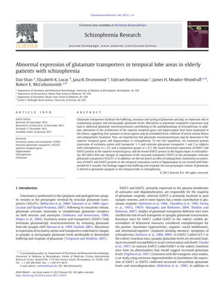
We examined expression of EAAT1-3 proteins in the superior
temporal gyrus and hippocampus (Fig. 1). EAAT1 [F (1, 26)=40.2,
Pb0.01] and EAAT2 [F (1, 25)=32.4, Pb0.01], but not EAAT3, were
significantly decreased in the superior temporal gyrus in subjects
with schizophrenia (Fig. 1A). We also found that EAAT2 [F (1, 20)=
7.40, Pb0.02], but not EAAT1 or EAAT3, was significantly decreased
in the hippocampus in subjects with schizophrenia (Fig. 1B).
In order to determine whether changes in transporter expression
extend to the presynaptic component of glutamate synapses, we ex-
amined expression of VGLUT1-2 proteins in the superior temporal
gyrus and hippocampus (Fig. 2). We didn't detect any changes in
expression of VGLUT1 and VGLUT2 proteins in the superior tem-
poral gyrus (Fig. 2A) or hippocampus (Fig. 2B) in subjects with
schizophrenia.
Expression of β-tubulin protein in schizophrenia subjects was not
changed compared to comparison subjects. No significant difference
was found in subjects with schizophrenia on antipsychotic medica-
tions at the time of death, compared to subjects off medications for
at least 6 weeks (data not shown).
3.2. Antipsychotic rat studies
The potential effects of antipsychotic medication on the expres-
sion of EAAT1 and EAAT2 protein were explored in rats treated with
haloperidol for 9 months. There were no changes in expression
of EAAT1 and EAAT2 proteins in rats treated with haloperidol
(28.5 mg/kg) compared to rats administrated vehicle in the temporal
association cortex (Fig. 3A) or hippocampus (Fig. 3B). No significant
differences in β-tubulin expression were detected in our animal
studies.
4. Discussion
In this study, we examined the expression of glutamate transporter
proteins in temporal lobe areas in elderly patients with schizophrenia.
Decreased expression of EAAT2 protein was found in the STG and
hippocampus, while expression of EAAT1 protein was decreased
in the STG in schizophrenia. Decreased expression of EAAT1 and/or
EAAT2 protein may have profound effects of glutamate neurotrans-
mission. These transporters are expressed at high levels on astrocytic
membranes near excitatory synapses, where they either bind and re-
lease glutamate or bind and transport glutamate into the cell. Since
the transport efficiency of bound glutamate is about 0.5, the trans-
porters act as a buffer to limit glutamate spillover to extrasynaptic
regions (Tzingounis and Wadiche, 2007). The transporters rapidly re-
move glutamate from the perisynaptic space, maintaining low basal
levels of glutamate in the synaptic cleft and limiting the pool of gluta-
mate available to spill out of the synapse. A decrease in this glutamate
buffering and reuptake capacity may lead to spillover of glutamate, in-
creasing glutamate levels in extrasynaptic microenvironments where
the level of glutamate is tightly regulated (Bridges et al., 2012).
Increased extrasynaptic glutamate may impact neurotransmission in
a number of ways (Danbolt, 2001; Tzingounis and Wadiche, 2007). In-
creased extrasynaptic glutmate levels could activate extrasynaptic glu-
tamate receptors on neurons and astrocytes, modulating the responses
of these cells. For example, activation of presynaptic metabotropic
receptors (that are localized to areas outside of the synaptic cleft) de-
creases glutamate release from the presynaptic terminal (Moghaddam
and Adams, 1998). A decrease in astrocytic glutamate reuptake may
also impact the activation of postsynaptic receptors within the synaptic
cleft; prolonged exposure to higher glutamate levels, as well as in-
creased baseline glutamate levels within the synapse, may impact
the physiology of ionotropic glutamate receptor activiation, disrupting
molecular correlates of learning and memory, including long-term
potentiation or depression (Tzingounis and Wadiche, 2007). Finally,
extrasynaptic glutamate may also spillover into adjacent synapses and
activate receptors in those synapses, leading to a loss of input specificity
(Tzingounis and Wadiche, 2007; Bridges et al., 2012).
Changes in EAAT1 expression and glycosylation have previously
been reported in schizophrenia. We found decreased expression of
EAAT1 protein in the DLPFC, as well as a change in EAAT1 glycosylation
consistent with decreased localization of EAAT1 to the plasma mem-
brane in the same region (Bauer et al., 2008, 2010). In contrast to
these protein studies, increased levels of EAAT1 mRNA were found in
the anterior cingulate cortex (ACC) and thalamus (Smith et al., 2001;
Bauer et al., 2008; Rao et al., 2012). Taken together, these data suggest
a profound abnormality of EAAT1 protein expression, with a compensa-
tory increase in EAAT1 mRNA expression in schizophrenia.
Since EAAT1 is mainly expressed in astrocytes, and not neurons,
throughout the central nervous system, our data suggest an abnormal-
ity of astrocytes in schizophrenia (Lehre et al., 1995; Bar-Peled et al.,
1997; Schmitt et al., 1997). Consistent with this hypothesis, we found
decreased expression of glial fibrillary acidic protein (GFAP), an astro-
cyte marker, in the frontal cortex in this illness (Johnston-Wilson et
al., 2000; Steffek et al., 2008). These data raise the question of whether
changes in EAAT1 protein expression are due to a decrease in EAAT1
3D. Shan et al. / Schizophrenia Research 144 (2013) 1–8
4. protein levels in astrocytes, or an overall decrease in the number of as-
trocytes where the remaining astrocytes are expressing normal levels of
EAAT1 protein. Several studies have found decreased astroglial cell
numbers in the PFC (Cotter et al., 2002; Rajkowska et al., 2002), ACC
(Cotter et al., 2001a, 2001b; Stark et al., 2004) and hippocampus
(Falkai and Bogerts, 1986) in schizophrenia, supporting the hypothesis
that decreases in EAAT1-2 protein expression may be secondary to di-
minished numbers of astrocytes.
Similar to EAAT1, EAAT2 protein expression is widely distrib-
uted throughout the brain in astroglial cell bodies and processes
(Rothstein et al., 1994; Chaudhry et al., 1995). While there is some
overlap of expression of EAAT1 and EAAT2 in the same astrocytes,
these molecules are generally expressed in different populations
of astroglia (Rothstein et al., 1996; Tanaka et al., 1997). Consistent
with our findings, decreased EAAT2 mRNA expression was found in
the hippocampus in schizophrenia (Ohnuma et al., 2000). In contrast
to the hippocampus, divergent findings have been reported for the
neocortex. In the frontal cortex, one study found decreased EAAT2
mRNA using in situ hybridization (ISH) (Ohnuma et al., 1998), while
another found increased EAAT2 mRNA with QPCR (Matute et al.,
2005) and three others found no changes using QPCR or ISH (Lauriat
et al., 2006; Bauer et al., 2008; Rao et al., 2012). These conflicting data
may be attributable to differences in methodology, tissue source and
subject demographics, as well as differential treatment with antipsy-
chotic medications.
In subjects from the same brain collection as the present study, we
have previously measured EAAT2 protein expression in the DLPFC
and ACC and detected no changes (Bauer et al., 2008). However,
while we did not find changes in EAAT2 protein levels in these two
regions, we did find an increase in G-protein pathway suppressor-1
(GPS1) in the frontal cortex (Bauer et al., 2008). GPS1 decreases
EAAT2 mediated glutamate reuptake through a direct protein–protein
interaction by regulating surface trafficking through a leucine zipper-
like motif (Watanabe et al., 2003).
EAAT-mediated glial reuptake of glutamate may also be regulated
by posttranslational modifications including N-linked glycosylation
(Conradt et al., 1995; Raunser et al., 2005). N-linked glycosylation of
EAATs is associated with trafficking from the ER to plasma membrane,
increased stability at the plasma membrane, and increased glutamate
reuptake (Trotti et al., 2001). We found an alteration in EAAT2 glyco-
sylation that may reflect retention of EAAT2 in the endoplasmic
reticulum, which might indicate decreased trafficking of EAAT2 to
the plasma membrane (Bauer et al., 2010).
Our previous findings of increased GPS1 protein expression and
altered EAAT2 glycosylation suggest that while total EAAT2 levels
are normal in the frontal cortex, there may in fact still be a decrease
in EAAT2 mediated glutamate reuptake via mechanisms other than
a change in regional EAAT2 protein expression. Taken together with
our findings of decreased EAAT2 protein levels in the hippocampus
and STG, these results suggest that there are pervasive abnormalities
of EAAT2 protein expression and trafficking in schizophrenia, via
region-specific mechanisms, leading to impaired glutamate reuptake
in temporal and frontal brain regions.
We did not detect changes in EAAT3 protein expression in the
STG or hippocampus in schizophrenia. We previously reported in-
creased expression of EAAT3 protein and mRNA in the ACC, while
other studies have found changes in EAAT3 mRNA expression
in the frontal cortex (increased), DLPFC (no change) and striatum
EAAT1
0
2
3
4
1
EAAT1:β-Tubulin
EAAT2
0
1
2
EAAT2:β-Tubulin
0.0
0.1
0.2
0.3
EAAT3:β-Tubulin
* *
superior temporal gyrus
EAAT3
A
hippocampusB
EAAT1
0.0
2.5
5.0
7.5
EAAT1:β-Tubulin
EAAT2:β-Tubulin
Schizophrenia
Control
EAAT2
0
1
2
EAAT3
0.0
0.1
0.2
0.3
0.4
0.5
EAAT3:β-Tubulin
*
Schizophrenia
Control
EAAT
β-Tubulin
EAAT
β-Tubulin
Fig. 1. Western blot analysis of EAAT1-3 normalized to β-tubulin in the (A) superior temporal gyrus (STG) and (B) hippocampus. Data are shown as mean±standard deviation.
*Pb0.05. A) EAAT1 and EAAT2 were significantly decreased in subjects with schizophrenia. No change was observed for EAAT3. B) EAAT2 was significantly decreased in subjects
with schizophrenia. No changes were observed for EAAT1 or EAAT3. Abbreviation: excitatory amino acid transporter (EAAT).
4 D. Shan et al. / Schizophrenia Research 144 (2013) 1–8
5. (decreased) (McCullumsmith and Meador-Woodruff, 2002; Lauriat et
al., 2006; Nudmamud-Thanoi et al., 2007; Bauer et al., 2008; Horiuchi
et al., 2012; Rao et al., 2012). Our lack of findings for EAAT3 protein
levels suggests that neuronal glutamate reuptake may be preserved in
these regions in schizophrenia. However, EAAT3 protein is localized to
regulatable cytosolic pools that may be rapidly mobilized to the plasma
membrane in response to elevated synaptic glutamate levels (Kugler
and Schmitt, 1999). Such a mechanism would suggest that EAAT3 activ-
ity may be regulated by modulating EAAT3 localization, rather than
increasing or decreasing levels of EAAT3 protein in the cell. Thus,
the possibility remains that there may be an alteration in EAAT3-
mediated glutamate reuptake without a change in total EAAT3 protein
levels.
In contrast to EAAT1-2, we did not find changes in the expression
of VGLUT1-2 protein levels in the STG or hippocampus. Alterations in
synaptic activity can be induced via regulation of the amount of glu-
tamate released from presynaptic terminals, and VGLUT-mediated
packaging of glutamate is a central control point for normal synaptic
activity (Bellocchio et al., 2000; Takamori et al., 2000; Shigeri et al.,
2004). Thus, measurement of VGLUT protein levels may indicate the
relative strength of presynaptic innervation for a given brain region
(Pothos et al., 2000; Daniels et al., 2004; Wilson et al., 2005; Daniels
et al., 2006). We previously examined VGLUT mRNA expression in
the hippocampal subfields in tissues from a different brain collection,
and found no changes in VGLUT1 or VGLUT2 transcript expression. A
different study, using tissues from a different source, found reduced
VGLUT1 mRNA in a younger cohort with schizophrenia (Eastwood
and Harrison, 2005; Uezato et al., 2009). In the present study we
did not find an association between age and our dependent measures,
while the study using the younger subjects reported an association
between VGLUT1 expression and age, which may account for these
divergent findings.
Since antipsychotic medications are known to influence glutamate
transporter expression, we used two complimentary approaches to
evaluate this potential confounding factor (Schneider et al., 1998;
De Souza et al., 1999; Melone et al., 2001). Our secondary analyses
showed no differences in expression of EAAT1 and EAAT2 proteins
between medicated patients and patients who had been free of anti-
psychotic medications for at least 6 weeks prior to death. We also in-
vestigated the effects of haloperidol on the expression of EAAT1 and
EAAT2 protein levels in the rat brain. We chose haloperidol because
the vast majority of our subjects who were on medications were
receiving a typical antipsychotic. Consistent with our human data,
following 9 months of treatment with haloperidol we did not detect
changes in EAAT1 or EAAT2 protein expression in the rat brain.
Taken together, these data suggest our findings are not due to treat-
ment with antipsychotic medication.
The temporal lobe plays a central role in many symptoms as-
sociated with schizophrenia, including altered cognitive function,
oversensitization to sensory stimuli, and auditory hallucinations
(Bruder et al., 1999; Boyer et al., 2007; Brady et al., 2010; Fujimoto
et al., 2012). Structural and functional abnormalities of STG and
hippocampus have been reported in this illness, while quantitative
investigations have demonstrated reductions in neuronal size, corti-
cal glial cell number, as well as changes in neuronal density (Benes et
al., 1991; Bruder et al., 1999; Rajarethinam et al., 2000; Heckers,
2001; Cotter et al., 2002; Heckers and Konradi, 2002; Rajarethinam
et al., 2004; Uranova et al., 2004; Steen et al., 2006; Boyer et al.,
A
hippocampus
B
VGLUT1
0.00
0.25
0.50
0.75
1.00
VGLUT1:β-Tubulin
VGLUT2
0.0
0.5
1.0
1.5
VGLUT2:β-Tubulin
VGLUT1
VGLUT
0
1
2
3
VGLUT1:β-Tubulin VGLUT2
0
1
2
3
VGLUT2:β-Tubulin
superior temporal gyrus
Schizophrenia
Control
Schizophrenia
Control
β-Tubulin
VGLUT
β-Tubulin
Fig. 2. Western blot analysis of VGLUT1-2 normalized to β-tubulin in the (A) superior temporal gyrus (STG) and (B) hippocampus. Data are shown as mean±standard
deviation. VGLUT1 and VGLUT2 were not changed in the (A) superior temporal gyrus or (B) hippocampus in subjects with schizophrenia. Abbreviation: vesicular glutamate
transporter (VGLUT).
5D. Shan et al. / Schizophrenia Research 144 (2013) 1–8
6. 2007). Consistent with loss of volume and cellular pathology, a
number of postmortem studies found altered expression of synaptic
proteins in the temporal lobe, including decreased expression of
complexin, synaptophysin, SNAP-25, VGLUT1 and several ionotropic
glutamate receptor subunits (Young et al., 1998; Eastwood and
Harrison, 2000; Sokolov et al., 2000; Harrison et al., 2003; Eastwood
and Harrison, 2005). Taken together with our findings, these data
support the hypothesis that there are profound abnormalities of glu-
tamate neurotransmission in excitatory circuits in the temporal lobe
in schizophrenia.
In summary, we found that expression of astroglial glutamate
transporters was abnormal in temporal lobe areas in schizophrenia.
These data suggest a deficit of glutamate reuptake capacity that
may lead to alterations in excitatory neurotransmission. We did
not find any changes in expression of the neuronal glutamate trans-
porter EAAT3 or the neuronal vesicular glutamate transporters
VGLUT1 and VGLUT2, suggesting that aspects of neuronal function
are preserved in the STG and hippocampus in this illness. Our find-
ings indicate a critical role for glial glutamate reuptake in severe
mental illness and suggest that elements of the glutamate reuptake
pathway may be high yield targets for development of novel treat-
ments for this illness.
Supplementary data to this article can be found online at http://
dx.doi.org/10.1016/j.schres.2012.12.019.
Role of the funding source
The study sponsors did not have any role in study design; in the collection, analy-
sis, and interpretation of data.
Contributors
Robert E. McCullumsmith and James H. Meador-Woodruff designed the study and
provided intellectual contributions. Vahram Haroutunian provided the tissue for the
study. Dan Shan helped to design the study, performed the experiments, performed
the statistical analyses, and wrote the first draft of the manuscript. Elizabeth K. Lucas
and Jana B. Drummond performed the experiments and the statistical analyses. All
authors contributed and have approved the final manuscript.
Conflict of interest
Dr James H Meador-Woodruff receives an honorarium as editor of
Neuropsychopharmacology. The other authors declare no conflict of interest.
Acknowledgments
This work was supported by MH53327 and MH88752 (JMW), MH064673 and
MH066392 (VH), MH074016, MH094445 and Doris Duke Clinical Scientist Award
(REM).
References
Bar-Peled, O., Ben-Hur, H., Biegon, A., Groner, Y., Dewhurst, S., Furuta, A., Rothstein, J.D.,
1997. Distribution of glutamate transporter subtypes during human brain develop-
ment. J. Neurochem. 69 (6), 2571–2580.
Bauer, D., Gupta, D., Harotunian, V., Meador-Woodruff, J.H., McCullumsmith, R.E., 2008.
Abnormal expression of glutamate transporter and transporter interacting mole-
cules in prefrontal cortex in elderly patients with schizophrenia. Schizophr. Res.
104 (1–3), 108–120.
Bauer, D., Haroutunian, V., Meador-Woodruff, J.H., McCullumsmith, R.E., 2010. Abnormal
glycosylation of EAAT1 and EAAT2 in prefrontal cortex of elderly patients with
schizophrenia. Schizophr. Res. 117 (1), 92–98.
Bellocchio, E.E., Reimer, R.J., Fremeau Jr., R.T., Edwards, R.H., 2000. Uptake of glutamate
into synaptic vesicles by an inorganic phosphate transporter. Science 289 (5481),
957–960.
Benes, F.M., Sorensen, I., Bird, E.D., 1991. Reduced neuronal size in posterior hippocam-
pus of schizophrenic patients. Schizophr. Bull. 17 (4), 597–608.
Haloperidol
Control
temporal association cortex
0
1
2
3
0
25
50
75
0
1
2
3
4
hippocampus
0
10
20
30
B
A
EAAT1
EAAT1
EAAT2
EAAT2
Haloperidol
Control
EAAT1:β-Tubulin
EAAT2:β-TubulinEAAT2:β-Tubulin
EAAT1:β-Tubulin
EAAT
β-Tubulin
EAAT
β-Tubulin
Fig. 3. Western blot analysis of EAAT1-2 normalized to β-tubulin in the (A) temporal association cortex (TAC) and (B) hippocampus in rats treated with haloperidol (28.5 mg/kg) or
vehicle. Data are shown as mean±standard deviation. There were no differences in EAAT1 or EAAT2 protein expression. Abbreviation: excitatory amino acid transporter (EAAT).
6 D. Shan et al. / Schizophrenia Research 144 (2013) 1–8
7. Bogerts, B., Ashtari, M., Degreef, G., Alvir, J.M., Bilder, R.M., Lieberman, J.A., 1990. Re-
duced temporal limbic structure volumes on magnetic resonance images in first
episode schizophrenia. Psychiatry Res. 35 (1), 1–13.
Boyer, P., Phillips, J.L., Rousseau, F.L., Ilivitsky, S., 2007. Hippocampal abnormalities and
memory deficits: new evidence of a strong pathophysiological link in schizophrenia.
Brain Res. Rev. 54 (1), 92–112.
Brady, A.M., Saul, R.D., Wiest, M.K., 2010. Selective deficits in spatial working memory
in the neonatal ventral hippocampal lesion rat model of schizophrenia. Neurophar-
macology 59 (7–8), 605–611.
Bridges, R., Lutgen, V., Lobner, D., Baker, D.A., 2012. Thinking outside the cleft to under-
stand synaptic activity: contribution of the cystine-glutamate antiporter (System xc-)
to normal and pathological glutamatergic signaling. Pharmacol. Rev. 64 (3),
780–802.
Bruder, G., Kayser, J., Tenke, C., Amador, X., Friedman, M., Sharif, Z., Gorman, J., 1999.
Left temporal lobe dysfunction in schizophrenia: event-related potential and be-
havioral evidence from phonetic and tonal dichotic listening tasks. Arch. Gen. Psy-
chiatry 56 (3), 267–276.
Chaudhry, F.A., Lehre, K.P., van Lookeren Campagne, M., Ottersen, O.P., Danbolt, N.C.,
Storm-Mathisen, J., 1995. Glutamate transporters in glial plasma membranes:
highly differentiated localizations revealed by quantitative ultrastructural immu-
nocytochemistry. Neuron 15 (3), 711–720.
Conradt, M., Storck, T., Stoffel, W., 1995. Localization of N-glycosylation sites
and functional role of the carbohydrate units of GLAST-1, a cloned rat brain
L-glutamate/L-aspartate transporter. Eur. J. Biochem. 229 (3), 682–687.
Cotter, D., Mackay, D., Landau, S., Kerwin, R., Everall, I., 2001a. Reduced glial cell density
and neuronal size in the anterior cingulate cortex in major depressive disorder.
Arch. Gen. Psychiatry 58 (6), 545–553.
Cotter, D.R., Pariante, C.M., Everall, I.P., 2001b. Glial cell abnormalities in major psychiatric
disorders: the evidence and implications. Brain Res. Bull. 55 (5), 585–595.
Cotter, D., Mackay, D., Chana, G., Beasley, C., Landau, S., Everall, I.P., 2002. Reduced neu-
ronal size and glial cell density in area 9 of the dorsolateral prefrontal cortex in
subjects with major depressive disorder. Cereb. Cortex 12 (4), 386–394.
Danbolt, N.C., 2001. Glutamate uptake. Prog. Neurobiol. 65 (1), 1–105.
Daniels, R.W., Collins, C.A., Gelfand, M.V., Dant, J., Brooks, E.S., Krantz, D.E., DiAntonio,
A., 2004. Increased expression of the Drosophila vesicular glutamate transporter
leads to excess glutamate release and a compensatory decrease in quantal content.
J. Neurosci. 24 (46), 10466–10474.
Daniels, R.W., Collins, C.A., Chen, K., Gelfand, M.V., Featherstone, D.E., DiAntonio, A.,
2006. A single vesicular glutamate transporter is sufficient to fill a synaptic vesicle.
Neuron 49 (1), 11–16.
De Souza, I.E., McBean, G.J., Meredith, G.E., 1999. Chronic haloperidol treatment im-
pairs glutamate transport in the rat striatum. Eur. J. Pharmacol. 382 (2), 139–142.
Eastwood, S.L., Harrison, P.J., 2000. Hippocampal synaptic pathology in schizophrenia,
bipolar disorder and major depression: a study of complexin mRNAs. Mol. Psychi-
atry 5 (4), 425–432.
Eastwood, S.L., Harrison, P.J., 2005. Decreased expression of vesicular glutamate trans-
porter 1 and complexin II mRNAs in schizophrenia: further evidence for a synaptic
pathology affecting glutamate neurons. Schizophr. Res. 73 (2–3), 159–172.
Egan, M.F., Straub, R.E., Goldberg, T.E., Yakub, I., Callicott, J.H., Hariri, A.R., Mattay, V.S.,
Bertolino, A., Hyde, T.M., Shannon-Weickert, C., Akil, M., Crook, J., Vakkalanka, R.K.,
Balkissoon, R., Gibbs, R.A., Kleinman, J.E., Weinberger, D.R., 2004. Variation in GRM3
affects cognition, prefrontal glutamate, and risk for schizophrenia. Proc. Natl. Acad.
Sci. U. S. A. 101 (34), 12604–12609.
Falkai, P., Bogerts, B., 1986. Cell loss in the hippocampus of schizophrenics. Eur. Arch.
Psychiatry Neurol. Sci. 236 (3), 154–161.
Fatemi, S.H., Earle, J.A., Stary, J.M., Lee, S., Sedgewick, J., 2001. Altered levels of the syn-
aptosomal associated protein SNAP-25 in hippocampus of subjects with mood dis-
orders and schizophrenia. Neuroreport 12 (15), 3257–3262.
Fujimoto, T., Okumura, E., Takeuchi, K., Kodabashi, A., Tanaka, H., Otsubo, T., Nakamura, K.,
Sekine, M., Kamiya, S., Higashi, Y., Tsuji, M., Shimooki, S., Tamura, T., 2012. Changes in
event-related desynchronization and synchronization during the auditory oddball
task in schizophrenia patients. Open Neuroimaging J. 6, 26–36.
Furuta, A., Martin, L.J., Lin, C.L., Dykes-Hoberg, M., Rothstein, J.D., 1997a. Cellular and syn-
aptic localization of the neuronal glutamate transporters excitatory amino acid trans-
porter 3 and 4. Neuroscience 81 (4), 1031–1042.
Furuta, A., Rothstein, J.D., Martin, L.J., 1997b. Glutamate transporter protein subtypes are
expressed differentially during rat CNS development. J. Neurosci. 17 (21), 8363–8375.
Harrison, P.J., Law, A.J., Eastwood, S.L., 2003. Glutamate receptors and transporters in
the hippocampus in schizophrenia. Ann. N. Y. Acad. Sci. 1003, 94–101.
Heckers, S., 2001. Neuroimaging studies of the hippocampus in schizophrenia. Hippo-
campus 11 (5), 520–528.
Heckers, S., Konradi, C., 2002. Hippocampal neurons in schizophrenia. J. Neural Transm.
109 (5–6), 891–905.
Hollmann, M., Heinemann, S., 1994. Cloned glutamate receptors. Annu. Rev. Neurosci.
17, 31–108.
Horiuchi, Y., Iida, S., Koga, M., Ishiguro, H., Iijima, Y., Inada, T., Watanabe, Y., Someya, T.,
Ujike, H., Iwata, N., Ozaki, N., Kunugi, H., Tochigi, M., Itokawa, M., Arai, M., Niizato,
K., Iritani, S., Kakita, A., Takahashi, H., Nawa, H., Arinami, T., 2012. Association of
SNPs linked to increased expression of SLC1A1 with schizophrenia. Am. J. Med.
Genet. B Neuropsychiatr. Genet. 159B (1), 30–37.
Johnston-Wilson, N.L., Sims, C.D., Hofmann, J.P., Anderson, L., Shore, A.D., Torrey, E.F.,
Yolken, R.H., 2000. Disease-specific alterations in frontal cortex brain proteins in
schizophrenia, bipolar disorder, and major depressive disorder. The Stanley Neuro-
pathology Consortium. Mol. Psychiatry 5 (2), 142–149.
Karlsson, R.M., Tanaka, K., Heilig, M., Holmes, A., 2008. Loss of glial glutamate and
aspartate transporter (excitatory amino acid transporter 1) causes locomotor
hyperactivity and exaggerated responses to psychotomimetics: rescue by haloper-
idol and metabotropic glutamate 2/3 agonist. Biol. Psychiatry 64 (9), 810–814.
Karlsson, R.M., Tanaka, K., Saksida, L.M., Bussey, T.J., Heilig, M., Holmes, A., 2009. As-
sessment of glutamate transporter GLAST (EAAT1)-deficient mice for phenotypes
relevant to the negative and executive/cognitive symptoms of schizophrenia.
Neuropsychopharmacology 34 (6), 1578–1589.
Kugler, P., Schmitt, A., 1999. Glutamate transporter EAAC1 is expressed in neurons and
glial cells in the rat nervous system. Glia 27 (2), 129–142.
Lauriat, T.L., Dracheva, S., Chin, B., Schmeidler, J., McInnes, L.A., Haroutunian, V.,
2006. Quantitative analysis of glutamate transporter mRNA expression in pre-
frontal and primary visual cortex in normal and schizophrenic brain. Neurosci-
ence 137 (3), 843–851.
Le Corre, S., Harper, C.G., Lopez, P., Ward, P., Catts, S., 2000. Increased levels of expres-
sion of an NMDARI splice variant in the superior temporal gyrus in schizophrenia.
Neuroreport 11 (5), 983–986.
Lehre, K.P., Levy, L.M., Ottersen, O.P., Storm-Mathisen, J., Danbolt, N.C., 1995. Differen-
tial expression of two glial glutamate transporters in the rat brain: quantitative
and immunocytochemical observations. J. Neurosci. 15 (3 Pt 1), 1835–1853.
Liguz-Lecznar, M., Skangiel-Kramska, J., 2007. Vesicular glutamate transporters
(VGLUTs): the three musketeers of glutamatergic system. Acta Neurobiol. Exp.
(Wars) 67 (3), 207–218.
Lipska, B.K., 2004. Using animal models to test a neurodevelopmental hypothesis of
schizophrenia. J. Psychiatry Neurosci. 29 (4), 282–286.
Maragakis, N.J., Rothstein, J.D., 2004. Glutamate transporters: animal models to neuro-
logic disease. Neurobiol. Dis. 15 (3), 461–473.
Masson, J., Sagne, C., Hamon, M., El Mestikawy, S., 1999. Neurotransmitter transporters
in the central nervous system. Pharmacol. Rev. 51 (3), 439–464.
Matute, C., Melone, M., Vallejo-Illarramendi, A., Conti, F., 2005. Increased expression of
the astrocytic glutamate transporter GLT-1 in the prefrontal cortex of schizophrenics.
Glia 49 (3), 451–455.
McCullumsmith, R.E., Meador-Woodruff, J.H., 2002. Striatal excitatory amino acid trans-
porter transcript expression in schizophrenia, bipolar disorder, and major depressive
disorder. Neuropsychopharmacology 26 (3), 368–375.
Melone, M., Vitellaro-Zuccarello, L., Vallejo-Illarramendi, A., Perez-Samartin, A., Matute,
C., Cozzi, A., Pellegrini-Giampietro, D.E., Rothstein, J.D., Conti, F., 2001. The expression
of glutamate transporter GLT-1 in the rat cerebral cortex is down-regulated by the
antipsychotic drug clozapine. Mol. Psychiatry 6 (4), 380–386.
Moghaddam, B., Adams, B.W., 1998. Reversal of phencyclidine effects by a group II
metabotropic glutamate receptor agonist in rats. Science 281 (5381), 1349–1352.
Nelson, M.D., Saykin, A.J., Flashman, L.A., Riordan, H.J., 1998. Hippocampal volume
reduction in schizophrenia as assessed by magnetic resonance imaging: a meta-
analytic study. Arch. Gen. Psychiatry 55 (5), 433–440.
Nudmamud-Thanoi, S., Piyabhan, P., Harte, M.K., Cahir, M., Reynolds, G.P., 2007. Deficits
of neuronal glutamatergic markers in the caudate nucleus in schizophrenia. J. Neural
Transm. Suppl. (72), 281–285.
Ohnuma, T., Augood, S.J., Arai, H., McKenna, P.J., Emson, P.C., 1998. Expression of the
human excitatory amino acid transporter 2 and metabotropic glutamate recep-
tors 3 and 5 in the prefrontal cortex from normal individuals and patients with
schizophrenia. Brain Res. Mol. Brain Res. 56 (1–2), 207–217.
Ohnuma, T., Tessler, S., Arai, H., Faull, R.L., McKenna, P.J., Emson, P.C., 2000. Gene expres-
sion of metabotropic glutamate receptor 5 and excitatory amino acid transporter 2 in
the schizophrenic hippocampus. Brain Res. Mol. Brain Res. 85 (1–2), 24–31.
Oni-Orisan, A., Kristiansen, L.V., Haroutunian, V., Meador-Woodruff, J.H., McCullumsmith,
R.E., 2008. Altered vesicular glutamate transporter expression in the anterior cingu-
late cortex in schizophrenia. Biol. Psychiatry 63 (8), 766–775.
Peghini, P., Janzen, J., Stoffel, W., 1997. Glutamate transporter EAAC-1-deficient
mice develop dicarboxylic aminoaciduria and behavioral abnormalities but no
neurodegeneration. EMBO J. 16 (13), 3822–3832.
Pothos, E.N., Larsen, K.E., Krantz, D.E., Liu, Y., Haycock, J.W., Setlik, W., Gershon, M.D.,
Edwards, R.H., Sulzer, D., 2000. Synaptic vesicle transporter expression regulates
vesicle phenotype and quantal size. J. Neurosci. 20 (19), 7297–7306.
Rajarethinam, R.P., DeQuardo, J.R., Nalepa, R., Tandon, R., 2000. Superior temporal gyrus
in schizophrenia: a volumetric magnetic resonance imaging study. Schizophr. Res.
41 (2), 303–312.
Rajarethinam, R., Sahni, S., Rosenberg, D.R., Keshavan, M.S., 2004. Reduced superior
temporal gyrus volume in young offspring of patients with schizophrenia. Am.
J. Psychiatry 161 (6), 1121–1124.
Rajkowska, G., Miguel-Hidalgo, J.J., Makkos, Z., Meltzer, H., Overholser, J., Stockmeier,
C., 2002. Layer-specific reductions in GFAP-reactive astroglia in the dorsolateral
prefrontal cortex in schizophrenia. Schizophr. Res. 57 (2–3), 127–138.
Rao, J.S., Kellom, M., Reese, E.A., Rapoport, S.I., Kim, H.W., 2012. Dysregulated glutamate
and dopamine transporters in postmortem frontal cortex from bipolar and schizo-
phrenic patients. J. Affect. Disord. 136 (1–2), 63–71.
Raunser, S., Haase, W., Bostina, M., Parcej, D.N., Kuhlbrandt, W., 2005. High-yield expres-
sion, reconstitution and structure of the recombinant, fully functional glutamate
transporter GLT-1 from Rattus norvegicus. J. Mol. Biol. 351 (3), 598–613.
Rothstein, J.D., Martin, L., Levey, A.I., Dykes-Hoberg, M., Jin, L., Wu, D., Nash, N., Kuncl,
R.W., 1994. Localization of neuronal and glial glutamate transporters. Neuron 13
(3), 713–725.
Rothstein, J.D., Dykes-Hoberg, M., Pardo, C.A., Bristol, L.A., Jin, L., Kuncl, R.W., Kanai, Y.,
Hediger, M.A., Wang, Y., Schielke, J.P., Welty, D.F., 1996. Knockout of glutamate
transporters reveals a major role for astroglial transport in excitotoxicity and clear-
ance of glutamate. Neuron 16 (3), 675–686.
Schmitt, A., Asan, E., Puschel, B., Kugler, P., 1997. Cellular and regional distribution of
the glutamate transporter GLAST in the CNS of rats: nonradioactive in situ hybrid-
ization and comparative immunocytochemistry. J. Neurosci. 17 (1), 1–10.
7D. Shan et al. / Schizophrenia Research 144 (2013) 1–8
8. Schneider, J.S., Wade, T., Lidsky, T.I., 1998. Chronic neuroleptic treatment alters expression
of glial glutamate transporter GLT-1 mRNA in the striatum. Neuroreport 9 (1), 133–136.
Schroeder, H., Grecksch, G., Becker, A., Bogerts, B., Hoellt, V., 1999. Alterations of the
dopaminergic and glutamatergic neurotransmission in adult rats with postnatal
ibotenic acid hippocampal lesion. Psychopharmacology (Berl.) 145 (1), 61–66.
Sheldon, A.L., Robinson, M.B., 2007. The role of glutamate transporters in neurodegen-
erative diseases and potential opportunities for intervention. Neurochem. Int. 51
(6–7), 333–355.
Shigeri, Y., Seal, R.P., Shimamoto, K., 2004. Molecular pharmacology of glutamate trans-
porters, EAATs and VGLUTs. Brain Res. Brain Res. Rev. 45 (3), 250–265.
Smith, R.E., Haroutunian, V., Davis, K.L., Meador-Woodruff, J.H., 2001. Expression of
excitatory amino acid transporter transcripts in the thalamus of subjects with
schizophrenia. Am. J. Psychiatry 158 (9), 1393–1399.
Sokolov, B.P., Tcherepanov, A.A., Haroutunian, V., Davis, K.L., 2000. Levels of mRNAs
encoding synaptic vesicle and synaptic plasma membrane proteins in the temporal
cortex of elderly schizophrenic patients. Biol. Psychiatry 48 (3), 184–196.
Stark, A.K., Uylings, H.B., Sanz-Arigita, E., Pakkenberg, B., 2004. Glial cell loss in the
anterior cingulate cortex, a subregion of the prefrontal cortex, in subjects with
schizophrenia. Am. J. Psychiatry 161 (5), 882–888.
Steen, R.G., Mull, C., McClure, R., Hamer, R.M., Lieberman, J.A., 2006. Brain volume in
first-episode schizophrenia: systematic review and meta-analysis of magnetic res-
onance imaging studies. Br. J. Psychiatry 188, 510–518.
Steffek, A.E., McCullumsmith, R.E., Haroutunian, V., Meador-Woodruff, J.H., 2008. Cortical
expression of glial fibrillary acidic protein and glutamine synthetase is decreased in
schizophrenia. Schizophr. Res. 103 (1–3), 71–82.
Takamori, S., Rhee, J.S., Rosenmund, C., Jahn, R., 2000. Identification of a vesicular glu-
tamate transporter that defines a glutamatergic phenotype in neurons. Nature 407
(6801), 189–194.
Tanaka, K., Watase, K., Manabe, T., Yamada, K., Watanabe, M., Takahashi, K., Iwama, H.,
Nishikawa, T., Ichihara, N., Kikuchi, T., Okuyama, S., Kawashima, N., Hori, S., Takimoto,
M., Wada, K., 1997. Epilepsy and exacerbation of brain injury in mice lacking the gluta-
mate transporter GLT-1. Science 276 (5319), 1699–1702.
Trotti, D., Aoki, M., Pasinelli, P., Berger, U.V., Danbolt, N.C., Brown Jr., R.H., Hediger, M.A.,
2001. Amyotrophic lateral sclerosis-linked glutamate transporter mutant has
impaired glutamate clearance capacity. J. Biol. Chem. 276 (1), 576–582.
Tseng, K.Y., Chambers, R.A., Lipska, B.K., 2009. The neonatal ventral hippocampal lesion
as a heuristic neurodevelopmental model of schizophrenia. Behav. Brain Res. 204
(2), 295–305.
Tzingounis, A.V., Wadiche, J.I., 2007. Glutamate transporters: confining runaway exci-
tation by shaping synaptic transmission. Nat. Rev. Neurosci. 8 (12), 935–947.
Uezato, A., Meador-Woodruff, J.H., McCullumsmith, R.E., 2009. Vesicular glutamate
transporter mRNA expression in the medial temporal lobe in major depressive dis-
order, bipolar disorder, and schizophrenia. Bipolar Disord. 11 (7), 711–725.
Uranova, N.A., Vostrikov, V.M., Orlovskaya, D.D., Rachmanova, V.I., 2004. Oligodendrog-
lial density in the prefrontal cortex in schizophrenia and mood disorders: a study
from the Stanley Neuropathology Consortium. Schizophr. Res. 67 (2–3), 269–275.
Walsh, T., McClellan, J.M., McCarthy, S.E., Addington, A.M., Pierce, S.B., Cooper, G.M.,
Nord, A.S., Kusenda, M., Malhotra, D., Bhandari, A., Stray, S.M., Rippey, C.F.,
Roccanova, P., Makarov, V., Lakshmi, B., Findling, R.L., Sikich, L., Stromberg, T.,
Merriman, B., Gogtay, N., Butler, P., Eckstrand, K., Noory, L., Gochman, P., Long, R.,
Chen, Z., Davis, S., Baker, C., Eichler, E.E., Meltzer, P.S., Nelson, S.F., Singleton, A.B.,
Lee, M.K., Rapoport, J.L., King, M.C., Sebat, J., 2008. Rare structural variants disrupt
multiple genes in neurodevelopmental pathways in schizophrenia. Science 320
(5875), 539–543.
Watanabe, et al., 2003. GPS1, interacting protein with GLT-1. Program No 37216 2003
SFN Abstract.
Wilson, N.R., Kang, J., Hueske, E.V., Leung, T., Varoqui, H., Murnick, J.G., Erickson, J.D.,
Liu, G., 2005. Presynaptic regulation of quantal size by the vesicular glutamate
transporter VGLUT1. J. Neurosci. 25 (26), 6221–6234.
Young, C.E., Arima, K., Xie, J., Hu, L., Beach, T.G., Falkai, P., Honer, W.G., 1998. SNAP-25 def-
icit and hippocampal connectivity in schizophrenia. Cereb. Cortex 8 (3), 261–268.
8 D. Shan et al. / Schizophrenia Research 144 (2013) 1–8