
Recommended
More Related Content
What's hot
Similar to P6 binocular loupe
Similar to P6 binocular loupe (20)
Recently uploaded
Recently uploaded (20)
P6 binocular loupe
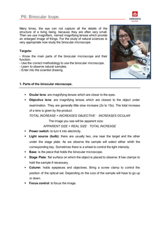
- 1. P6: Binocular loupe. Many times, the eye can not capture all the details of the structure of a living being, because they are often very small. Then we use magnifiers, named magnifying lenses which provide an enlarged image of things. For the study of natural sciences is very appropriate now study the binocular microscope. Targets: - Know the main parts of the binocular microscope and their function. - Use the correct methodology to use the binocular microscope. - Learn to observe natural samples. - Enter into the scientist drawing. 1. Parts of the binocular microscope. Ocular lens: are magnifying lenses which are closer to the eyes. Objective lens: are magnifying lenses which are closest to the object under examination. They are generally little slow increase (2x to 15x). The total increase of a lens is given by the product: TOTAL INCREASE = INCREASES OBJECTIVE · INCREASES OCULAR The image you see will be apparent size: APPARENT SIZE = REAL SIZE · TOTAL INCREASE Power switch: to turn it into electricity. Light source (bulb): there are usually two, one near the target and the other under the stage plate. As we observe the sample will select either whith the corresponding key. Sometimes there is a wheel to control the light intensity. Base: is the piece that holds the binocular microscope. Stage Plate: flat surface on which the object is placed to observe. It has clamps to hold the sample if necessary. Column: holds eyepieces and objectives. Bring a screw clamp to control the position of the optical set. Depending on the size of the sample will have to go up or down. Focus control: to focus the image.
- 2. Exercises: 1. Fill in the photography the names of different parts of the binocular microscope: 2. What is the total increase of a binocular microscope? 3. What is the total increase of a binocular eyepiece that has a 2x and 15x objective? 4. We note a fly larva 7 mm long with a lens that has some objective and ocular lens 7,5x and 2x. What will be apparent length? 2. Correct use of binocular microscope. The binocular microscope is a delicate instrument. We must use it properly and treat it carefully. Transport: a) Carry it with both hands. With one hand we take the column, and let rest the base of the loupe on the palm of the other hand. b) always move it in a vertical position to avoid falling or the eye of the base plate. Working position a) The screws should be well primed to prevent the fall of the optical system. b) Move laterally tubes eye to find the right distance between the pupils of your eyes.
- 3. c) Put the object on the observation deck, but not directly on it. Place it in a petri dish or in a small bowl. d) Moving the screw set the objective lens about 2 cm of the object. This operation should be done looking laterally. e) Look through the ocular and go up through the optical system focus screw until the object had clearly focused. f) Once the work is finished clean the stage plate and keep the lens in your box using (remember!) both hands. Exercici: Observe different things, starting with your fingers. Then choose some of the objects of following list and try to draw them. Calculate the total increase in each one. increases: increases: increases: increases: increases: increases: Objects: small flowers, a piece of cloth, the tip of your ball pen, an scourer, a leaf, a dirty toothbrush, a small insect or a worm (if you find someone in a flower...) Glossary: to note a screw a leaf