SlideShare a Scribd company logo
1 of 6
Download to read offline
Schwannoma of the Ulnar Nerve: A Case and Review of the Literature

                                    Ashley Boustany, MS II
                          West Virginia University School of Medicine

                                  W. Thomas McClellan, M.D.
                                       Plastic Surgeon
                                       Private Practice

Abstract

        We report a case of a large ulnar nerve schwannoma, a rare type of soft tissue neoplasm.
Diagnostic pearls are described to facilitate a more accurate and timely diagnosis. These
characteristics include mobility, Tinel’s sign, MRI target sign, S100 histological staining, Antoni
patterns, and others. With a correct diagnosis, the tumor can be extirpated with preservation of
nerve function and a low risk of recurrence.

Introduction

        Schwannomas are the most common type of tumor arising in peripheral nerves. (1)
However, peripheral nerve tumors are rare, representing less than 8% of soft tissue neoplasms.
Schwannomas are non-invasive tumors arising from peripheral nerve sheaths and are
encapsulated by epineurium. (2) There is a 2:1 occurrence of upper limb to lower limb
schwannomas, generally on the volar surface. There is no predisposition for sex or race, but they
usually develop in 30-60 year olds. (1) Schwannomas are often misdiagnosed due to their
indistinct signs and symptoms, which may lead to detrimental neurologic deficits if approached
incorrectly. It has been shown that less than a quarter of diagnostic tests provide an accurate
diagnosis of schwannoma. (3)

Case

         We present a 53 year old right hand dominant female with a slowly enlarging mass
within her proximal volar right forearm (Figure 1). The mass had been present for a minimum of
two years and had become more painful and noticeable over time. Dull, intermittent pain was
reported at rest over the mass, and was moderately sharper during flexion of the fingers or
bumping of the forearm. The level of discomfort increasingly hindered her ability to perform
activities of daily living.
         She was initially treated conservatively at an outside facility for a ruptured forearm
muscle belly. Subsequently, her symptoms worsened and a CT guided biopsy of the suspected
mass was performed. The patient reported the biopsy was extremely painful and caused an
exacerbation of her forearm pain. Histopathology demonstrated a paucicellular specimen with
very rare fragments of fibrous tissue that was insufficient for diagnosis.
Figure 1. Preoperative: The dotted
                                                     line indicates the edge of the
                                                     palpable tumor. Additionally, the
                                                     olecranon, medial epicondyle, and
                                                     path of the ulnar nerve are
                                                     identified.


        She was referred to our hand surgery service following the inconclusive biopsy. The 5 cm
firm mass was mobile perpendicular to the nerve axis and immobile along the parallel axis. A
very sensitive and positive Tinel’s sign radiated in the ulnar distribution, with dysthesias directly
over the mass and along the dorsal ulnar distribution of her hand. Semmes Weinstein testing
indicated diminished light touch and hypersensitivity to sharp touch in the same region.
However, her strength of the intrinsic muscles of the right hand was 5/5 without evidence of
weakness or loss of range of motion.
        An MRI was performed and showed a 5.0 cm x 3.0 cm x 4.6 cm diameter mass
displacing the flexor digitorum superficialis and palmaris longus muscles (Figure 2). The mass
had smooth margins but displayed a complex heterogeneous signal. There was no evidence of
invasion of adjoining muscles or bones. With gadolinium injection, an intense irregular
enhancement was seen. A capsule surrounded the eccentrically placed mass. Images also
indicated the presence of a hemorrhagic center related to the needle biopsy. MRI diagnosis was
inconclusive, but the differential included, schwannoma, malignant histiocytoma,
neurofibrosarcoma, or soft tissue malignancy.
                                                                             Figure 2. MRI: T1
                                                                             weighted MRI of the
                                                                             right upper extremity
                                                                             showing a large well
                                                                             demarcated           but
                                                                             heterogeneous mass.
                                                                             The MRI lacked the
                                                                             traditional      “target
                                                                             sign”       that      is
                                                                             commonly associated
                                                                             with schwannomas.
Figure 3. Intraoperative:
                                                                          The proximal and distal
                                                                          ulnar nerve are identified
                                                                          with vascular loops. Note
                                                                          how the fascicles of the
                                                                          ulnar nerve are splayed
                                                                          out over the tumor.
                                                                          Identification of these
                                                                          fascicles is crucial to
                                                                          determine      a      safe
                                                                          longitudinal entry point
                                                                          through the epineurium.


        She was taken to the operating room for exploration. Extirpation of the tumor was
performed via a longitudinal access incision through the epineurium and careful separation of the
nerve and tumor (Figure 3). The tumor measured 4.0 cm x 3.0 cm x 1.9 cm (Figure 4). It showed
a hypo-cellular tissue with Schwann cells strongly positive for S100 protein by immunostain
technique (Figure 5). Antoni B patterns predominated with few areas suggestive of Antoni A
patterns. Incision of the tumor also revealed hemorrhage and thrombosis consistent with prior
needle aspiration. Three months following surgery the patient has retained full ulnar motor and
sensory function as well as improving sensory paresthesias in her dorsal ulnar division.


                                                                Figure 4. Tumor: Gross
                                                                appearance of the Schwannoma
                                                                following removal from within
                                                                the ulnar nerve.
Figure 5. Histopathology:
                                                                   A hypo-cellular tissue with
                                                                   Schwann cells staining
                                                                   strongly positive for S100
                                                                   protein.


Discussion

        Diagnosis of a schwannoma in the preoperative period is challenging because of the slow
growth and paucity of symptoms. Diagnostic accuracy is crucial to maintaining the integrity of
the nerve involved and to properly plan the appropriate surgical intervention. Often these tumors
present as palpable masses, tender to displacement without muscles weakness. Tinel’s sign is
positive in the majority of cases. The tumors are transversely mobile but immobile
longitudinally, likely due to their nested intraneural location. Schwannomas share many features
with other soft tissue tumors and are frequently misdiagnosed due to similarities. Differential
diagnosis should include neurofibroma, ganglion cysts, malignant tumors, lipomas, and
xanthomas. (1,4) Neurofibromas, in particular, cannot be distinguished from schwannomas on
physical examination. The symptoms appear to be nonspecific, which adds to difficulty in
diagnosis. (5) They may be differentiated on MRI or fine needle aspiration. Malignant masses
exhibit more distinct signs but are often mistaken for schwannomas in early stages of diagnosis.
Unlike schwannomas, malignant tumors often have immobility, firmness, constant pain at rest,
and motor weakness. (6) Weakness may occur if the benign tumor exceeds 2.5 cm and can be
location dependent. Kehoe et al (1995) analyzed 88 peripheral nerve tumors, where only one
was correctly diagnosed as a schwannoma preoperatively.
        Gadolinium enhanced T1-weighted and T2-weighted MRIs are particularly useful in
diagnosing schwannomas. Koga et al (2007) found the presence of the target sign to be 100%
specific and 59% sensitive for the tumors. The target sign is the contrasting central and
peripheral intensities demonstrated on the images. Histological analysis credits the central
hyperintensities and hypointensities on T1 gadolinium and T2, respectively, to Antoni A cells.
 Antoni A patterns are of low cellular concentration. Antoni B areas are of high cell
concentration and correspond to peripheral intensities on MRI and CT. (8)
        Histological staining reveals a strongly positive S100 protein that is specific for
schwannomas and helps to rule out neurofibromas. (5,9) Imaging shows the tumors as round or
oval, eccentrically located in relation to the nerve, encapsulated, isolated, and non-invasive. In
comparison, neurofibromas are non-encapsulated and intimately surround the nerve. They cannot
be surgically removed without damaging the connected nerve, often necessitating nerve grafting
to repair functioning. (2,4,5) Schwannomas, on the other hand, can be separated surgically from
the nerve fascicles avoiding neurologic deficits. (4) This emphasizes the importance of a correct
preoperative diagnosis. Despite the structural differences of soft tissue tumors, they are difficult
to distinguish with imaging.
        Fine needle aspiration tends to be extremely painful in cases of schwannomas, and
hemorrhaging may result with temporary worsening of symptoms. The results are frequently
inconclusive, but are helpful to exclude ganglion cysts. (3) Domanksi et al (2006) aspirated 116
different schwannomas, and results were not sufficient for diagnosis for about 44% of the cases.
        Extirpation of the intraneural schwannoma can be challenging. Sterile tourniquet
dissection is recommended and assists in visualization. Loupe magnification and or use of the
operating microscope is highly recommended. Identification of the nerve proximal and distal to
the tumor is the first important step to reducing injury and traction neuropraxia. Identification of
the individual splayed nerve fascicles as they spread over of the tumor is critical in determining
the entry through epineurium. A longitudinal incision is created between the splayed fascicles
down to the tumor sheath. Once the outer layer of the tumor is identified, a plane can be
developed between the more superficial fascicles and the tumor wall. Slow, deliberate,
circumferential dissection with a “peanut” and Littler scissors facilitates delivery of the tumor.
Once the tumor is removed, the nerve is inspected for injury, the tourniquet is released, and
precise hemostasis is achieved. Repair of the epineurium is not required and the longitudinally
split muscle is repaired loosely over the nerve. Drains are optional, bulky dressing is preferred,
and immediate post operative hand therapy is instituted.
        It is uncommon for schwannomas to recur in identical locations. (1) Das et al (2007)
found that surgical removal of schwannomas was successful in alleviating preoperative
symptoms while maintaining nerve functioning in 89% of their cases.

Conclusion

        Schwannomas are rare peripheral nerve tumors that have important diagnostic and
radiographic features. These tumors are transversely mobile and longitudinally immobile, have a
positive Tinel’s sign, and exertional dysathesias or pain. MRI typically reveals the target sign of
biphasic contrast of peripheral and central regions and distinct encapsulation displacing the
intimately associated nerve fascicles. Surgical resection must be approached with caution to
protect nerve function and continuity. Surgical resection is associated with good outcomes. The
recurrence rate is low.


References

1. Ozdemir O, Kurt C, et.al. Schwannomas of the hand and wrist: long-term results and review of
literature. Journal of Orthopaedic Surgery. 2005;13(3):267-272.

2. Lin J, Martel W. Cross-sectional imaging of peripheral nerve sheath tumors: characteristic
signs on CT, MR imaging, and sonography. Amer Journ Roentgenology. 2001 Jan.;176:75-82.

3. Rockwell G, Achilleas T, et. al. Schwannoma of the hand and wrist. Plast Reconstr Surg. 2003
Mar.;111(3):1227-1232.
4. Sandberg K, Nilsson J, et. al. Tumors of peripheral nerves in the upper extremity: A 22-year
epidemiological study. Scand J Plast Reconstr Hand Surg. 2009 Sep.;43:43-49.

5. Adani R, Baccarani A, et. al. Schwannomas of the upper extremity: diagnosis and treatment.
Chir Orani Mov. 2008 Sep.;92:85-88.

6. Ogose A, Hotta T, et. al. Tumors of peripheral nerves: correlation of symptoms, clinical signs,
imaging features, and histologic diagnosis. Skeletal Radiol. 1999 Jan.;28:183-188.

7. Kehoe N, Reid R, et. al. Solitary benign peripheral-nerve tumors: review of 32 years’
experience. J Bone Joint Surg. 1995 Oct.;77B:497-500.

8. Koga H, Matsumoto S, et. al. Definition of the target sign and its use for diagnosis of
schwannomas. Clinical Orthopaedics and Related Research. 2007 Aug.;464:224-229.

9. Das S, Ganju A, et. al. Tumors of the brachial plexus. Neurosurg. 2007June;22(6):E26.

10. Domanski H, Akerman M, et. al. Fine Needle Aspiration of Neurilemoma (Schwannoma). A
clinicocytopahologic study of 116 patients. Diagnostic Cytopathology. 2006;34:403-412.

More Related Content

What's hot

Leprous neuritis management by aseem
Leprous neuritis management by aseemLeprous neuritis management by aseem
Leprous neuritis management by aseem
Dr. Aseem Sharma
 
4. humerus fractures
4. humerus fractures4. humerus fractures
4. humerus fractures
Fahad Zakwan
 

What's hot (20)

Odontoid and hangman fracture
Odontoid and hangman fractureOdontoid and hangman fracture
Odontoid and hangman fracture
 
Knee joint xray views
Knee joint xray views Knee joint xray views
Knee joint xray views
 
Congenital Diseases of the Spine
Congenital Diseases of the SpineCongenital Diseases of the Spine
Congenital Diseases of the Spine
 
endospine easygo system.pptx
endospine easygo system.pptxendospine easygo system.pptx
endospine easygo system.pptx
 
Facial reanimation
Facial reanimationFacial reanimation
Facial reanimation
 
Vestibular schwanoma
Vestibular schwanoma Vestibular schwanoma
Vestibular schwanoma
 
Leprous neuritis management by aseem
Leprous neuritis management by aseemLeprous neuritis management by aseem
Leprous neuritis management by aseem
 
Arms and Axilla Liposuction.pptx
Arms and Axilla Liposuction.pptxArms and Axilla Liposuction.pptx
Arms and Axilla Liposuction.pptx
 
scar management - nonsurgical
scar management - nonsurgicalscar management - nonsurgical
scar management - nonsurgical
 
L17 forefoot fxs
L17 forefoot fxsL17 forefoot fxs
L17 forefoot fxs
 
elbow and wrist and hand fracture with management
elbow and wrist and hand fracture with managementelbow and wrist and hand fracture with management
elbow and wrist and hand fracture with management
 
Surgical treatment chronic sinusitis
Surgical treatment chronic sinusitisSurgical treatment chronic sinusitis
Surgical treatment chronic sinusitis
 
4. humerus fractures
4. humerus fractures4. humerus fractures
4. humerus fractures
 
Z plasty
Z plasty Z plasty
Z plasty
 
Pharyngocutaneous-fistula (1).pptx
Pharyngocutaneous-fistula (1).pptxPharyngocutaneous-fistula (1).pptx
Pharyngocutaneous-fistula (1).pptx
 
Spinal Surgery 2019
Spinal Surgery 2019Spinal Surgery 2019
Spinal Surgery 2019
 
Acute & chronic om
Acute & chronic omAcute & chronic om
Acute & chronic om
 
New advances in spine surgery
New advances in spine surgeryNew advances in spine surgery
New advances in spine surgery
 
Scapula Pain
Scapula PainScapula Pain
Scapula Pain
 
Acoustic Neuroma
Acoustic NeuromaAcoustic Neuroma
Acoustic Neuroma
 

Similar to Schwannoma of the ulnar nerve

A large malignant peripheral nerve sheath tumour (mpsnt) of radial nerve
A large malignant peripheral nerve sheath tumour (mpsnt) of radial nerveA large malignant peripheral nerve sheath tumour (mpsnt) of radial nerve
A large malignant peripheral nerve sheath tumour (mpsnt) of radial nerve
dhanabir thangjam
 
Brain tumor neuroimaging 4 17th may 02
Brain tumor neuroimaging   4 17th may 02Brain tumor neuroimaging   4 17th may 02
Brain tumor neuroimaging 4 17th may 02
PS Deb
 
Acs0403 Chest Wall Mass
Acs0403 Chest Wall MassAcs0403 Chest Wall Mass
Acs0403 Chest Wall Mass
medbookonline
 
Pinched nerve-shoulder
Pinched nerve-shoulderPinched nerve-shoulder
Pinched nerve-shoulder
SoulderPain
 
Intramuscular myxoma of the hypothenar muscles.pdf
Intramuscular myxoma of the hypothenar muscles.pdfIntramuscular myxoma of the hypothenar muscles.pdf
Intramuscular myxoma of the hypothenar muscles.pdf
Prof Freih Abu Hassan البروفيسور فريح ابوحسان
 

Similar to Schwannoma of the ulnar nerve (20)

A large malignant peripheral nerve sheath tumour (mpsnt) of radial nerve
A large malignant peripheral nerve sheath tumour (mpsnt) of radial nerveA large malignant peripheral nerve sheath tumour (mpsnt) of radial nerve
A large malignant peripheral nerve sheath tumour (mpsnt) of radial nerve
 
Case record...Neurofibromatosis type 2
Case record...Neurofibromatosis type 2Case record...Neurofibromatosis type 2
Case record...Neurofibromatosis type 2
 
peripheral nerve tumors.pptx
peripheral nerve tumors.pptxperipheral nerve tumors.pptx
peripheral nerve tumors.pptx
 
Neuro ultrasound in carpal tunnel syndrome
Neuro ultrasound in carpal tunnel syndromeNeuro ultrasound in carpal tunnel syndrome
Neuro ultrasound in carpal tunnel syndrome
 
Basics of msk ultrasound By Dr. Raham Bacha
Basics of msk ultrasound  By Dr. Raham BachaBasics of msk ultrasound  By Dr. Raham Bacha
Basics of msk ultrasound By Dr. Raham Bacha
 
Short case...brain stem glioma
Short case...brain stem gliomaShort case...brain stem glioma
Short case...brain stem glioma
 
Quiste coloide gigante
Quiste coloide giganteQuiste coloide gigante
Quiste coloide gigante
 
Cervical Sympathetic chain ganglioneuroma : case report and review of literature
Cervical Sympathetic chain ganglioneuroma : case report and review of literatureCervical Sympathetic chain ganglioneuroma : case report and review of literature
Cervical Sympathetic chain ganglioneuroma : case report and review of literature
 
Management of bilateral_brachial_artery PRS
Management of bilateral_brachial_artery PRS Management of bilateral_brachial_artery PRS
Management of bilateral_brachial_artery PRS
 
Brain tumor neuroimaging 4 17th may 02
Brain tumor neuroimaging   4 17th may 02Brain tumor neuroimaging   4 17th may 02
Brain tumor neuroimaging 4 17th may 02
 
Non-Communicating Spinal Extradural Arachnoid Cyst: Rare Case with Review of ...
Non-Communicating Spinal Extradural Arachnoid Cyst: Rare Case with Review of ...Non-Communicating Spinal Extradural Arachnoid Cyst: Rare Case with Review of ...
Non-Communicating Spinal Extradural Arachnoid Cyst: Rare Case with Review of ...
 
Lep141 144
Lep141 144Lep141 144
Lep141 144
 
Lep141 144
Lep141 144Lep141 144
Lep141 144
 
Caso
CasoCaso
Caso
 
Acs0403 Chest Wall Mass
Acs0403 Chest Wall MassAcs0403 Chest Wall Mass
Acs0403 Chest Wall Mass
 
Case record...Dysembryoplastic neuroepithelial tumor
Case record...Dysembryoplastic neuroepithelial tumorCase record...Dysembryoplastic neuroepithelial tumor
Case record...Dysembryoplastic neuroepithelial tumor
 
Schwanomma of the Ulnar Nerve
Schwanomma of the Ulnar NerveSchwanomma of the Ulnar Nerve
Schwanomma of the Ulnar Nerve
 
2d international conference Hemangioblastoma Supratentorial [Autoguardado].pptx
2d international conference Hemangioblastoma Supratentorial [Autoguardado].pptx2d international conference Hemangioblastoma Supratentorial [Autoguardado].pptx
2d international conference Hemangioblastoma Supratentorial [Autoguardado].pptx
 
Pinched nerve-shoulder
Pinched nerve-shoulderPinched nerve-shoulder
Pinched nerve-shoulder
 
Intramuscular myxoma of the hypothenar muscles.pdf
Intramuscular myxoma of the hypothenar muscles.pdfIntramuscular myxoma of the hypothenar muscles.pdf
Intramuscular myxoma of the hypothenar muscles.pdf
 

More from W. Thomas McClellan, MD FACS

More from W. Thomas McClellan, MD FACS (20)

McClellan Innovator and BioEntrepreneur
McClellan Innovator and BioEntrepreneurMcClellan Innovator and BioEntrepreneur
McClellan Innovator and BioEntrepreneur
 
IV3000 Presentation ASPS September 2016 Slideshare
IV3000 Presentation ASPS September 2016 SlideshareIV3000 Presentation ASPS September 2016 Slideshare
IV3000 Presentation ASPS September 2016 Slideshare
 
IV3000 Dressing for Fingertip Injuries
IV3000 Dressing for Fingertip InjuriesIV3000 Dressing for Fingertip Injuries
IV3000 Dressing for Fingertip Injuries
 
Top 8 traits of the BioEntrepreneur
Top 8 traits of the BioEntrepreneurTop 8 traits of the BioEntrepreneur
Top 8 traits of the BioEntrepreneur
 
Unilateral Forehead Paralysis Following Operative Repair of Facial Trauma: A ...
Unilateral Forehead Paralysis Following Operative Repair of Facial Trauma: A ...Unilateral Forehead Paralysis Following Operative Repair of Facial Trauma: A ...
Unilateral Forehead Paralysis Following Operative Repair of Facial Trauma: A ...
 
Acquired Anterior Thoracic Lung Herniation and Repair: A Rare Case and Discus...
Acquired Anterior Thoracic Lung Herniation and Repair: A Rare Case and Discus...Acquired Anterior Thoracic Lung Herniation and Repair: A Rare Case and Discus...
Acquired Anterior Thoracic Lung Herniation and Repair: A Rare Case and Discus...
 
Metacarpal fractures
Metacarpal fracturesMetacarpal fractures
Metacarpal fractures
 
IV 3000: An Innovative Fingertip Dressing
IV 3000: An Innovative Fingertip DressingIV 3000: An Innovative Fingertip Dressing
IV 3000: An Innovative Fingertip Dressing
 
Radial ArterioVenous Fistula with pictures
Radial ArterioVenous Fistula with picturesRadial ArterioVenous Fistula with pictures
Radial ArterioVenous Fistula with pictures
 
Basal joint arthritis presentation
Basal joint arthritis presentationBasal joint arthritis presentation
Basal joint arthritis presentation
 
Osteoarthritis of the_wrist from Mayo Clinic
Osteoarthritis of the_wrist from Mayo ClinicOsteoarthritis of the_wrist from Mayo Clinic
Osteoarthritis of the_wrist from Mayo Clinic
 
Dupuytrens contracture presentation
Dupuytrens contracture presentationDupuytrens contracture presentation
Dupuytrens contracture presentation
 
WVExecutive Article Tom McClellan, MD FACS
WVExecutive Article Tom McClellan, MD FACSWVExecutive Article Tom McClellan, MD FACS
WVExecutive Article Tom McClellan, MD FACS
 
Prospective Pilot Study: Figure 8 FlatWire Sternal Closure System
Prospective Pilot Study: Figure 8 FlatWire Sternal Closure System Prospective Pilot Study: Figure 8 FlatWire Sternal Closure System
Prospective Pilot Study: Figure 8 FlatWire Sternal Closure System
 
Eyelid Reconstruction CME Article Dr. McClellan
Eyelid Reconstruction CME Article Dr. McClellanEyelid Reconstruction CME Article Dr. McClellan
Eyelid Reconstruction CME Article Dr. McClellan
 
Reducing Pathogen Transmission in a Hospital Setting. Handshake verses Fistbu...
Reducing Pathogen Transmission in a Hospital Setting. Handshake verses Fistbu...Reducing Pathogen Transmission in a Hospital Setting. Handshake verses Fistbu...
Reducing Pathogen Transmission in a Hospital Setting. Handshake verses Fistbu...
 
The Inframammary Crease
The Inframammary CreaseThe Inframammary Crease
The Inframammary Crease
 
Breast Augmentation / Breast Implants : An Informed Consent Presentation
Breast Augmentation / Breast Implants : An Informed Consent PresentationBreast Augmentation / Breast Implants : An Informed Consent Presentation
Breast Augmentation / Breast Implants : An Informed Consent Presentation
 
The Lazy Lateral Incision: An Innovative approach to mastectomy
The Lazy Lateral Incision: An Innovative approach to mastectomyThe Lazy Lateral Incision: An Innovative approach to mastectomy
The Lazy Lateral Incision: An Innovative approach to mastectomy
 
Forehead Reconstruction Using an A to T
Forehead Reconstruction Using an A to TForehead Reconstruction Using an A to T
Forehead Reconstruction Using an A to T
 

Recently uploaded

Premium Call Girls Nagpur {9xx000xx09} ❤️VVIP POOJA Call Girls in Nagpur Maha...
Premium Call Girls Nagpur {9xx000xx09} ❤️VVIP POOJA Call Girls in Nagpur Maha...Premium Call Girls Nagpur {9xx000xx09} ❤️VVIP POOJA Call Girls in Nagpur Maha...
Premium Call Girls Nagpur {9xx000xx09} ❤️VVIP POOJA Call Girls in Nagpur Maha...
Sheetaleventcompany
 
Goa Call Girl Service 📞9xx000xx09📞Just Call Divya📲 Call Girl In Goa No💰Advanc...
Goa Call Girl Service 📞9xx000xx09📞Just Call Divya📲 Call Girl In Goa No💰Advanc...Goa Call Girl Service 📞9xx000xx09📞Just Call Divya📲 Call Girl In Goa No💰Advanc...
Goa Call Girl Service 📞9xx000xx09📞Just Call Divya📲 Call Girl In Goa No💰Advanc...
Sheetaleventcompany
 
Jaipur Call Girl Service 📞9xx000xx09📞Just Call Divya📲 Call Girl In Jaipur No💰...
Jaipur Call Girl Service 📞9xx000xx09📞Just Call Divya📲 Call Girl In Jaipur No💰...Jaipur Call Girl Service 📞9xx000xx09📞Just Call Divya📲 Call Girl In Jaipur No💰...
Jaipur Call Girl Service 📞9xx000xx09📞Just Call Divya📲 Call Girl In Jaipur No💰...
Sheetaleventcompany
 
Whitefield { Call Girl in Bangalore ₹7.5k Pick Up & Drop With Cash Payment 63...
Whitefield { Call Girl in Bangalore ₹7.5k Pick Up & Drop With Cash Payment 63...Whitefield { Call Girl in Bangalore ₹7.5k Pick Up & Drop With Cash Payment 63...
Whitefield { Call Girl in Bangalore ₹7.5k Pick Up & Drop With Cash Payment 63...
dishamehta3332
 
👉 Chennai Sexy Aunty’s WhatsApp Number 👉📞 7427069034 👉📞 Just📲 Call Ruhi Colle...
👉 Chennai Sexy Aunty’s WhatsApp Number 👉📞 7427069034 👉📞 Just📲 Call Ruhi Colle...👉 Chennai Sexy Aunty’s WhatsApp Number 👉📞 7427069034 👉📞 Just📲 Call Ruhi Colle...
👉 Chennai Sexy Aunty’s WhatsApp Number 👉📞 7427069034 👉📞 Just📲 Call Ruhi Colle...
rajnisinghkjn
 
Premium Call Girls Dehradun {8854095900} ❤️VVIP ANJU Call Girls in Dehradun U...
Premium Call Girls Dehradun {8854095900} ❤️VVIP ANJU Call Girls in Dehradun U...Premium Call Girls Dehradun {8854095900} ❤️VVIP ANJU Call Girls in Dehradun U...
Premium Call Girls Dehradun {8854095900} ❤️VVIP ANJU Call Girls in Dehradun U...
Sheetaleventcompany
 
Nagpur Call Girl Service 📞9xx000xx09📞Just Call Divya📲 Call Girl In Nagpur No💰...
Nagpur Call Girl Service 📞9xx000xx09📞Just Call Divya📲 Call Girl In Nagpur No💰...Nagpur Call Girl Service 📞9xx000xx09📞Just Call Divya📲 Call Girl In Nagpur No💰...
Nagpur Call Girl Service 📞9xx000xx09📞Just Call Divya📲 Call Girl In Nagpur No💰...
Sheetaleventcompany
 
💚Call Girls In Amritsar 💯Anvi 📲🔝8725944379🔝Amritsar Call Girl No💰Advance Cash...
💚Call Girls In Amritsar 💯Anvi 📲🔝8725944379🔝Amritsar Call Girl No💰Advance Cash...💚Call Girls In Amritsar 💯Anvi 📲🔝8725944379🔝Amritsar Call Girl No💰Advance Cash...
💚Call Girls In Amritsar 💯Anvi 📲🔝8725944379🔝Amritsar Call Girl No💰Advance Cash...
Sheetaleventcompany
 
👉 Amritsar Call Girls 👉📞 8725944379 👉📞 Just📲 Call Ruhi Call Girl Near Me Amri...
👉 Amritsar Call Girls 👉📞 8725944379 👉📞 Just📲 Call Ruhi Call Girl Near Me Amri...👉 Amritsar Call Girls 👉📞 8725944379 👉📞 Just📲 Call Ruhi Call Girl Near Me Amri...
👉 Amritsar Call Girls 👉📞 8725944379 👉📞 Just📲 Call Ruhi Call Girl Near Me Amri...
Sheetaleventcompany
 

Recently uploaded (20)

Premium Call Girls Nagpur {9xx000xx09} ❤️VVIP POOJA Call Girls in Nagpur Maha...
Premium Call Girls Nagpur {9xx000xx09} ❤️VVIP POOJA Call Girls in Nagpur Maha...Premium Call Girls Nagpur {9xx000xx09} ❤️VVIP POOJA Call Girls in Nagpur Maha...
Premium Call Girls Nagpur {9xx000xx09} ❤️VVIP POOJA Call Girls in Nagpur Maha...
 
Goa Call Girl Service 📞9xx000xx09📞Just Call Divya📲 Call Girl In Goa No💰Advanc...
Goa Call Girl Service 📞9xx000xx09📞Just Call Divya📲 Call Girl In Goa No💰Advanc...Goa Call Girl Service 📞9xx000xx09📞Just Call Divya📲 Call Girl In Goa No💰Advanc...
Goa Call Girl Service 📞9xx000xx09📞Just Call Divya📲 Call Girl In Goa No💰Advanc...
 
Call Girls Bangalore - 450+ Call Girl Cash Payment 💯Call Us 🔝 6378878445 🔝 💃 ...
Call Girls Bangalore - 450+ Call Girl Cash Payment 💯Call Us 🔝 6378878445 🔝 💃 ...Call Girls Bangalore - 450+ Call Girl Cash Payment 💯Call Us 🔝 6378878445 🔝 💃 ...
Call Girls Bangalore - 450+ Call Girl Cash Payment 💯Call Us 🔝 6378878445 🔝 💃 ...
 
Shazia Iqbal 2024 - Bioorganic Chemistry.pdf
Shazia Iqbal 2024 - Bioorganic Chemistry.pdfShazia Iqbal 2024 - Bioorganic Chemistry.pdf
Shazia Iqbal 2024 - Bioorganic Chemistry.pdf
 
Jaipur Call Girl Service 📞9xx000xx09📞Just Call Divya📲 Call Girl In Jaipur No💰...
Jaipur Call Girl Service 📞9xx000xx09📞Just Call Divya📲 Call Girl In Jaipur No💰...Jaipur Call Girl Service 📞9xx000xx09📞Just Call Divya📲 Call Girl In Jaipur No💰...
Jaipur Call Girl Service 📞9xx000xx09📞Just Call Divya📲 Call Girl In Jaipur No💰...
 
💰Call Girl In Bangalore☎️63788-78445💰 Call Girl service in Bangalore☎️Bangalo...
💰Call Girl In Bangalore☎️63788-78445💰 Call Girl service in Bangalore☎️Bangalo...💰Call Girl In Bangalore☎️63788-78445💰 Call Girl service in Bangalore☎️Bangalo...
💰Call Girl In Bangalore☎️63788-78445💰 Call Girl service in Bangalore☎️Bangalo...
 
ANATOMY AND PHYSIOLOGY OF RESPIRATORY SYSTEM.pptx
ANATOMY AND PHYSIOLOGY OF RESPIRATORY SYSTEM.pptxANATOMY AND PHYSIOLOGY OF RESPIRATORY SYSTEM.pptx
ANATOMY AND PHYSIOLOGY OF RESPIRATORY SYSTEM.pptx
 
Intramuscular & Intravenous Injection.pptx
Intramuscular & Intravenous Injection.pptxIntramuscular & Intravenous Injection.pptx
Intramuscular & Intravenous Injection.pptx
 
tongue disease lecture Dr Assadawy legacy
tongue disease lecture Dr Assadawy legacytongue disease lecture Dr Assadawy legacy
tongue disease lecture Dr Assadawy legacy
 
Whitefield { Call Girl in Bangalore ₹7.5k Pick Up & Drop With Cash Payment 63...
Whitefield { Call Girl in Bangalore ₹7.5k Pick Up & Drop With Cash Payment 63...Whitefield { Call Girl in Bangalore ₹7.5k Pick Up & Drop With Cash Payment 63...
Whitefield { Call Girl in Bangalore ₹7.5k Pick Up & Drop With Cash Payment 63...
 
👉 Chennai Sexy Aunty’s WhatsApp Number 👉📞 7427069034 👉📞 Just📲 Call Ruhi Colle...
👉 Chennai Sexy Aunty’s WhatsApp Number 👉📞 7427069034 👉📞 Just📲 Call Ruhi Colle...👉 Chennai Sexy Aunty’s WhatsApp Number 👉📞 7427069034 👉📞 Just📲 Call Ruhi Colle...
👉 Chennai Sexy Aunty’s WhatsApp Number 👉📞 7427069034 👉📞 Just📲 Call Ruhi Colle...
 
Kolkata Call Girls Shobhabazar 💯Call Us 🔝 8005736733 🔝 💃 Top Class Call Gir...
Kolkata Call Girls Shobhabazar  💯Call Us 🔝 8005736733 🔝 💃  Top Class Call Gir...Kolkata Call Girls Shobhabazar  💯Call Us 🔝 8005736733 🔝 💃  Top Class Call Gir...
Kolkata Call Girls Shobhabazar 💯Call Us 🔝 8005736733 🔝 💃 Top Class Call Gir...
 
Premium Call Girls Dehradun {8854095900} ❤️VVIP ANJU Call Girls in Dehradun U...
Premium Call Girls Dehradun {8854095900} ❤️VVIP ANJU Call Girls in Dehradun U...Premium Call Girls Dehradun {8854095900} ❤️VVIP ANJU Call Girls in Dehradun U...
Premium Call Girls Dehradun {8854095900} ❤️VVIP ANJU Call Girls in Dehradun U...
 
Nagpur Call Girl Service 📞9xx000xx09📞Just Call Divya📲 Call Girl In Nagpur No💰...
Nagpur Call Girl Service 📞9xx000xx09📞Just Call Divya📲 Call Girl In Nagpur No💰...Nagpur Call Girl Service 📞9xx000xx09📞Just Call Divya📲 Call Girl In Nagpur No💰...
Nagpur Call Girl Service 📞9xx000xx09📞Just Call Divya📲 Call Girl In Nagpur No💰...
 
❤️Chandigarh Escorts Service☎️9814379184☎️ Call Girl service in Chandigarh☎️ ...
❤️Chandigarh Escorts Service☎️9814379184☎️ Call Girl service in Chandigarh☎️ ...❤️Chandigarh Escorts Service☎️9814379184☎️ Call Girl service in Chandigarh☎️ ...
❤️Chandigarh Escorts Service☎️9814379184☎️ Call Girl service in Chandigarh☎️ ...
 
💚Call Girls In Amritsar 💯Anvi 📲🔝8725944379🔝Amritsar Call Girl No💰Advance Cash...
💚Call Girls In Amritsar 💯Anvi 📲🔝8725944379🔝Amritsar Call Girl No💰Advance Cash...💚Call Girls In Amritsar 💯Anvi 📲🔝8725944379🔝Amritsar Call Girl No💰Advance Cash...
💚Call Girls In Amritsar 💯Anvi 📲🔝8725944379🔝Amritsar Call Girl No💰Advance Cash...
 
Ahmedabad Call Girls Book Now 9630942363 Top Class Ahmedabad Escort Service A...
Ahmedabad Call Girls Book Now 9630942363 Top Class Ahmedabad Escort Service A...Ahmedabad Call Girls Book Now 9630942363 Top Class Ahmedabad Escort Service A...
Ahmedabad Call Girls Book Now 9630942363 Top Class Ahmedabad Escort Service A...
 
Genuine Call Girls Hyderabad 9630942363 Book High Profile Call Girl in Hydera...
Genuine Call Girls Hyderabad 9630942363 Book High Profile Call Girl in Hydera...Genuine Call Girls Hyderabad 9630942363 Book High Profile Call Girl in Hydera...
Genuine Call Girls Hyderabad 9630942363 Book High Profile Call Girl in Hydera...
 
Most Beautiful Call Girl in Chennai 7427069034 Contact on WhatsApp
Most Beautiful Call Girl in Chennai 7427069034 Contact on WhatsAppMost Beautiful Call Girl in Chennai 7427069034 Contact on WhatsApp
Most Beautiful Call Girl in Chennai 7427069034 Contact on WhatsApp
 
👉 Amritsar Call Girls 👉📞 8725944379 👉📞 Just📲 Call Ruhi Call Girl Near Me Amri...
👉 Amritsar Call Girls 👉📞 8725944379 👉📞 Just📲 Call Ruhi Call Girl Near Me Amri...👉 Amritsar Call Girls 👉📞 8725944379 👉📞 Just📲 Call Ruhi Call Girl Near Me Amri...
👉 Amritsar Call Girls 👉📞 8725944379 👉📞 Just📲 Call Ruhi Call Girl Near Me Amri...
 

Schwannoma of the ulnar nerve

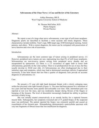
  • 1. Schwannoma of the Ulnar Nerve: A Case and Review of the Literature Ashley Boustany, MS II West Virginia University School of Medicine W. Thomas McClellan, M.D. Plastic Surgeon Private Practice Abstract We report a case of a large ulnar nerve schwannoma, a rare type of soft tissue neoplasm. Diagnostic pearls are described to facilitate a more accurate and timely diagnosis. These characteristics include mobility, Tinel’s sign, MRI target sign, S100 histological staining, Antoni patterns, and others. With a correct diagnosis, the tumor can be extirpated with preservation of nerve function and a low risk of recurrence. Introduction Schwannomas are the most common type of tumor arising in peripheral nerves. (1) However, peripheral nerve tumors are rare, representing less than 8% of soft tissue neoplasms. Schwannomas are non-invasive tumors arising from peripheral nerve sheaths and are encapsulated by epineurium. (2) There is a 2:1 occurrence of upper limb to lower limb schwannomas, generally on the volar surface. There is no predisposition for sex or race, but they usually develop in 30-60 year olds. (1) Schwannomas are often misdiagnosed due to their indistinct signs and symptoms, which may lead to detrimental neurologic deficits if approached incorrectly. It has been shown that less than a quarter of diagnostic tests provide an accurate diagnosis of schwannoma. (3) Case We present a 53 year old right hand dominant female with a slowly enlarging mass within her proximal volar right forearm (Figure 1). The mass had been present for a minimum of two years and had become more painful and noticeable over time. Dull, intermittent pain was reported at rest over the mass, and was moderately sharper during flexion of the fingers or bumping of the forearm. The level of discomfort increasingly hindered her ability to perform activities of daily living. She was initially treated conservatively at an outside facility for a ruptured forearm muscle belly. Subsequently, her symptoms worsened and a CT guided biopsy of the suspected mass was performed. The patient reported the biopsy was extremely painful and caused an exacerbation of her forearm pain. Histopathology demonstrated a paucicellular specimen with very rare fragments of fibrous tissue that was insufficient for diagnosis.
  • 2. Figure 1. Preoperative: The dotted line indicates the edge of the palpable tumor. Additionally, the olecranon, medial epicondyle, and path of the ulnar nerve are identified. She was referred to our hand surgery service following the inconclusive biopsy. The 5 cm firm mass was mobile perpendicular to the nerve axis and immobile along the parallel axis. A very sensitive and positive Tinel’s sign radiated in the ulnar distribution, with dysthesias directly over the mass and along the dorsal ulnar distribution of her hand. Semmes Weinstein testing indicated diminished light touch and hypersensitivity to sharp touch in the same region. However, her strength of the intrinsic muscles of the right hand was 5/5 without evidence of weakness or loss of range of motion. An MRI was performed and showed a 5.0 cm x 3.0 cm x 4.6 cm diameter mass displacing the flexor digitorum superficialis and palmaris longus muscles (Figure 2). The mass had smooth margins but displayed a complex heterogeneous signal. There was no evidence of invasion of adjoining muscles or bones. With gadolinium injection, an intense irregular enhancement was seen. A capsule surrounded the eccentrically placed mass. Images also indicated the presence of a hemorrhagic center related to the needle biopsy. MRI diagnosis was inconclusive, but the differential included, schwannoma, malignant histiocytoma, neurofibrosarcoma, or soft tissue malignancy. Figure 2. MRI: T1 weighted MRI of the right upper extremity showing a large well demarcated but heterogeneous mass. The MRI lacked the traditional “target sign” that is commonly associated with schwannomas.
  • 3. Figure 3. Intraoperative: The proximal and distal ulnar nerve are identified with vascular loops. Note how the fascicles of the ulnar nerve are splayed out over the tumor. Identification of these fascicles is crucial to determine a safe longitudinal entry point through the epineurium. She was taken to the operating room for exploration. Extirpation of the tumor was performed via a longitudinal access incision through the epineurium and careful separation of the nerve and tumor (Figure 3). The tumor measured 4.0 cm x 3.0 cm x 1.9 cm (Figure 4). It showed a hypo-cellular tissue with Schwann cells strongly positive for S100 protein by immunostain technique (Figure 5). Antoni B patterns predominated with few areas suggestive of Antoni A patterns. Incision of the tumor also revealed hemorrhage and thrombosis consistent with prior needle aspiration. Three months following surgery the patient has retained full ulnar motor and sensory function as well as improving sensory paresthesias in her dorsal ulnar division. Figure 4. Tumor: Gross appearance of the Schwannoma following removal from within the ulnar nerve.
  • 4. Figure 5. Histopathology: A hypo-cellular tissue with Schwann cells staining strongly positive for S100 protein. Discussion Diagnosis of a schwannoma in the preoperative period is challenging because of the slow growth and paucity of symptoms. Diagnostic accuracy is crucial to maintaining the integrity of the nerve involved and to properly plan the appropriate surgical intervention. Often these tumors present as palpable masses, tender to displacement without muscles weakness. Tinel’s sign is positive in the majority of cases. The tumors are transversely mobile but immobile longitudinally, likely due to their nested intraneural location. Schwannomas share many features with other soft tissue tumors and are frequently misdiagnosed due to similarities. Differential diagnosis should include neurofibroma, ganglion cysts, malignant tumors, lipomas, and xanthomas. (1,4) Neurofibromas, in particular, cannot be distinguished from schwannomas on physical examination. The symptoms appear to be nonspecific, which adds to difficulty in diagnosis. (5) They may be differentiated on MRI or fine needle aspiration. Malignant masses exhibit more distinct signs but are often mistaken for schwannomas in early stages of diagnosis. Unlike schwannomas, malignant tumors often have immobility, firmness, constant pain at rest, and motor weakness. (6) Weakness may occur if the benign tumor exceeds 2.5 cm and can be location dependent. Kehoe et al (1995) analyzed 88 peripheral nerve tumors, where only one was correctly diagnosed as a schwannoma preoperatively. Gadolinium enhanced T1-weighted and T2-weighted MRIs are particularly useful in diagnosing schwannomas. Koga et al (2007) found the presence of the target sign to be 100% specific and 59% sensitive for the tumors. The target sign is the contrasting central and peripheral intensities demonstrated on the images. Histological analysis credits the central hyperintensities and hypointensities on T1 gadolinium and T2, respectively, to Antoni A cells. Antoni A patterns are of low cellular concentration. Antoni B areas are of high cell concentration and correspond to peripheral intensities on MRI and CT. (8) Histological staining reveals a strongly positive S100 protein that is specific for schwannomas and helps to rule out neurofibromas. (5,9) Imaging shows the tumors as round or oval, eccentrically located in relation to the nerve, encapsulated, isolated, and non-invasive. In comparison, neurofibromas are non-encapsulated and intimately surround the nerve. They cannot be surgically removed without damaging the connected nerve, often necessitating nerve grafting to repair functioning. (2,4,5) Schwannomas, on the other hand, can be separated surgically from
  • 5. the nerve fascicles avoiding neurologic deficits. (4) This emphasizes the importance of a correct preoperative diagnosis. Despite the structural differences of soft tissue tumors, they are difficult to distinguish with imaging. Fine needle aspiration tends to be extremely painful in cases of schwannomas, and hemorrhaging may result with temporary worsening of symptoms. The results are frequently inconclusive, but are helpful to exclude ganglion cysts. (3) Domanksi et al (2006) aspirated 116 different schwannomas, and results were not sufficient for diagnosis for about 44% of the cases. Extirpation of the intraneural schwannoma can be challenging. Sterile tourniquet dissection is recommended and assists in visualization. Loupe magnification and or use of the operating microscope is highly recommended. Identification of the nerve proximal and distal to the tumor is the first important step to reducing injury and traction neuropraxia. Identification of the individual splayed nerve fascicles as they spread over of the tumor is critical in determining the entry through epineurium. A longitudinal incision is created between the splayed fascicles down to the tumor sheath. Once the outer layer of the tumor is identified, a plane can be developed between the more superficial fascicles and the tumor wall. Slow, deliberate, circumferential dissection with a “peanut” and Littler scissors facilitates delivery of the tumor. Once the tumor is removed, the nerve is inspected for injury, the tourniquet is released, and precise hemostasis is achieved. Repair of the epineurium is not required and the longitudinally split muscle is repaired loosely over the nerve. Drains are optional, bulky dressing is preferred, and immediate post operative hand therapy is instituted. It is uncommon for schwannomas to recur in identical locations. (1) Das et al (2007) found that surgical removal of schwannomas was successful in alleviating preoperative symptoms while maintaining nerve functioning in 89% of their cases. Conclusion Schwannomas are rare peripheral nerve tumors that have important diagnostic and radiographic features. These tumors are transversely mobile and longitudinally immobile, have a positive Tinel’s sign, and exertional dysathesias or pain. MRI typically reveals the target sign of biphasic contrast of peripheral and central regions and distinct encapsulation displacing the intimately associated nerve fascicles. Surgical resection must be approached with caution to protect nerve function and continuity. Surgical resection is associated with good outcomes. The recurrence rate is low. References 1. Ozdemir O, Kurt C, et.al. Schwannomas of the hand and wrist: long-term results and review of literature. Journal of Orthopaedic Surgery. 2005;13(3):267-272. 2. Lin J, Martel W. Cross-sectional imaging of peripheral nerve sheath tumors: characteristic signs on CT, MR imaging, and sonography. Amer Journ Roentgenology. 2001 Jan.;176:75-82. 3. Rockwell G, Achilleas T, et. al. Schwannoma of the hand and wrist. Plast Reconstr Surg. 2003 Mar.;111(3):1227-1232.
  • 6. 4. Sandberg K, Nilsson J, et. al. Tumors of peripheral nerves in the upper extremity: A 22-year epidemiological study. Scand J Plast Reconstr Hand Surg. 2009 Sep.;43:43-49. 5. Adani R, Baccarani A, et. al. Schwannomas of the upper extremity: diagnosis and treatment. Chir Orani Mov. 2008 Sep.;92:85-88. 6. Ogose A, Hotta T, et. al. Tumors of peripheral nerves: correlation of symptoms, clinical signs, imaging features, and histologic diagnosis. Skeletal Radiol. 1999 Jan.;28:183-188. 7. Kehoe N, Reid R, et. al. Solitary benign peripheral-nerve tumors: review of 32 years’ experience. J Bone Joint Surg. 1995 Oct.;77B:497-500. 8. Koga H, Matsumoto S, et. al. Definition of the target sign and its use for diagnosis of schwannomas. Clinical Orthopaedics and Related Research. 2007 Aug.;464:224-229. 9. Das S, Ganju A, et. al. Tumors of the brachial plexus. Neurosurg. 2007June;22(6):E26. 10. Domanski H, Akerman M, et. al. Fine Needle Aspiration of Neurilemoma (Schwannoma). A clinicocytopahologic study of 116 patients. Diagnostic Cytopathology. 2006;34:403-412.