NEONATAL RESUSCITATION GUIDE
•
2 likes•462 views

This document provides an overview of neonatal resuscitation. It defines neonatal resuscitation as interventions at birth to support breathing and circulation. It discusses the history of neonatal resuscitation and how techniques have developed over time. It also outlines the key steps and principles of resuscitation including initial assessment, positive pressure ventilation, chest compressions, and intubation. Special considerations for preterm infants are also addressed.

Recommended
More Related Content
What's hot
What's hot (20)
Similar to NEONATAL RESUSCITATION GUIDE
Similar to NEONATAL RESUSCITATION GUIDE (20)
Recently uploaded
Recently uploaded (20)
NEONATAL RESUSCITATION GUIDE
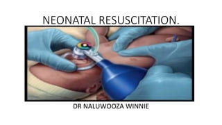
- 1. NEONATAL RESUSCITATION. DR NALUWOOZA WINNIE
- 2. Outline • Definition • History • Overview and Principles of resuscitation • Initial steps of resuscitation • Positive – pressure ventilation • Chest compressions • Endotracheal tube intubation and LMA insertion • Medications • Special considerations • Resuscitation of preterm babies • CPAP in delivery room • Warm transport.
- 3. Definition • Neonatal resuscitation is a set of interventions at the time of birth to support the establishment of breathing and circulation. (AAP) Levels. Simple interventions – drying and stimulation, suction with bulb syringe. Basic resuscitation – PPV (bag and mask), deep suction. Advanced resuscitation – chest compressions, Endotracheal Tube(ETT), medications and fluids.
- 4. Historical aspects. • For the past 45years,fetal anoxia was one of the most investigated conditions affecting the new born. • It was then realised that obstruction to the airway immediately following birth should be the first concern in newborn resuscitation. • 18th century Scottish obstetrician Blundell first used mechanical device for tracheal intubation in living newborn.
- 5. • In 1920, Joseph B. DeLee introduced simple rubber catheter and glass trap to clear upper airways and stomach. • In 1953 Apgar score was given by Varginia Apgar. She is also the first to catheterise Umbilical Artery in new born. Score 0 Score 1 Score 2 Appearance Blue all over Blue only at extremity No blue colouration Pulse No pulse <100b/m >100b/m Grimace No response to stimulation Grimace or feeble cry when stimulated Sneezing, coughing, or pulling away when stimulated Activity No movement Some movement Active movement Respiration No breathing Weak, slow or irregular breathing Strong cry
- 6. • In 1966 national guidelines for resuscitation of adults was recommended by National Academy of Science. • In 2000 the consensus document on advanced life support of the new born converted the previously published advisory statements into a set of guidelines. • These guideline were revised further in 2010, 2015 and latest revision being in 2020 and published in June 2021 (8th edition of the Neonatal Resuscitation Program) by AAP and AHA.
- 7. Statistics • Globally, 2 to 10 per 1000 term newborns faced perinatal asphyxia (inability of a newborn to initiate and sustain adequate respiration after delivery) , and the report of WHO 2014 indicated that birth asphyxia leads to about 4million neonatal deaths annually. • The incidence of birth asphyxia in most developed countries accounts for less than 1/1000 of newborn deaths contrary to developing countries where it ranges from 4.6/1000 to 26/1000 live births.
- 8. • More than 25.0% of the world’s newborn deaths have occurred in Africa, of which birth asphyxia accounts for 24.0%. • From 20 countries in the world with the highest risk of neonatal death, 75.0% are in Africa. (Guo A et al 2017) • The incidence of asphyxia in East, Central and southern Africa was 22.0% by 1993. (kinoti S- asphyxia of the new born in east, central and southern Africa) But by 2020, prevalence of perinatal asphyxia was at 18.0% and 9.0% in East and Central African countries respectively (Yinager Workineh et al, prevalence of perinatal asphyxia in East and Central Africa)
- 9. Why learn newborn resuscitation? • Uganda’s neonatal mortality rate (NMR) is 19 deaths per 1,000 live births with 30 deaths per 1,000 live births in rural areas and 31 deaths per 1,000live births in urban areas. (UNICEF facts sheet 2021) With birth asphyxia as the leading cause.
- 11. Overview and principles • Approximately 90% of newborns make smooth transition from intrauterine to extrauterine life requiring little or no assistance • 10% of newborns need some assistance • Only 1% require extensive resuscitation • We must always be prepared to resuscitate, as even some of those with no risk factors will require resuscitation.
- 12. Fetal transition. Before birth • Oxygen supply by placental membranes • No role of lungs- fluid filled alveoli and constricted arterioles due to low O2 on Fetal blood. • Thus the increased pulmonary resistance leads to shunting of blood from pulmonary artery to ductus arteriosus to aorta.
- 13. After birth • Baby cries - Takes first breath - Air enters alveoli - Alveolar fluid gets absorbed -Increased P02 relaxes pulmonary arterioles - Thus decreased PVR ( pulmonary Vascular Resistance). • Umbilical arteries constrict and clamp cord- closure of umbilical arteries and umbilical vein- increased SVR (Systemic Vascular Resistance) • Decreased PVR and increased SVR – functional closure of ductus arteriosus- increased blood flow into lungs – oxygenation – supply to body through aorta.
- 15. Signs of compromised new born These present as a consequence of interruption in transition. Low muscle tone Respiratory depression (apnoea/gasping ) Tachypnea bradycardia Hypotension Cyanosis Low oxygen saturation
- 16. Principles • The sequence of resuscitation in adults is C-A-B • But in newborns it remains A-B-C as the etiology of neonatal compromise is nearly always a breathing difficulty. • A- position and clear • B- stimulate breathe • C- assess HR and oxygenation • D-drugs
- 17. Preparing for birth • Review antepartum and intrapartum risk factors. • Must know – expected gestational age? – is the amniotic fluid clear? – number of babies expected? – any additional rick factors? • Personnel – at least 2. • Equipment Resuscitation table Sterile linen Suction apparatus Laryngoscope Ambu bag and face mask Oral airways Oxygen with flow meter and tubing Endotracheal tubes Sterile gloves
- 18. Miscellenous • Radiant warmer • Stethoscope • Adhesive tape • Syringe • Umbilical artery catheterisation tray • Umbilical catheter 3 and 5 fr • Feeding tube • Alcohol • Drugs
- 19. Neonatal Resuscitation Program flowchart
- 20. Initial evaluation • Term • Tone • Breathing or crying
- 21. What to do with a vigorous term newborn • Provide warmth – 36.5 – 37.5c place baby under radiant warmer uncovered. • Position the head and neck neck in neutral position, head in sniffing position. • Clear secretions, if needed bulb syringe or penguin sucker. • Dry • Stimulate
- 22. Re-evaluation • Respiration – rate 40 to 60b/m • Heart rate – auscultation or pulsations at the base of cord is felt. 6s * 10. > 100b/m • Oxygenation by oximeter
- 23. Airway • Reposition neck and head • Suction (suction catheter with/without laryngoscope guided). • Laryngeal mask airway.
- 24. • If breathing and heart rate is over 100b/m, but baby is still cyanotic: Acrocyanosis – bluish hue of hands and feet Central cyanosis – bluish hue of lips, tongue and torso Place pulse ox and provide supplemental oxygen by -flow-inflating bag and mask -Oxygen tubing -t-piece resuscitator -self inflating bag If cyanosis persists, provide positive pressure ventilation (PPV)
- 27. • Blow by oxygen using a T piece Mengo hospital NRP
- 28. Target pre-ductal SPO2after birth Minutes Target 1 min 60% -65% 2 min 65%-70% 3min 70%-75% 4 min 75%-80% 5 min 80%-85% 10 min 85%-95%
- 29. POSTIVE PRESSURE VENTILATION Indication • Apnoea • Gasping • HR <100b/m • O2 saturation below the target range despite free-flow oxygen or CPAP. Effective ventilation is the single most important step in CPR of compromised infant.
- 30. Equipment for PPV • Masks Rims -cushioned , non cushioned Shape -round, anatomically shaped Size -small, large Notably, mask should cover the tip of the chin, mouth and nose so as to create a seal.
- 31. • Devices Flow inflating bag T-piece resuscitator Self-inflating bag. Each of these have advantages and disadvantages over each other.
- 32. Self inflating bag Flow inflating bag T-piece Resuscitator PIP Doesn’t require compressed source for inflation of bag Requires compressed source for inflation of the bag Requires compressed gas source for inflating the bag Functions even without proper seal Doesn’t work without proper seal Doesn’t work without proper seal PIP How hard and long the bag is squeezed Flow of incoming gas and how hard and long the bag is squeezed Can be set exactly manually PEEP Only if additional valve is attached CPAP/free flow 02 Cannot be delivered Given by adjusting flow control valve Can be set exactly manually Safety features Pop off valve pressure gauge Pressure gauge Maximum pressure relief valve pressure gauge.
- 33. • Peak Inspiratory Pressure (PIP) The summation of the pressure generated by the ventilator to overcome airway resistance and alveolar resistance to attain peak inspiratory flow and to deliver desired tidal volume. • Positive End Expiratory Pressure (PEEP) Is the pressure applied by the ventilator at the end of each breath to ensure that the alveoli are not so prone to collapse. Reduces trauma to the alveoli, increases functional residual capacity which improves oxygenation, lesser pressure is needed to get the same volume of air into the lung as the alveoli are already open thus increasing compliance, ventilation/perfusion mismatches are improved.
- 34. CPAP • Its the pressure in the system at the end of spontaneous breath when a mask is held tightly on baby’s face but the bag isn’t being squeezed. NCPAP.
- 35. Starting ventilation Before beginning positive pressure ventilation: • Select appropriate-sized mask • Be sure airway is clear • Position baby’s head • Position yourself at baby’s head or side • Confirm equipment is attached to oxygen source.
- 36. Continued… • How much oxygen • >35weeks of gestation begin with 21% • <35weeks begin with 21-30% • Breaths should be given at 40 to 60 bpm • How much pressure? • Neopuff/neotee • PIP at 20-25cm H20 • PEEP at 5cm H2O.
- 38. Signs of effective ventilation • Signs of adequate ventilation Improved heart rate, color and muscle tone. • Signs of improvement in the newborn Improved heart rate, color, breathing, tone and saturation. Thus these parameters are reassessed during PPV.
- 39. After starting PPV • At 15seconds, and 30seconds from the start of PPV, the assistant should announce loudly; 1. Change in Heart rate (increasing, decreasing or the same) 2. chest movements (is moving or not moving) 3. +/- o2 saturation.
- 40. First assessment (after 15 seconds) • After 15seconds of PPV • Announce heart is increasing • Continue PPV • Second HR assessment after 15seconds of PPV • Announce heart rate not increasing, chest moving. • Continue PPV that moves the chest • Second HR assessment after another 15secs of PPV that moves the chest. • Announce HR not increasing, chest is not moving • Ventilation corrective steps until chest movements with PPV. • Intubate or laryngeal mask if necessary • Announce when chest is moving • Continue PPV that moves the chest • Second HR assessment after 30seconds of PPV that moves the chest. Increasing Not increasing chest moving Not increasing Chest not moving.
- 41. Second assessment (after 30 seconds) • After 30seconds of PPV that moves the chest • Continue PPV 40-60 breaths/min until spontaneous effort • Reassess ventilation • Ventilation corrective steps if necessary. • Reassess ventilation • Ventilation corrective steps if necessary • Insert an alternative airway • If no improvement 100% oxygen and chest compressions. At least 100bpm 60-99bpm <60bpm
- 42. Causes and solutions for inadequate chest expansions. Condition Actions Inadequate seal Reapply mask to face and lift jaw forward Blocked airway Reposition the head Check for secretions : suction if present. Ventilate with the newborn’s mouth slightly open. Not enough pressure Increase pressure until there is a perceptible chest movement. Consider endotracheal intubation.
- 43. Mr. SOPA CORRECTIVE STEPS Actions M Mask adjustment Reapply the mask, consider the 2 hand technique R Reposition airway Place head neutral or slightly extended Try PPV and reassess chest movements S Suction mouth and nose Use a bulb syringe or suction catheter. O Open mouth Open the mouth and lift the jaw forward. Try PPV and reassess chest movements P Pressure increase Increase pressure in 5 to 10cm H2O increments, maximum 40cm H2O. Try PPV and reassess chest movements A Alternative airway Place an endotracheal tube or laryngeal mask. Try PPV and assess chest movements and breath sounds
- 44. Orogastric tube • After several minutes of PPV or CPAP with a mask, gas enters the stomach and may interfere with ventilation or cause regurgitation and aspiration. • Place orogastric tube, suction gastric contents and leave it uncapped to act as a vent to the stomach.
- 45. Chest compressions. • Indicated when heart rate remains less than 60bpm despite 30seconds of effective PPV. • Need 2 people. • Ratio of 3:1 compression to ventilation ratio. (30breaths and 90compressions in one minute) • Place thumbs or fingers 2cm below the imaginary nipple line. • Apply pressure on the sternum during compressions, compressions depth is approximately one-third of the anterior posterior diameter of the chest. • Allow chest recoil and ventilation.
- 46. CPR with the thumbs technique.
- 48. Complications • Lacerations of the liver • Broken ribs • Pneumothorax. • Hemorrhage.
- 49. Reassessment. Check HR after 60seconds. If >60bpm, stop compressions but continue ventilation at 40 to 60bpm If <60 check quality of ventilation and compressions If still remains less than 60bpm, consider intubation if not already done, consider epinephrine either via ETT or insert umbilical catheter.
- 50. INTUBATION.
- 51. Endotracheal intubation Indications To improve ventilation after several minutes ineffective bag-and-mask ventilation To facilitate coordination of chest compressions and ventilation To administer epinephrine while establishing IV access. Special Extreme prematurity Surfactant administration Suspected diaphragmatic hernia. HR <100b/m in spite of PPV. HR <60b/m Baby is floppy, not crying and preterm.
- 52. Equipment. • Laryngoscope blade • 00 for extreme prematures • 0 for preterm newborns • 1 for term newborns • Laryngoscope, check blade, handle and functioning light. • Suction source, set at 80-100mm Hg, large suction catheter for mouth/pharynx, small catheter for endotracheal tube (5 or 6 F). • Endotracheal tube size Weight (g) Gestational Age (wks) Endotracheal Tube Size (mm ID) Below 1,000 Below 28 2.5 1,000n- 2,000 28-34 3.0 Greater than 2,000 Greater than 34 3.5
- 53. Depth of insertion. Add 6 to the baby’s weight, tip to lip. Weight Depth of insertion <750g 6cm 1kg 7cm 2kg 8cm 3kg 9cm 4kg 10cm
- 55. Steps for intubation 1. Preparation for insertion stabilize the newborn’s head in the sniffing position. deliver free-flow oxygen during the procedure. 2. Insertion of laryngoscope slide the laryngoscope over right side of the tongue, pushing the tongue to the left side of the mouth. advance blade until the tip lies just beyond the base of the tongue. 3. Lift blade lift the blade slightly on angle to ceiling, raising the entire blade not just the tip. Visualize pharyngeal area. Don’t rock.
- 56. 4. Visualize landmarks Vocal cords should appear as vertical stripes on each side of the glottis or as an inverted letter V. apply downward pressure on cricoid to help bring glottis into view. Suction for visualization, if necessary. 5. Inserting tube insert the tube following the curvature of the blade. Insert the tip of the endotracheal tube until the level of the vocal cords. Limit attempts to 30 seconds, and if the cords are closed, wait for them to open.
- 60. Confirmation of ETT placement. • Detecting exhaled CO2 and rapid rise in heart rate are primary methods. • Auscultate breath sounds during PPV, near both axillae. • Symmetrical chest movements. • Little or no air leak from mouth during PPV. • Decreased or absent air entry over the stomach • Chest X-ray.
- 61. Securing ETT in place.
- 63. If baby’s condition worsen after intubation The DOPE mnemonic D Displaced endotracheal tube O Obstructed endotracheal tube P Pneumothorax, collapsed lung E Equipment failure.
- 64. CPAP in the delivery room • Importance • CPAP if initiated early at the time of delivery, established adequate lung volume or functional residual capacity • It avoids the need for intubation • Reduces need for surfactant. • Ventilation also leads to volutrauma and lung inflammation. • It also prevents the injuries caused by mechanical ventilation which is more aggressive.
- 65. Indications for CPAP. Preterm neonates Term neonates Respiratory distress in the delivery room Moderate to severe respiratory distress Atelectasis Meconium aspiration syndrome Recurrent apnoea Large patent ductus arteriosus Pulmonary edema Neonatal pneumonia
- 68. Who needs CPAP immediately after birth. Silverman Anderson Score (SAS) • Mild 1 to 3 – supplemental O2 • moderate respiratory distress 4 to 6 – CPAP • Severe respiratory stress 7 to 10 – intubation Also babies weighing less than 1500grams. Score Upper chest retractions Lower chest retractions Xiphoid retraction Nasal flaring Grunting 0 None None None None None 1 Lag on inspiration Just visible Just visible Just visible Only with stethoscope 2 See-saw Marked Marked Marked Audible to naked ear.
- 69. DRUGS. Epinephrine • Indicated when heart rate remains <60b/m. • Not indicated before adequate ventilation is established • IV or endotracheal route, • 0.1 to 0.3ml/kg of 1:10,000 solution (0.3 to 1 ml/kg if given endotracheal) • As rapidly as possible. • Reassess after 1 minute, if HR <60bpm repeat dose every 3 – 5 minutes. And consider increasing dose to higher end of range.
- 70. Volume expanders • If no response to epinephrine especially with persistent bradycardia and shock is suspected like in placenta Previa/abruption. Normal saline is recommended but ringer’s lactate, or O Rh negative blood can be used. 10ml/kg via umbilical vein given over 5 to 10minutes.
- 71. Resuscitation of preterms • Thermoregulation: Increase delivery room temperature Radiant warmer Polyethylene plastic bag or wrap Preheated transport incubator Monitor baby’s temperature frequently. • Respiratory support Use oxygen blender, adjust oxygen concentration to target 85% - 95%. Use lower PIP 20-25cm of H20 during PPV Consider giving CPAP and/0r surfactant.
- 72. Post resuscitation care. • Monitor the baby’s temperature • Monitor blood glucose • Monitor the baby for apnoea and bradycardia. • Proper transport for admission including warm transport and maintained respiratory support. Discontinuation of resuscitation efforts may be appropriate after 10 minutes of absent heart rate following complete and adequate resuscitation efforts.
- 73. Warm transport • The incidence of hypothermia at time of admission to the NICU in VLBW newborns ranges from 31% to 78% (Watkinson Met al 20006 and zymankiewicz M et al 2003) • A study in Nsambya Hospital reported 87% of all preterms had hypothermia at the time f admission to newborn unit (Cheptoris and Nakibuuka et al 2016). • each 1c decrease in axillary temperature is associated with a 28-75% increase in neonatal mortality (abbot and Mullany LC et al 2010) Newborns loss heat through radiation, convection, evaporation and conduction.
- 74. • Warmilu Incu blanket • Insta warmer
- 75. Respiratory support while transporting. • Portable oxygen source • Portable CPAP/bubble • Portable pulse oximeter. • Suction apparatus.
- 77. • Warm transport and respiratory support by CPAP by Mengo Hospital NRT.
- 78. Doing the simple things effectively.
- 79. Thank you. Psalms 127: 1-2 Unless the Lord builds the house, its builders labour in vain. Unless the LORD watches over the city, the watchmen stand guard in vain. In vain you rise early and stay up late, toiling for food to eat – for he grants sleep to those he loves.
Editor's Notes
- ACOG and AAP, asphyxiated neonate – umbilical cord pH <7, Apgar score 0-3 at 5 mins, neonatal neurological manifestations (seizures, coma, hypotonia), multisystem organ dysfunction.
- Epinephrine increases systemic vascular resistance, increases coronary artery perfusion pressure and improves blood flow to myocardium and restores depleted ATP.