SlideShare a Scribd company logo
1 of 72
JSS Medical College, Mysuru
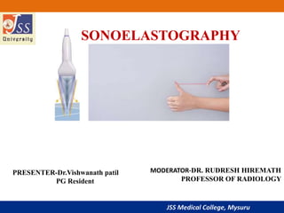
SONOELASTOGRAPHY
PRESENTER-Dr.Vishwanath patil
PG Resident
MODERATOR-DR. RUDRESH HIREMATH
PROFESSOR OF RADIOLOGY
JSS Medical College, Mysuru
Definition.
Principle.
Terminologies used in elastography.
Different elastography techniques.
1. Compression Elastography
2. ARFI
3. SWE
Depiction of elastograms.
JSS Medical College, Mysuru
DEFINITION
• It is non invasive novel technique of imaging & mapping the
stiffness or elasticity of tissues induced by compression.
• Elastography can be performed with ultrasound and MRI. In
ultrasound it is also known as sonoelastography.
JSS Medical College, Mysuru
PRINCIPLE
• Every tissue in the human body pertains some amount of
elastic properties. This elastic property get distorted or
hampered by the diseases, particularly by the malignant
lesions.
• This change in the elastic property of the diseased tissue is
used as principle in elastography.
• The technique is first described in 1987 by Krouskop et al.
JSS Medical College, Mysuru
Physicians & surgeons have
acquired clinical skill of
palpation of masses to detect
differences in tissue stiffness as
an aid to diagnosis.
We radiologist lack this as
we stick to our ultrasound
machines and probes.
JSS Medical College, Mysuru
So what do we call
this technique as ?
We can call elastography as VIRTUAL PALPATION technique
designed for radiologists to differentiate stiffer tissues from
softer one.
JSS Medical College, Mysuru
The advantage we have with this technique is we can
virtually palpate the masses situated deep, which is
not available for manual palpation.
JSS Medical College, Mysuru
Terminologies used in elastography
• STRESS
• STRAIN
• ELASTICITY
• VISCOSITY.
• VISCOELASTICITY.
• YOUNG’S MODULUS
JSS Medical College, Mysuru
STRESS: Stress is defined as force per unit area
Stress perpendicular to the object
shortening of object
Stress parallel to the object
distortion of object.
JSS Medical College, Mysuru
STRAIN: Amount of Change or deformation in the size and
shape of an object subjected to stress is strain.
LONGITUDINALSTRAIN- change in the
length of an object.
SHEAR STRAIN- change in the angles of
an object
• Stress parallel
to the object
• Stress perpendicular
to the object
JSS Medical College, Mysuru
• The property of materials to return back to the its original form after stress
is removed is known as elasticity.
Elasticity
JSS Medical College, Mysuru
Viscosity
• Viscosity is the measure of resistance of a fluid when it
undergoes shear stress or tensile stress.
JSS Medical College, Mysuru
VISCOELASTICITY
• This is property of materials to exhibit both viscous and elastic
properties
JSS Medical College, Mysuru
YOUNG'S MODULUS
• Tissue stiffness can be quantified using the a value known as
Young's Modulus.
• Ratio of the compression force applied to tissue (stress) and the
resulting tissue deformation (strain).
• Young’s modulus(elasticity) = Stress/Strain.
• This is measured in kilopascals (kPa).
JSS Medical College, Mysuru
Typical values of elasticity in different types of tissues .
JSS Medical College, Mysuru
DIFFERENT ELASTOGRAPHY
TECHNIQUES,
JSS Medical College, Mysuru
FREE HAND SONOELASTOGRAPHY OR
COMPRESSION ELASTOGRAPHY OR STATIC
ELASTOGRAPHY
• Is based on the signals acquired before and after tissue
displacement induced by the manual probe compression.
JSS Medical College, Mysuru
STRAIN ELASTOGRAPHY
• Manual compression is applied by probe over the mass lesion
in the direction perpendicular to the tissue surface.
Radiofrequency signals are obtained from the tissues before
and after compression.
• Tiny changes produced by the manual compression induces
change in the phase of radiofrequency echoes. The amount of
shift in the signals equals the amount of tissue displacement at
the point in the image frame.
JSS Medical College, Mysuru
Pre
compression
RF A LINE
Post
compression
RF A LINE
JSS Medical College, Mysuru
• Tissue displacements are registered and converted into B mode
images.
• A dedicated software is then used to calculate the relative
difference in tissue movement from one frame to another and
then estimates tissue deformation.
JSS Medical College, Mysuru
• PRE
COMPRESSION
RF A LINE
POST
COMPRESSION
RF A LINE
REGISTERED AND
CONVERTED INTO B
MODE IMAGES
REGISTERED AND
CONVERTED INTO B
MODE IMAGES
Dedicated software to
calculate difference in tissue
movements from one frame to
another
Elastograms
JSS Medical College, Mysuru
What to look for in elastographic images ?
• In case of sonoelastographic images, lesion brightness,
uniformity and the size ratio helps to differentiate benign from
malignant lesions.
JSS Medical College, Mysuru
Display of elastogram
 Images are displayed as darker and brighter images.
 Color scale images which are superimposed on B mode
images.
 Strain ratio – calculated to compare two area of different
stiffness.
JSS Medical College, Mysuru
• Images are displayed as darker and brighter images.
• Harder areas exhibit less tissue displacement, displaying darker
strain images while soft areas exhibit more tissue displacement
and brighter strain images.
JSS Medical College, Mysuru
• In benign tumors, the transverse diameters measured on
elastographic images are almost always the same as or smaller
than the diameters of the tumors seen on B-mode images,
whereas the diameters of malignant tumors on elastographic
images are larger than those seen on B-mode images.
JSS Medical College, Mysuru
Infiltrative ductal carcinoma – depicting dark strain images with surrounding normal
tissue revealing bright strain.
Lesion is larger in elastography images compared to the normal B mode ultrasound.
JSS Medical College, Mysuru
Cysts have a typical “bull’s eye” appearance (smaller size,
white center, black peripheral circle) (Siemens machines) or
characteristic noise pattern(philips). (Philips).
JSS Medical College, Mysuru
B. Color scale images which are superimposed on B
mode images.
 On ultrasound machines, a color elastograms is displayed on
the screens.
 Itoh proposed an elasticity score which is compared to the BI-
RADS.
JSS Medical College, Mysuru
 Itoh et al, evaluated the color pattern of images in both the
hypoechoic lesion and in the surrounding breast tissue.
 An elasticity score on a five-point scale was attributed :
 The risk of malignancy increases from Score 1 (a benign
lesion) to score 5 (malignant lesion).
JSS Medical College, Mysuru
A score of 1 ~ an entirely green lesion (even strain throughout
the lesion),
JSS Medical College, Mysuru
A score of 2 ~ strain in most of the hypoechoic lesion, with some
strain free areas (a mosaic pattern of green and blue),
JSS Medical College, Mysuru
A score of 3 ~ strain at the periphery of the hypoechoic lesion, with
sparing of the center of the lesion (the peripheral part of the lesion in
green, and the central part in blue),
JSS Medical College, Mysuru
A score of 4 indicated no strain throughout the hypoechoic lesion (the entire
lesion in blue, but the area surrounding it is not included).
JSS Medical College, Mysuru
A score of 5 indicated no strain in the entire hypoechoic lesion or in
the surrounding area (both the lesion and surrounding area are
blue).
JSS Medical College, Mysuru
Mosaic Green & blue appearance- benign- Fibro
adenoma
JSS Medical College, Mysuru
Homogenously blue appearance on elastograms- suggestive of
malignancy – infiltrative ductal carcinoma.
JSS Medical College, Mysuru
PHILIPS MACHINES :
• In color mode, malignant lesions are depicted in blue whereas benign
lesions appear in red (soft).
• Cystic lesions have a typical three-color mixed appearance (red, green,
blue).
• Finally, with a specific software, a pure cystic content appears in yellow,
and can be differentiated from a dense cyst exhibiting a blue center.
JSS Medical College, Mysuru
THREE COLOR MIXED APPERANCE SEEN IN BENIGN
CYSTIC LESION
JSS Medical College, Mysuru
C. Strain ratio – calculated to compare two area of different
stiffness.
 Two Regions of Interest (ROI) are simultaneously set in the center of the lesion,
and in the surrounding breast tissue.
 Elasticity values are shown on compression-decompression time curves.
JSS Medical College, Mysuru
Strain ratio
 The higher the ratio between the two curves at time , greater
the stiffness, and the higher the risk of malignancy, especially
for a lesion with little elasticity.
 Conversely, a benign lesion shows similar elasticity values on
the time/intensity curves as the surrounding tissue.
JSS Medical College, Mysuru
High difference between the compression and decompression time curves
JSS Medical College, Mysuru
Similar the compression and decompression time curves
JSS Medical College, Mysuru
• Limitations :
 Pressure on the probe should be very light much lighter than for a normal
ultrasound acquisition.
 The technique is qualitative imaging of tissue stiffness.
 Can be utilized as semi quantitative technique with the evaluation of strain
ratio.
JSS Medical College, Mysuru
Transient Elastography (TE):
• A system developed and commonly used for liver fibrosis
• Mechanical piston within a ultrasound transducer is used to apply a push to
the skin over an intercostal space.
• The speed of the produced shear waves into the liver, along the direction of
the ultrasound beam, is measured in a way similar to M-mode.
JSS Medical College, Mysuru
ARFI TECHNOLOGY: ACOUSTIC RADIATION
FORCE IMAGING
• Advantage of being objective and independent of the sonographer.
JSS Medical College, Mysuru
PRINCIPLE
Ultrasound scanners are used to generate short-duration acoustic
radiation forces that impart small (1–10 micrometer) localized
displacements in the tissue, the response of which is observed
using conventional B-mode imaging that correlates with local
stiffness.
No requirement of external compression.
JSS Medical College, Mysuru
• Velocity of the shear wave depends on tissue stiffness, so it is possible to
apply ARFI technology to evaluate deep tissue stiffness.
• This technology was first available on abdominal probes and mostly used
to evaluate the degree of fibrosis in a cirrhotic liver.
• ARFI technology is now available on high-frequency (9 MHz) probes, and
can be used for breast imaging.
JSS Medical College, Mysuru
• Although it is independent of the operator, it is still important
to apply very light pressure to the probe, because high pressure
on a probe can affect the compression and elasticity of breast
tissues, and modify stiffness values.
JSS Medical College, Mysuru
Virtual Touch Imaging (VTI):
• Series of acoustic push pulse/detection pulse sequences: Acoustic push
pulse is transmitted to compress tissue than detection pulses are used to
track the amount of displacement on axis to the push pulse within the ROI.
• The relative tissue displacements are mapped to the image.
JSS Medical College, Mysuru
Infiltrative ductal carcinoma- black elastogram indicative of
malignancy
VTIB MODE
JSS Medical College, Mysuru
Virtual Touch Quantification (VTQ):
• Push pulse (orange) generates shear waves (blue) through a user-placed region of
interest.Detection pulses (green) track displacement vs. time off axis to the acoustic
push pulse. Time to Peak (TTP) is measured along each detection beam and speed
is computed. The speed of shear wave propagation is related to tissue stiffness.
JSS Medical College, Mysuru
Virtual Touch Quantification (VTQ):
 It provides the quantitative numerical measurement of the lesion.
 The shear waves propagate faster in stiffer tissue than in the soft tissues.
 In most of the malignant breast lesions, speed is usually higher than 2 m/s.
JSS Medical College, Mysuru
Speed of shear wave
JSS Medical College, Mysuru
Advantages
 More homogenous and
better contrast than
compression elastography.
 Elastography of deeper
tissue can be performed
(liver).
Disadvantages
 Physiological and
transducer motion can
degrade the image quality.
 Tissues deeper than 10 cms
cannot be accurately
assessed.
JSS Medical College, Mysuru
SWE: SHEAR WAVE ELASTICITY: SUPERSONIC IMAGING
• Features of this technique are similar to those of ARFI technology.
• Supersonic speed is a rate of travel of an object that exceeds the speed of
the sound.
JSS Medical College, Mysuru
Shear wave Elastography
• Supersonic imagine is the company which has developed & utilizing this
technique in their machines (Aixplorer).
• FDA APPROVED IN 2013
JSS Medical College, Mysuru
Shear Wave Elastography
• Impulsive Acoustic radiation force of a focused ultrasound beam.
• Shear wave propagation perpendicular to the axial displacement caused by
ultrasound wave
JSS Medical College, Mysuru
Shear Wave Elastography
• A very fast (5000 frames /sec) US acquisition sequence is used to capture
the propagation of shear waves.
JSS Medical College, Mysuru
Shear Wave Elastography
 The displacement induced creates a shear wave that provides information
about the local viscoelastic properties of the tissue. Hence providing the
quantitative information.
 Stiffness information is given in kPa.
 A color scale linked to the value in kPa which ranges from 0 to 240 kPa is
also available
JSS Medical College, Mysuru
Cystic lesion showing 0 kPa and blue color on color code.
JSS Medical College, Mysuru
• Optimal cut-off value to differentiate benign and malignant breast lesions
in recent studies, was 80.17 kPa.
JSS Medical College, Mysuru
Advantages
 More objective measurement.
 Direct assessment of elasticity &
quantitative measurements are
provided.
Disadvantages
• Assessment of superficial
structures may be difficult, as
certain depth of ultrasound
penetration is needed for shear
waves to be produced.
JSS Medical College, Mysuru
Uses
• Differentiating malignant and benign neoplasms (especially breast)
• Identifying early traumatic changes in muscles and tendons
• Aiding in deciding the biopsy site more accurately, reducing negative
biopsy rates assessing liver fibrosis
• Assessing liver steatosis (eg non-alcoholic fatty liver disease and
steatohepatitis).
• Splenic stiffness >9kPa correlates with portal hypertension.
JSS Medical College, Mysuru
Uses
• Some studies show that the biopsy rate could be reduced in case of
BIRADS 3-4a benign lesions in women with a high risk of breast cancer .
JSS Medical College, Mysuru
Recent Advances
• Spatially Modulated Ultrasound Radiation Force (SMURF):propagation of shear
waves generated at separated excitation locations is monitored at a single tracking location.
• Shear Wave Spectroscopy
JSS Medical College, Mysuru
SUMMARY
JSS Medical College, Mysuru
SUMMARY
JSS Medical College, Mysuru
SUMMARY
• ShearWave Elastography is the result of the exploration of a new type of
wave – the shear wave - by a revolutionary new architecture which enables
quantification of soft tissue elasticity in real time.
JSS Medical College, Mysuru
References
• 1. Sigrist R, Liau J, Kaffas A, Chammas M, Willmann J. Ultrasound Elastography: Review of Techniques and Clinical
Applications. Theranostics. 2017;7(5):1303-1329.
• 2.Gallotti A, D’Onofrio M, Romanini L, Cantisani V, Pozzi Mucelli R. Acoustic radiation force impulse (ARFI) ultrasound
imaging of solid focal liver lesions. Eur J Radiol 2012;81(March (3)):451–5.
• 3.A. Arcidiacono, A. Corazza, S. Perugin Bernardi, R. Sartoris, D. Orlandi, E. Silvestri; Genova/IT, Real-time Shear Wave
and Strain Sonoelastography in muscles and tendons.
• 4.Itoh A, Ueno E, Tohno E, et al. Breast disease: clinical application of US elastography for diagnosis. Radiology
2006;239(May (2)):341–50.
•
JSS Medical College, Mysuru
• THANK YOU….,

More Related Content

What's hot

Role of Imaging in Male Infertility
Role of Imaging in Male InfertilityRole of Imaging in Male Infertility
Role of Imaging in Male InfertilityNiranjan Chavan
 
Mri breast,indication,contra,prtocol,techniq,curves
Mri  breast,indication,contra,prtocol,techniq,curvesMri  breast,indication,contra,prtocol,techniq,curves
Mri breast,indication,contra,prtocol,techniq,curvesDr;Mohamed alkenawy
 
CT ARTIFACTS.pptx
CT ARTIFACTS.pptxCT ARTIFACTS.pptx
CT ARTIFACTS.pptxdypradio
 
Perfusion MRI (DSC and DCE perfusion techniques) for radiology residents
Perfusion MRI (DSC and DCE perfusion techniques) for radiology residentsPerfusion MRI (DSC and DCE perfusion techniques) for radiology residents
Perfusion MRI (DSC and DCE perfusion techniques) for radiology residentsRiham Dessouky
 
Diffusion weighted imaging
Diffusion  weighted imagingDiffusion  weighted imaging
Diffusion weighted imagingSharath Raj
 
TRIPLE PHASE CT AND ITS TECHNIQUE BY VANI PUSHPA (2).pptx
TRIPLE PHASE CT AND ITS TECHNIQUE BY VANI PUSHPA (2).pptxTRIPLE PHASE CT AND ITS TECHNIQUE BY VANI PUSHPA (2).pptx
TRIPLE PHASE CT AND ITS TECHNIQUE BY VANI PUSHPA (2).pptxVANI PUSHPA MUDAVATH
 
Strain Elastography for Breast
Strain Elastography for BreastStrain Elastography for Breast
Strain Elastography for BreastAqib Umair
 
Gradient echo pulse sequence swastik
Gradient echo pulse sequence  swastikGradient echo pulse sequence  swastik
Gradient echo pulse sequence swastikBipul Poudel
 
Information regarding e lora for AERB registration
Information regarding e lora for AERB registrationInformation regarding e lora for AERB registration
Information regarding e lora for AERB registrationDr Jitu Lal Meena
 
CT perfusion physics and its application in Neuroimaging
CT perfusion physics and its application in NeuroimagingCT perfusion physics and its application in Neuroimaging
CT perfusion physics and its application in NeuroimagingDr.Suhas Basavaiah
 
CT Physics
CT PhysicsCT Physics
CT PhysicsRMLIMS
 
Breast ultrasound
Breast ultrasoundBreast ultrasound
Breast ultrasoundairwave12
 
Ct image quality artifacts and it remedy
Ct image quality artifacts and it remedyCt image quality artifacts and it remedy
Ct image quality artifacts and it remedyYashawant Yadav
 
Presentation1.pptx, diffusion weighted imaging in brain tumour.
Presentation1.pptx, diffusion weighted imaging in brain tumour.Presentation1.pptx, diffusion weighted imaging in brain tumour.
Presentation1.pptx, diffusion weighted imaging in brain tumour.Abdellah Nazeer
 
Recent advances in Mammography
Recent advances in MammographyRecent advances in Mammography
Recent advances in MammographyDr.Suhas Basavaiah
 
Ultrasound Elastography: Principles, techniques & clinical applications
Ultrasound Elastography: Principles, techniques & clinical applications Ultrasound Elastography: Principles, techniques & clinical applications
Ultrasound Elastography: Principles, techniques & clinical applications Abhishek Kaushik
 

What's hot (20)

Role of Imaging in Male Infertility
Role of Imaging in Male InfertilityRole of Imaging in Male Infertility
Role of Imaging in Male Infertility
 
CT artifact
CT artifact CT artifact
CT artifact
 
Mri breast,indication,contra,prtocol,techniq,curves
Mri  breast,indication,contra,prtocol,techniq,curvesMri  breast,indication,contra,prtocol,techniq,curves
Mri breast,indication,contra,prtocol,techniq,curves
 
CT ARTIFACTS.pptx
CT ARTIFACTS.pptxCT ARTIFACTS.pptx
CT ARTIFACTS.pptx
 
Perfusion MRI (DSC and DCE perfusion techniques) for radiology residents
Perfusion MRI (DSC and DCE perfusion techniques) for radiology residentsPerfusion MRI (DSC and DCE perfusion techniques) for radiology residents
Perfusion MRI (DSC and DCE perfusion techniques) for radiology residents
 
Diffusion weighted imaging
Diffusion  weighted imagingDiffusion  weighted imaging
Diffusion weighted imaging
 
TRIPLE PHASE CT AND ITS TECHNIQUE BY VANI PUSHPA (2).pptx
TRIPLE PHASE CT AND ITS TECHNIQUE BY VANI PUSHPA (2).pptxTRIPLE PHASE CT AND ITS TECHNIQUE BY VANI PUSHPA (2).pptx
TRIPLE PHASE CT AND ITS TECHNIQUE BY VANI PUSHPA (2).pptx
 
Strain Elastography for Breast
Strain Elastography for BreastStrain Elastography for Breast
Strain Elastography for Breast
 
MAMMOGRAM
MAMMOGRAM MAMMOGRAM
MAMMOGRAM
 
Gradient echo pulse sequence swastik
Gradient echo pulse sequence  swastikGradient echo pulse sequence  swastik
Gradient echo pulse sequence swastik
 
Information regarding e lora for AERB registration
Information regarding e lora for AERB registrationInformation regarding e lora for AERB registration
Information regarding e lora for AERB registration
 
CT perfusion physics and its application in Neuroimaging
CT perfusion physics and its application in NeuroimagingCT perfusion physics and its application in Neuroimaging
CT perfusion physics and its application in Neuroimaging
 
Breast elastography
Breast elastographyBreast elastography
Breast elastography
 
CT Physics
CT PhysicsCT Physics
CT Physics
 
Breast ultrasound
Breast ultrasoundBreast ultrasound
Breast ultrasound
 
Ct image quality artifacts and it remedy
Ct image quality artifacts and it remedyCt image quality artifacts and it remedy
Ct image quality artifacts and it remedy
 
Presentation1.pptx, diffusion weighted imaging in brain tumour.
Presentation1.pptx, diffusion weighted imaging in brain tumour.Presentation1.pptx, diffusion weighted imaging in brain tumour.
Presentation1.pptx, diffusion weighted imaging in brain tumour.
 
Recent advances in Mammography
Recent advances in MammographyRecent advances in Mammography
Recent advances in Mammography
 
MRI BREAST PPT
MRI BREAST PPTMRI BREAST PPT
MRI BREAST PPT
 
Ultrasound Elastography: Principles, techniques & clinical applications
Ultrasound Elastography: Principles, techniques & clinical applications Ultrasound Elastography: Principles, techniques & clinical applications
Ultrasound Elastography: Principles, techniques & clinical applications
 

Similar to SONOELASTOGRAPHY

Elastography
ElastographyElastography
ElastographyThoi Pham
 
MRI-Magnetic Resonance Elastography (MRE) -Avinesh Shrestha
MRI-Magnetic Resonance Elastography (MRE) -Avinesh ShresthaMRI-Magnetic Resonance Elastography (MRE) -Avinesh Shrestha
MRI-Magnetic Resonance Elastography (MRE) -Avinesh ShresthaAvinesh Shrestha
 
эндоэластография пж 2012
эндоэластография пж 2012эндоэластография пж 2012
эндоэластография пж 2012ivansemenenko
 
Breast imaging modalities
Breast  imaging modalitiesBreast  imaging modalities
Breast imaging modalitiesMadhu Reddy
 
043 thermo elastography
043 thermo elastography043 thermo elastography
043 thermo elastographySHAPE Society
 
Cosgrove SWE reproducibility release from multicenter study
Cosgrove SWE reproducibility release from multicenter studyCosgrove SWE reproducibility release from multicenter study
Cosgrove SWE reproducibility release from multicenter studyNasos Papapostolou
 
Tomosynthesis & Dexa Scan (BMD)- Its Application & Techniques
Tomosynthesis & Dexa Scan (BMD)- Its Application & Techniques  Tomosynthesis & Dexa Scan (BMD)- Its Application & Techniques
Tomosynthesis & Dexa Scan (BMD)- Its Application & Techniques Nitish Virmani
 
In vivo assessment of the biomechanical properties of the uterine
In vivo assessment of the biomechanical properties of the uterineIn vivo assessment of the biomechanical properties of the uterine
In vivo assessment of the biomechanical properties of the uterineLuis Carlos Murillo Valencia
 
Clinical Neuroscience News
Clinical Neuroscience NewsClinical Neuroscience News
Clinical Neuroscience NewsJulie Altebrando
 
Ultrasound elastography of breast and prostate lesions
Ultrasound elastography of breast and prostate lesionsUltrasound elastography of breast and prostate lesions
Ultrasound elastography of breast and prostate lesionsRajesh Venunath
 
Compressive behavior of soft muscle tissues
Compressive behavior of soft muscle tissuesCompressive behavior of soft muscle tissues
Compressive behavior of soft muscle tissuesVivek Kumar
 
Biomechanical & Basic Principles CMF Osteosynthesis.pptx
Biomechanical & Basic Principles CMF Osteosynthesis.pptxBiomechanical & Basic Principles CMF Osteosynthesis.pptx
Biomechanical & Basic Principles CMF Osteosynthesis.pptxCWSScape
 

Similar to SONOELASTOGRAPHY (20)

Overview of the uses of sonoelastography in Gynecology
Overview of the uses of sonoelastography in GynecologyOverview of the uses of sonoelastography in Gynecology
Overview of the uses of sonoelastography in Gynecology
 
Elastography
ElastographyElastography
Elastography
 
MRI-Magnetic Resonance Elastography (MRE) -Avinesh Shrestha
MRI-Magnetic Resonance Elastography (MRE) -Avinesh ShresthaMRI-Magnetic Resonance Elastography (MRE) -Avinesh Shrestha
MRI-Magnetic Resonance Elastography (MRE) -Avinesh Shrestha
 
эндоэластография пж 2012
эндоэластография пж 2012эндоэластография пж 2012
эндоэластография пж 2012
 
Short case...Spinal metastasis
Short case...Spinal metastasisShort case...Spinal metastasis
Short case...Spinal metastasis
 
Breast imaging modalities
Breast  imaging modalitiesBreast  imaging modalities
Breast imaging modalities
 
Thermo elastography
Thermo elastographyThermo elastography
Thermo elastography
 
043 thermo elastography
043 thermo elastography043 thermo elastography
043 thermo elastography
 
043 thermo elastography
043 thermo elastography043 thermo elastography
043 thermo elastography
 
Cosgrove SWE reproducibility release from multicenter study
Cosgrove SWE reproducibility release from multicenter studyCosgrove SWE reproducibility release from multicenter study
Cosgrove SWE reproducibility release from multicenter study
 
Tomosynthesis & Dexa Scan (BMD)- Its Application & Techniques
Tomosynthesis & Dexa Scan (BMD)- Its Application & Techniques  Tomosynthesis & Dexa Scan (BMD)- Its Application & Techniques
Tomosynthesis & Dexa Scan (BMD)- Its Application & Techniques
 
In vivo assessment of the biomechanical properties of the uterine
In vivo assessment of the biomechanical properties of the uterineIn vivo assessment of the biomechanical properties of the uterine
In vivo assessment of the biomechanical properties of the uterine
 
Stiff elbow by KRR
Stiff elbow by KRRStiff elbow by KRR
Stiff elbow by KRR
 
Shear wave Elastography of Liver
Shear wave Elastography of Liver Shear wave Elastography of Liver
Shear wave Elastography of Liver
 
Elastography
ElastographyElastography
Elastography
 
Clinical Neuroscience News
Clinical Neuroscience NewsClinical Neuroscience News
Clinical Neuroscience News
 
Ultrasound elastography of breast and prostate lesions
Ultrasound elastography of breast and prostate lesionsUltrasound elastography of breast and prostate lesions
Ultrasound elastography of breast and prostate lesions
 
Compressive behavior of soft muscle tissues
Compressive behavior of soft muscle tissuesCompressive behavior of soft muscle tissues
Compressive behavior of soft muscle tissues
 
Liver Fibrosis - SWE and MRE
Liver Fibrosis - SWE and MRE Liver Fibrosis - SWE and MRE
Liver Fibrosis - SWE and MRE
 
Biomechanical & Basic Principles CMF Osteosynthesis.pptx
Biomechanical & Basic Principles CMF Osteosynthesis.pptxBiomechanical & Basic Principles CMF Osteosynthesis.pptx
Biomechanical & Basic Principles CMF Osteosynthesis.pptx
 

More from vishwanath0908

VP pulmonary thmboembolism.pptx
VP pulmonary thmboembolism.pptxVP pulmonary thmboembolism.pptx
VP pulmonary thmboembolism.pptxvishwanath0908
 
Solitary pulmonary nodule vp.pptx
Solitary pulmonary nodule vp.pptxSolitary pulmonary nodule vp.pptx
Solitary pulmonary nodule vp.pptxvishwanath0908
 
Pancreatic Endocrine Tumor imaging2.pptx
Pancreatic Endocrine Tumor imaging2.pptxPancreatic Endocrine Tumor imaging2.pptx
Pancreatic Endocrine Tumor imaging2.pptxvishwanath0908
 
hepatic infections and inflammatory lesions VP (1).pptx
hepatic infections and inflammatory lesions VP (1).pptxhepatic infections and inflammatory lesions VP (1).pptx
hepatic infections and inflammatory lesions VP (1).pptxvishwanath0908
 
SPINAL dysraphism VP,,.pptx
SPINAL dysraphism VP,,.pptxSPINAL dysraphism VP,,.pptx
SPINAL dysraphism VP,,.pptxvishwanath0908
 
Ovarian cancer ppt VP.pptx
Ovarian cancer ppt VP.pptxOvarian cancer ppt VP.pptx
Ovarian cancer ppt VP.pptxvishwanath0908
 
Liver Doppler made simple JC
Liver Doppler made simple JCLiver Doppler made simple JC
Liver Doppler made simple JCvishwanath0908
 
Hysterosalphyngography
HysterosalphyngographyHysterosalphyngography
Hysterosalphyngographyvishwanath0908
 
Filters and beam restrictors
Filters  and beam restrictorsFilters  and beam restrictors
Filters and beam restrictorsvishwanath0908
 
Hip ultrasonography in infants and children
Hip ultrasonography in infants  and  childrenHip ultrasonography in infants  and  children
Hip ultrasonography in infants and childrenvishwanath0908
 

More from vishwanath0908 (13)

MRI KNEE.pptx
MRI KNEE.pptxMRI KNEE.pptx
MRI KNEE.pptx
 
VP pulmonary thmboembolism.pptx
VP pulmonary thmboembolism.pptxVP pulmonary thmboembolism.pptx
VP pulmonary thmboembolism.pptx
 
Solitary pulmonary nodule vp.pptx
Solitary pulmonary nodule vp.pptxSolitary pulmonary nodule vp.pptx
Solitary pulmonary nodule vp.pptx
 
Pancreatic Endocrine Tumor imaging2.pptx
Pancreatic Endocrine Tumor imaging2.pptxPancreatic Endocrine Tumor imaging2.pptx
Pancreatic Endocrine Tumor imaging2.pptx
 
hepatic infections and inflammatory lesions VP (1).pptx
hepatic infections and inflammatory lesions VP (1).pptxhepatic infections and inflammatory lesions VP (1).pptx
hepatic infections and inflammatory lesions VP (1).pptx
 
SPINAL dysraphism VP,,.pptx
SPINAL dysraphism VP,,.pptxSPINAL dysraphism VP,,.pptx
SPINAL dysraphism VP,,.pptx
 
Ovarian cancer ppt VP.pptx
Ovarian cancer ppt VP.pptxOvarian cancer ppt VP.pptx
Ovarian cancer ppt VP.pptx
 
Liver Doppler made simple JC
Liver Doppler made simple JCLiver Doppler made simple JC
Liver Doppler made simple JC
 
Hysterosalphyngography
HysterosalphyngographyHysterosalphyngography
Hysterosalphyngography
 
Filters and beam restrictors
Filters  and beam restrictorsFilters  and beam restrictors
Filters and beam restrictors
 
Mammography technique
Mammography techniqueMammography technique
Mammography technique
 
Grids
GridsGrids
Grids
 
Hip ultrasonography in infants and children
Hip ultrasonography in infants  and  childrenHip ultrasonography in infants  and  children
Hip ultrasonography in infants and children
 

Recently uploaded

Kesar Bagh Call Girl Price 9548273370 , Lucknow Call Girls Service
Kesar Bagh Call Girl Price 9548273370 , Lucknow Call Girls ServiceKesar Bagh Call Girl Price 9548273370 , Lucknow Call Girls Service
Kesar Bagh Call Girl Price 9548273370 , Lucknow Call Girls Servicemakika9823
 
Aspirin presentation slides by Dr. Rewas Ali
Aspirin presentation slides by Dr. Rewas AliAspirin presentation slides by Dr. Rewas Ali
Aspirin presentation slides by Dr. Rewas AliRewAs ALI
 
Bangalore Call Girls Hebbal Kempapura Number 7001035870 Meetin With Bangalor...
Bangalore Call Girls Hebbal Kempapura Number 7001035870  Meetin With Bangalor...Bangalore Call Girls Hebbal Kempapura Number 7001035870  Meetin With Bangalor...
Bangalore Call Girls Hebbal Kempapura Number 7001035870 Meetin With Bangalor...narwatsonia7
 
Bangalore Call Girls Marathahalli 📞 9907093804 High Profile Service 100% Safe
Bangalore Call Girls Marathahalli 📞 9907093804 High Profile Service 100% SafeBangalore Call Girls Marathahalli 📞 9907093804 High Profile Service 100% Safe
Bangalore Call Girls Marathahalli 📞 9907093804 High Profile Service 100% Safenarwatsonia7
 
Bangalore Call Girls Majestic 📞 9907093804 High Profile Service 100% Safe
Bangalore Call Girls Majestic 📞 9907093804 High Profile Service 100% SafeBangalore Call Girls Majestic 📞 9907093804 High Profile Service 100% Safe
Bangalore Call Girls Majestic 📞 9907093804 High Profile Service 100% Safenarwatsonia7
 
VIP Call Girls Pune Vrinda 9907093804 Short 1500 Night 6000 Best call girls S...
VIP Call Girls Pune Vrinda 9907093804 Short 1500 Night 6000 Best call girls S...VIP Call Girls Pune Vrinda 9907093804 Short 1500 Night 6000 Best call girls S...
VIP Call Girls Pune Vrinda 9907093804 Short 1500 Night 6000 Best call girls S...Miss joya
 
CALL ON ➥9907093804 🔝 Call Girls Baramati ( Pune) Girls Service
CALL ON ➥9907093804 🔝 Call Girls Baramati ( Pune)  Girls ServiceCALL ON ➥9907093804 🔝 Call Girls Baramati ( Pune)  Girls Service
CALL ON ➥9907093804 🔝 Call Girls Baramati ( Pune) Girls ServiceMiss joya
 
Russian Call Girls in Pune Tanvi 9907093804 Short 1500 Night 6000 Best call g...
Russian Call Girls in Pune Tanvi 9907093804 Short 1500 Night 6000 Best call g...Russian Call Girls in Pune Tanvi 9907093804 Short 1500 Night 6000 Best call g...
Russian Call Girls in Pune Tanvi 9907093804 Short 1500 Night 6000 Best call g...Miss joya
 
Low Rate Call Girls Patna Anika 8250192130 Independent Escort Service Patna
Low Rate Call Girls Patna Anika 8250192130 Independent Escort Service PatnaLow Rate Call Girls Patna Anika 8250192130 Independent Escort Service Patna
Low Rate Call Girls Patna Anika 8250192130 Independent Escort Service Patnamakika9823
 
♛VVIP Hyderabad Call Girls Chintalkunta🖕7001035870🖕Riya Kappor Top Call Girl ...
♛VVIP Hyderabad Call Girls Chintalkunta🖕7001035870🖕Riya Kappor Top Call Girl ...♛VVIP Hyderabad Call Girls Chintalkunta🖕7001035870🖕Riya Kappor Top Call Girl ...
♛VVIP Hyderabad Call Girls Chintalkunta🖕7001035870🖕Riya Kappor Top Call Girl ...astropune
 
Russian Call Girls in Pune Riya 9907093804 Short 1500 Night 6000 Best call gi...
Russian Call Girls in Pune Riya 9907093804 Short 1500 Night 6000 Best call gi...Russian Call Girls in Pune Riya 9907093804 Short 1500 Night 6000 Best call gi...
Russian Call Girls in Pune Riya 9907093804 Short 1500 Night 6000 Best call gi...Miss joya
 
Bangalore Call Girls Nelamangala Number 7001035870 Meetin With Bangalore Esc...
Bangalore Call Girls Nelamangala Number 7001035870  Meetin With Bangalore Esc...Bangalore Call Girls Nelamangala Number 7001035870  Meetin With Bangalore Esc...
Bangalore Call Girls Nelamangala Number 7001035870 Meetin With Bangalore Esc...narwatsonia7
 
Call Girls Cuttack Just Call 9907093804 Top Class Call Girl Service Available
Call Girls Cuttack Just Call 9907093804 Top Class Call Girl Service AvailableCall Girls Cuttack Just Call 9907093804 Top Class Call Girl Service Available
Call Girls Cuttack Just Call 9907093804 Top Class Call Girl Service AvailableDipal Arora
 
(👑VVIP ISHAAN ) Russian Call Girls Service Navi Mumbai🖕9920874524🖕Independent...
(👑VVIP ISHAAN ) Russian Call Girls Service Navi Mumbai🖕9920874524🖕Independent...(👑VVIP ISHAAN ) Russian Call Girls Service Navi Mumbai🖕9920874524🖕Independent...
(👑VVIP ISHAAN ) Russian Call Girls Service Navi Mumbai🖕9920874524🖕Independent...Taniya Sharma
 
CALL ON ➥9907093804 🔝 Call Girls Hadapsar ( Pune) Girls Service
CALL ON ➥9907093804 🔝 Call Girls Hadapsar ( Pune)  Girls ServiceCALL ON ➥9907093804 🔝 Call Girls Hadapsar ( Pune)  Girls Service
CALL ON ➥9907093804 🔝 Call Girls Hadapsar ( Pune) Girls ServiceMiss joya
 
Premium Call Girls Cottonpet Whatsapp 7001035870 Independent Escort Service
Premium Call Girls Cottonpet Whatsapp 7001035870 Independent Escort ServicePremium Call Girls Cottonpet Whatsapp 7001035870 Independent Escort Service
Premium Call Girls Cottonpet Whatsapp 7001035870 Independent Escort Servicevidya singh
 
Call Girls Service Jaipur Grishma WhatsApp ❤8445551418 VIP Call Girls Jaipur
Call Girls Service Jaipur Grishma WhatsApp ❤8445551418 VIP Call Girls JaipurCall Girls Service Jaipur Grishma WhatsApp ❤8445551418 VIP Call Girls Jaipur
Call Girls Service Jaipur Grishma WhatsApp ❤8445551418 VIP Call Girls Jaipurparulsinha
 
Call Girls Horamavu WhatsApp Number 7001035870 Meeting With Bangalore Escorts
Call Girls Horamavu WhatsApp Number 7001035870 Meeting With Bangalore EscortsCall Girls Horamavu WhatsApp Number 7001035870 Meeting With Bangalore Escorts
Call Girls Horamavu WhatsApp Number 7001035870 Meeting With Bangalore Escortsvidya singh
 
Call Girl Number in Vashi Mumbai📲 9833363713 💞 Full Night Enjoy
Call Girl Number in Vashi Mumbai📲 9833363713 💞 Full Night EnjoyCall Girl Number in Vashi Mumbai📲 9833363713 💞 Full Night Enjoy
Call Girl Number in Vashi Mumbai📲 9833363713 💞 Full Night Enjoybabeytanya
 
VIP Russian Call Girls in Varanasi Samaira 8250192130 Independent Escort Serv...
VIP Russian Call Girls in Varanasi Samaira 8250192130 Independent Escort Serv...VIP Russian Call Girls in Varanasi Samaira 8250192130 Independent Escort Serv...
VIP Russian Call Girls in Varanasi Samaira 8250192130 Independent Escort Serv...Neha Kaur
 

Recently uploaded (20)

Kesar Bagh Call Girl Price 9548273370 , Lucknow Call Girls Service
Kesar Bagh Call Girl Price 9548273370 , Lucknow Call Girls ServiceKesar Bagh Call Girl Price 9548273370 , Lucknow Call Girls Service
Kesar Bagh Call Girl Price 9548273370 , Lucknow Call Girls Service
 
Aspirin presentation slides by Dr. Rewas Ali
Aspirin presentation slides by Dr. Rewas AliAspirin presentation slides by Dr. Rewas Ali
Aspirin presentation slides by Dr. Rewas Ali
 
Bangalore Call Girls Hebbal Kempapura Number 7001035870 Meetin With Bangalor...
Bangalore Call Girls Hebbal Kempapura Number 7001035870  Meetin With Bangalor...Bangalore Call Girls Hebbal Kempapura Number 7001035870  Meetin With Bangalor...
Bangalore Call Girls Hebbal Kempapura Number 7001035870 Meetin With Bangalor...
 
Bangalore Call Girls Marathahalli 📞 9907093804 High Profile Service 100% Safe
Bangalore Call Girls Marathahalli 📞 9907093804 High Profile Service 100% SafeBangalore Call Girls Marathahalli 📞 9907093804 High Profile Service 100% Safe
Bangalore Call Girls Marathahalli 📞 9907093804 High Profile Service 100% Safe
 
Bangalore Call Girls Majestic 📞 9907093804 High Profile Service 100% Safe
Bangalore Call Girls Majestic 📞 9907093804 High Profile Service 100% SafeBangalore Call Girls Majestic 📞 9907093804 High Profile Service 100% Safe
Bangalore Call Girls Majestic 📞 9907093804 High Profile Service 100% Safe
 
VIP Call Girls Pune Vrinda 9907093804 Short 1500 Night 6000 Best call girls S...
VIP Call Girls Pune Vrinda 9907093804 Short 1500 Night 6000 Best call girls S...VIP Call Girls Pune Vrinda 9907093804 Short 1500 Night 6000 Best call girls S...
VIP Call Girls Pune Vrinda 9907093804 Short 1500 Night 6000 Best call girls S...
 
CALL ON ➥9907093804 🔝 Call Girls Baramati ( Pune) Girls Service
CALL ON ➥9907093804 🔝 Call Girls Baramati ( Pune)  Girls ServiceCALL ON ➥9907093804 🔝 Call Girls Baramati ( Pune)  Girls Service
CALL ON ➥9907093804 🔝 Call Girls Baramati ( Pune) Girls Service
 
Russian Call Girls in Pune Tanvi 9907093804 Short 1500 Night 6000 Best call g...
Russian Call Girls in Pune Tanvi 9907093804 Short 1500 Night 6000 Best call g...Russian Call Girls in Pune Tanvi 9907093804 Short 1500 Night 6000 Best call g...
Russian Call Girls in Pune Tanvi 9907093804 Short 1500 Night 6000 Best call g...
 
Low Rate Call Girls Patna Anika 8250192130 Independent Escort Service Patna
Low Rate Call Girls Patna Anika 8250192130 Independent Escort Service PatnaLow Rate Call Girls Patna Anika 8250192130 Independent Escort Service Patna
Low Rate Call Girls Patna Anika 8250192130 Independent Escort Service Patna
 
♛VVIP Hyderabad Call Girls Chintalkunta🖕7001035870🖕Riya Kappor Top Call Girl ...
♛VVIP Hyderabad Call Girls Chintalkunta🖕7001035870🖕Riya Kappor Top Call Girl ...♛VVIP Hyderabad Call Girls Chintalkunta🖕7001035870🖕Riya Kappor Top Call Girl ...
♛VVIP Hyderabad Call Girls Chintalkunta🖕7001035870🖕Riya Kappor Top Call Girl ...
 
Russian Call Girls in Pune Riya 9907093804 Short 1500 Night 6000 Best call gi...
Russian Call Girls in Pune Riya 9907093804 Short 1500 Night 6000 Best call gi...Russian Call Girls in Pune Riya 9907093804 Short 1500 Night 6000 Best call gi...
Russian Call Girls in Pune Riya 9907093804 Short 1500 Night 6000 Best call gi...
 
Bangalore Call Girls Nelamangala Number 7001035870 Meetin With Bangalore Esc...
Bangalore Call Girls Nelamangala Number 7001035870  Meetin With Bangalore Esc...Bangalore Call Girls Nelamangala Number 7001035870  Meetin With Bangalore Esc...
Bangalore Call Girls Nelamangala Number 7001035870 Meetin With Bangalore Esc...
 
Call Girls Cuttack Just Call 9907093804 Top Class Call Girl Service Available
Call Girls Cuttack Just Call 9907093804 Top Class Call Girl Service AvailableCall Girls Cuttack Just Call 9907093804 Top Class Call Girl Service Available
Call Girls Cuttack Just Call 9907093804 Top Class Call Girl Service Available
 
(👑VVIP ISHAAN ) Russian Call Girls Service Navi Mumbai🖕9920874524🖕Independent...
(👑VVIP ISHAAN ) Russian Call Girls Service Navi Mumbai🖕9920874524🖕Independent...(👑VVIP ISHAAN ) Russian Call Girls Service Navi Mumbai🖕9920874524🖕Independent...
(👑VVIP ISHAAN ) Russian Call Girls Service Navi Mumbai🖕9920874524🖕Independent...
 
CALL ON ➥9907093804 🔝 Call Girls Hadapsar ( Pune) Girls Service
CALL ON ➥9907093804 🔝 Call Girls Hadapsar ( Pune)  Girls ServiceCALL ON ➥9907093804 🔝 Call Girls Hadapsar ( Pune)  Girls Service
CALL ON ➥9907093804 🔝 Call Girls Hadapsar ( Pune) Girls Service
 
Premium Call Girls Cottonpet Whatsapp 7001035870 Independent Escort Service
Premium Call Girls Cottonpet Whatsapp 7001035870 Independent Escort ServicePremium Call Girls Cottonpet Whatsapp 7001035870 Independent Escort Service
Premium Call Girls Cottonpet Whatsapp 7001035870 Independent Escort Service
 
Call Girls Service Jaipur Grishma WhatsApp ❤8445551418 VIP Call Girls Jaipur
Call Girls Service Jaipur Grishma WhatsApp ❤8445551418 VIP Call Girls JaipurCall Girls Service Jaipur Grishma WhatsApp ❤8445551418 VIP Call Girls Jaipur
Call Girls Service Jaipur Grishma WhatsApp ❤8445551418 VIP Call Girls Jaipur
 
Call Girls Horamavu WhatsApp Number 7001035870 Meeting With Bangalore Escorts
Call Girls Horamavu WhatsApp Number 7001035870 Meeting With Bangalore EscortsCall Girls Horamavu WhatsApp Number 7001035870 Meeting With Bangalore Escorts
Call Girls Horamavu WhatsApp Number 7001035870 Meeting With Bangalore Escorts
 
Call Girl Number in Vashi Mumbai📲 9833363713 💞 Full Night Enjoy
Call Girl Number in Vashi Mumbai📲 9833363713 💞 Full Night EnjoyCall Girl Number in Vashi Mumbai📲 9833363713 💞 Full Night Enjoy
Call Girl Number in Vashi Mumbai📲 9833363713 💞 Full Night Enjoy
 
VIP Russian Call Girls in Varanasi Samaira 8250192130 Independent Escort Serv...
VIP Russian Call Girls in Varanasi Samaira 8250192130 Independent Escort Serv...VIP Russian Call Girls in Varanasi Samaira 8250192130 Independent Escort Serv...
VIP Russian Call Girls in Varanasi Samaira 8250192130 Independent Escort Serv...
 

SONOELASTOGRAPHY

  • 1. JSS Medical College, Mysuru SONOELASTOGRAPHY PRESENTER-Dr.Vishwanath patil PG Resident MODERATOR-DR. RUDRESH HIREMATH PROFESSOR OF RADIOLOGY
  • 2. JSS Medical College, Mysuru Definition. Principle. Terminologies used in elastography. Different elastography techniques. 1. Compression Elastography 2. ARFI 3. SWE Depiction of elastograms.
  • 3. JSS Medical College, Mysuru DEFINITION • It is non invasive novel technique of imaging & mapping the stiffness or elasticity of tissues induced by compression. • Elastography can be performed with ultrasound and MRI. In ultrasound it is also known as sonoelastography.
  • 4. JSS Medical College, Mysuru PRINCIPLE • Every tissue in the human body pertains some amount of elastic properties. This elastic property get distorted or hampered by the diseases, particularly by the malignant lesions. • This change in the elastic property of the diseased tissue is used as principle in elastography. • The technique is first described in 1987 by Krouskop et al.
  • 5. JSS Medical College, Mysuru Physicians & surgeons have acquired clinical skill of palpation of masses to detect differences in tissue stiffness as an aid to diagnosis. We radiologist lack this as we stick to our ultrasound machines and probes.
  • 6. JSS Medical College, Mysuru So what do we call this technique as ? We can call elastography as VIRTUAL PALPATION technique designed for radiologists to differentiate stiffer tissues from softer one.
  • 7. JSS Medical College, Mysuru The advantage we have with this technique is we can virtually palpate the masses situated deep, which is not available for manual palpation.
  • 8. JSS Medical College, Mysuru Terminologies used in elastography • STRESS • STRAIN • ELASTICITY • VISCOSITY. • VISCOELASTICITY. • YOUNG’S MODULUS
  • 9. JSS Medical College, Mysuru STRESS: Stress is defined as force per unit area Stress perpendicular to the object shortening of object Stress parallel to the object distortion of object.
  • 10. JSS Medical College, Mysuru STRAIN: Amount of Change or deformation in the size and shape of an object subjected to stress is strain. LONGITUDINALSTRAIN- change in the length of an object. SHEAR STRAIN- change in the angles of an object • Stress parallel to the object • Stress perpendicular to the object
  • 12. JSS Medical College, Mysuru • The property of materials to return back to the its original form after stress is removed is known as elasticity. Elasticity
  • 13. JSS Medical College, Mysuru Viscosity • Viscosity is the measure of resistance of a fluid when it undergoes shear stress or tensile stress.
  • 14. JSS Medical College, Mysuru VISCOELASTICITY • This is property of materials to exhibit both viscous and elastic properties
  • 15. JSS Medical College, Mysuru YOUNG'S MODULUS • Tissue stiffness can be quantified using the a value known as Young's Modulus. • Ratio of the compression force applied to tissue (stress) and the resulting tissue deformation (strain). • Young’s modulus(elasticity) = Stress/Strain. • This is measured in kilopascals (kPa).
  • 16. JSS Medical College, Mysuru Typical values of elasticity in different types of tissues .
  • 17. JSS Medical College, Mysuru DIFFERENT ELASTOGRAPHY TECHNIQUES,
  • 18. JSS Medical College, Mysuru FREE HAND SONOELASTOGRAPHY OR COMPRESSION ELASTOGRAPHY OR STATIC ELASTOGRAPHY • Is based on the signals acquired before and after tissue displacement induced by the manual probe compression.
  • 19. JSS Medical College, Mysuru STRAIN ELASTOGRAPHY • Manual compression is applied by probe over the mass lesion in the direction perpendicular to the tissue surface. Radiofrequency signals are obtained from the tissues before and after compression. • Tiny changes produced by the manual compression induces change in the phase of radiofrequency echoes. The amount of shift in the signals equals the amount of tissue displacement at the point in the image frame.
  • 20. JSS Medical College, Mysuru Pre compression RF A LINE Post compression RF A LINE
  • 21. JSS Medical College, Mysuru • Tissue displacements are registered and converted into B mode images. • A dedicated software is then used to calculate the relative difference in tissue movement from one frame to another and then estimates tissue deformation.
  • 22. JSS Medical College, Mysuru • PRE COMPRESSION RF A LINE POST COMPRESSION RF A LINE REGISTERED AND CONVERTED INTO B MODE IMAGES REGISTERED AND CONVERTED INTO B MODE IMAGES Dedicated software to calculate difference in tissue movements from one frame to another Elastograms
  • 23. JSS Medical College, Mysuru What to look for in elastographic images ? • In case of sonoelastographic images, lesion brightness, uniformity and the size ratio helps to differentiate benign from malignant lesions.
  • 24. JSS Medical College, Mysuru Display of elastogram  Images are displayed as darker and brighter images.  Color scale images which are superimposed on B mode images.  Strain ratio – calculated to compare two area of different stiffness.
  • 25. JSS Medical College, Mysuru • Images are displayed as darker and brighter images. • Harder areas exhibit less tissue displacement, displaying darker strain images while soft areas exhibit more tissue displacement and brighter strain images.
  • 27. JSS Medical College, Mysuru • In benign tumors, the transverse diameters measured on elastographic images are almost always the same as or smaller than the diameters of the tumors seen on B-mode images, whereas the diameters of malignant tumors on elastographic images are larger than those seen on B-mode images.
  • 28. JSS Medical College, Mysuru Infiltrative ductal carcinoma – depicting dark strain images with surrounding normal tissue revealing bright strain. Lesion is larger in elastography images compared to the normal B mode ultrasound.
  • 29. JSS Medical College, Mysuru Cysts have a typical “bull’s eye” appearance (smaller size, white center, black peripheral circle) (Siemens machines) or characteristic noise pattern(philips). (Philips).
  • 30. JSS Medical College, Mysuru B. Color scale images which are superimposed on B mode images.  On ultrasound machines, a color elastograms is displayed on the screens.  Itoh proposed an elasticity score which is compared to the BI- RADS.
  • 31. JSS Medical College, Mysuru  Itoh et al, evaluated the color pattern of images in both the hypoechoic lesion and in the surrounding breast tissue.  An elasticity score on a five-point scale was attributed :  The risk of malignancy increases from Score 1 (a benign lesion) to score 5 (malignant lesion).
  • 32. JSS Medical College, Mysuru A score of 1 ~ an entirely green lesion (even strain throughout the lesion),
  • 33. JSS Medical College, Mysuru A score of 2 ~ strain in most of the hypoechoic lesion, with some strain free areas (a mosaic pattern of green and blue),
  • 34. JSS Medical College, Mysuru A score of 3 ~ strain at the periphery of the hypoechoic lesion, with sparing of the center of the lesion (the peripheral part of the lesion in green, and the central part in blue),
  • 35. JSS Medical College, Mysuru A score of 4 indicated no strain throughout the hypoechoic lesion (the entire lesion in blue, but the area surrounding it is not included).
  • 36. JSS Medical College, Mysuru A score of 5 indicated no strain in the entire hypoechoic lesion or in the surrounding area (both the lesion and surrounding area are blue).
  • 37. JSS Medical College, Mysuru Mosaic Green & blue appearance- benign- Fibro adenoma
  • 38. JSS Medical College, Mysuru Homogenously blue appearance on elastograms- suggestive of malignancy – infiltrative ductal carcinoma.
  • 39. JSS Medical College, Mysuru PHILIPS MACHINES : • In color mode, malignant lesions are depicted in blue whereas benign lesions appear in red (soft). • Cystic lesions have a typical three-color mixed appearance (red, green, blue). • Finally, with a specific software, a pure cystic content appears in yellow, and can be differentiated from a dense cyst exhibiting a blue center.
  • 40. JSS Medical College, Mysuru THREE COLOR MIXED APPERANCE SEEN IN BENIGN CYSTIC LESION
  • 41. JSS Medical College, Mysuru C. Strain ratio – calculated to compare two area of different stiffness.  Two Regions of Interest (ROI) are simultaneously set in the center of the lesion, and in the surrounding breast tissue.  Elasticity values are shown on compression-decompression time curves.
  • 42. JSS Medical College, Mysuru Strain ratio  The higher the ratio between the two curves at time , greater the stiffness, and the higher the risk of malignancy, especially for a lesion with little elasticity.  Conversely, a benign lesion shows similar elasticity values on the time/intensity curves as the surrounding tissue.
  • 43. JSS Medical College, Mysuru High difference between the compression and decompression time curves
  • 44. JSS Medical College, Mysuru Similar the compression and decompression time curves
  • 45. JSS Medical College, Mysuru • Limitations :  Pressure on the probe should be very light much lighter than for a normal ultrasound acquisition.  The technique is qualitative imaging of tissue stiffness.  Can be utilized as semi quantitative technique with the evaluation of strain ratio.
  • 46. JSS Medical College, Mysuru Transient Elastography (TE): • A system developed and commonly used for liver fibrosis • Mechanical piston within a ultrasound transducer is used to apply a push to the skin over an intercostal space. • The speed of the produced shear waves into the liver, along the direction of the ultrasound beam, is measured in a way similar to M-mode.
  • 47. JSS Medical College, Mysuru ARFI TECHNOLOGY: ACOUSTIC RADIATION FORCE IMAGING • Advantage of being objective and independent of the sonographer.
  • 48. JSS Medical College, Mysuru PRINCIPLE Ultrasound scanners are used to generate short-duration acoustic radiation forces that impart small (1–10 micrometer) localized displacements in the tissue, the response of which is observed using conventional B-mode imaging that correlates with local stiffness. No requirement of external compression.
  • 49. JSS Medical College, Mysuru • Velocity of the shear wave depends on tissue stiffness, so it is possible to apply ARFI technology to evaluate deep tissue stiffness. • This technology was first available on abdominal probes and mostly used to evaluate the degree of fibrosis in a cirrhotic liver. • ARFI technology is now available on high-frequency (9 MHz) probes, and can be used for breast imaging.
  • 50. JSS Medical College, Mysuru • Although it is independent of the operator, it is still important to apply very light pressure to the probe, because high pressure on a probe can affect the compression and elasticity of breast tissues, and modify stiffness values.
  • 51. JSS Medical College, Mysuru Virtual Touch Imaging (VTI): • Series of acoustic push pulse/detection pulse sequences: Acoustic push pulse is transmitted to compress tissue than detection pulses are used to track the amount of displacement on axis to the push pulse within the ROI. • The relative tissue displacements are mapped to the image.
  • 52. JSS Medical College, Mysuru Infiltrative ductal carcinoma- black elastogram indicative of malignancy VTIB MODE
  • 53. JSS Medical College, Mysuru Virtual Touch Quantification (VTQ): • Push pulse (orange) generates shear waves (blue) through a user-placed region of interest.Detection pulses (green) track displacement vs. time off axis to the acoustic push pulse. Time to Peak (TTP) is measured along each detection beam and speed is computed. The speed of shear wave propagation is related to tissue stiffness.
  • 54. JSS Medical College, Mysuru Virtual Touch Quantification (VTQ):  It provides the quantitative numerical measurement of the lesion.  The shear waves propagate faster in stiffer tissue than in the soft tissues.  In most of the malignant breast lesions, speed is usually higher than 2 m/s.
  • 55. JSS Medical College, Mysuru Speed of shear wave
  • 56. JSS Medical College, Mysuru Advantages  More homogenous and better contrast than compression elastography.  Elastography of deeper tissue can be performed (liver). Disadvantages  Physiological and transducer motion can degrade the image quality.  Tissues deeper than 10 cms cannot be accurately assessed.
  • 57. JSS Medical College, Mysuru SWE: SHEAR WAVE ELASTICITY: SUPERSONIC IMAGING • Features of this technique are similar to those of ARFI technology. • Supersonic speed is a rate of travel of an object that exceeds the speed of the sound.
  • 58. JSS Medical College, Mysuru Shear wave Elastography • Supersonic imagine is the company which has developed & utilizing this technique in their machines (Aixplorer). • FDA APPROVED IN 2013
  • 59. JSS Medical College, Mysuru Shear Wave Elastography • Impulsive Acoustic radiation force of a focused ultrasound beam. • Shear wave propagation perpendicular to the axial displacement caused by ultrasound wave
  • 60. JSS Medical College, Mysuru Shear Wave Elastography • A very fast (5000 frames /sec) US acquisition sequence is used to capture the propagation of shear waves.
  • 61. JSS Medical College, Mysuru Shear Wave Elastography  The displacement induced creates a shear wave that provides information about the local viscoelastic properties of the tissue. Hence providing the quantitative information.  Stiffness information is given in kPa.  A color scale linked to the value in kPa which ranges from 0 to 240 kPa is also available
  • 62. JSS Medical College, Mysuru Cystic lesion showing 0 kPa and blue color on color code.
  • 63. JSS Medical College, Mysuru • Optimal cut-off value to differentiate benign and malignant breast lesions in recent studies, was 80.17 kPa.
  • 64. JSS Medical College, Mysuru Advantages  More objective measurement.  Direct assessment of elasticity & quantitative measurements are provided. Disadvantages • Assessment of superficial structures may be difficult, as certain depth of ultrasound penetration is needed for shear waves to be produced.
  • 65. JSS Medical College, Mysuru Uses • Differentiating malignant and benign neoplasms (especially breast) • Identifying early traumatic changes in muscles and tendons • Aiding in deciding the biopsy site more accurately, reducing negative biopsy rates assessing liver fibrosis • Assessing liver steatosis (eg non-alcoholic fatty liver disease and steatohepatitis). • Splenic stiffness >9kPa correlates with portal hypertension.
  • 66. JSS Medical College, Mysuru Uses • Some studies show that the biopsy rate could be reduced in case of BIRADS 3-4a benign lesions in women with a high risk of breast cancer .
  • 67. JSS Medical College, Mysuru Recent Advances • Spatially Modulated Ultrasound Radiation Force (SMURF):propagation of shear waves generated at separated excitation locations is monitored at a single tracking location. • Shear Wave Spectroscopy
  • 68. JSS Medical College, Mysuru SUMMARY
  • 69. JSS Medical College, Mysuru SUMMARY
  • 70. JSS Medical College, Mysuru SUMMARY • ShearWave Elastography is the result of the exploration of a new type of wave – the shear wave - by a revolutionary new architecture which enables quantification of soft tissue elasticity in real time.
  • 71. JSS Medical College, Mysuru References • 1. Sigrist R, Liau J, Kaffas A, Chammas M, Willmann J. Ultrasound Elastography: Review of Techniques and Clinical Applications. Theranostics. 2017;7(5):1303-1329. • 2.Gallotti A, D’Onofrio M, Romanini L, Cantisani V, Pozzi Mucelli R. Acoustic radiation force impulse (ARFI) ultrasound imaging of solid focal liver lesions. Eur J Radiol 2012;81(March (3)):451–5. • 3.A. Arcidiacono, A. Corazza, S. Perugin Bernardi, R. Sartoris, D. Orlandi, E. Silvestri; Genova/IT, Real-time Shear Wave and Strain Sonoelastography in muscles and tendons. • 4.Itoh A, Ueno E, Tohno E, et al. Breast disease: clinical application of US elastography for diagnosis. Radiology 2006;239(May (2)):341–50. •
  • 72. JSS Medical College, Mysuru • THANK YOU….,