2. A brain tumor or intracranial
neoplasm occurs when abnormal cells form
within the brain.There are two main types of
tumors: malignant or cancerous tumors
and benign tumors. Cancerous tumors can
be divided into primary tumors that start
within the brain, and secondary tumors that
have spread from somewhere else, known
as brain metastasis tumors.
INTRODUCTION
3.
4.
5.
6.
7.
8. The signs and symptoms of brain tumors are broad. People with
brain tumors will experience them no matter if the tumor is
benign (not cancerous) or cancerous.]Primary and secondary
brain tumors present with similar symptoms, with symptoms
depend on the location, size, and rate of growth of the tumor
Signs and symptoms
9. Despite the personality and behavior changes occur in
people with brain tumors, little research on such changes
has been done. A person's personality may be altered due
to the tumor damaging lobes of the brain. Since the
frontal, temporal, and parietal lobes control inhibition,
emotions, mood, judgment, reasoning, and behavior, a
primary or secondary tumor in that region can cause
inappropriate social behavior, temper tantrums,]laughing
at things which merit no laughter, and even psychological
symptoms such as depression and anxiety.
Behavior changes
10. Epidemiological studies are required to determine
risk factors.Aside from exposure to vinyl
chloride or ionizing radiation, there are no known
environmental factors associated with brain
tumors. Mutations and deletions of so-
called tumor suppressor genes, such as P53, are
thought to be the cause of some forms of brain
tumor. Inherited conditions, such asVon Hippel–
Lindau disease, multiple endocrine neoplasia,
and neurofibromatosis type 2 carry a high risk for
the development of brain tumors.
CAUSES
11. Human brains are surrounded by a system of connective tissue
membranes called meningesthat separate the brain from
the skull.
Path physiology(Meninges)
12. The brains of humans and other vertebrates are
composed of very soft tissue and a gelatin-like texture.
Living brain tissue has a pink tint in color on the outside
(grey matter), and nearly complete white on the inside
(white matter), with subtle variations in color.Three
separate brain areas make up most of the brain's
volume:
epencephalon (cerebral hemispheres or cerebrum)
me encephalon (midbrain)
cerebellum
Brain matter
13. The pons in the brainstem is a specific region that
consists of militated axons much like the spinal
cord.The thalamus and hypothalamus of
the diencephalon also consist of neuron and glial
cell tissue with the hypothesis (pituitary gland)
and pineal gland (which is glandular tissue)
attached at the bottom; tumors of
the pituitary and pineal gland are often benign.
The medulla oblongata is at the start of the spinal
cord and is composed mainly of neuron tissue
enveloped in Schwann cells and meanings tissue.
Spinal cord and other tissues
14.
15.
16.
17.
18.
19.
20.
21.
22.
23.
24.
25.
26.
27. The puns in the brainstem is a specific region that
consists of militated axons much like the spinal
cord.The thalamus and hypothalamus of
the diencephalon also consist of neuron and glial
cell tissue with the hypothesis (pituitary gland)
and pineal gland (which is glandular tissue)
attached at the bottom; tumors of
the pituitary and pineal gland are often benign.
The medulla oblongata is at the start of the spinal
cord and is composed mainly of neuron tissue
enveloped in Schwann cells and meanings tissue.
Spinal cord and other tissues
28. Most of the brain is separated from the blood by
the blood-brain barrier(BBB), which exerts a restrictive
controTherefore, many tracers that reach tumors
in the body very easily would only reach brain
tumors once there is a disruption of the BBB.
Thus the disruption of the BBB, which can be
detected by a MRI and CT, is regarded as the
main diagnostic indicator for malignant glooms,
meningiomas, and brain metastases' as to which
substances are allowed to pass.
Diagnosis
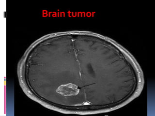
29. Benign brain tumors often show up as hypo dense
(darker than brain tissue) mass lesions on CT
scans. On MRI, they appear either hypo dense or
isointense (same intensity as brain tissue) onT1-
weightedscans, or hyper intense (brighter than
brain tissue) onT2-weighted MRI, although the
appearance is variable.
Contrast agent uptake, sometimes in
characteristic patterns, can be demonstrated on
either CT or MRI scans in most malignant primary
and metastatic brain tumors.
Imaging
30. Tumors have characteristics that allow determination
of malignancy and how they will evolve, and
determining these characteristics will allow the medical
team to determine the management plan.
Andalusia or dedifferentiation: loss of
differentiation of cells and of their orientation to
one another and blood vessels, a characteristic of
an plastic tumor tissue.
Pathology
31. Secondary tumors of the brain are metastatic and
have invaded the brain from cancers originating in
other organs.This means that a cancerous
neoplasm has developed in another organ
elsewhere in the body and that cancer cells have
leaked from that primary tumor and then entered
the lymphatic system and blood vessels. They then
circulate through the bloodstream, and are
deposited in the brain.There, these cells continue
growing and dividing, becoming another invasive
neoplasm of the primary cancer's tissue.
Secondary brain tumors