4. Lasers in Periodontics.ppt
•Download as PPT, PDF•
1 like•84 views

This document discusses lasers used in periodontics. It provides an overview of laser physics, types of lasers including diode, CO2, Nd:YAG and erbium lasers, and their applications in soft tissue procedures and osseous surgery. The benefits of lasers include less pain, better hemostasis and wound healing compared to conventional methods. Safety protocols must be followed when using lasers to prevent eye and tissue damage. Lasers are becoming more widely used in dentistry due to their advantages over traditional techniques.

Recommended
More Related Content
What's hot
What's hot (20)
Similar to 4. Lasers in Periodontics.ppt
Similar to 4. Lasers in Periodontics.ppt (20)
More from Viola Esther
More from Viola Esther (12)
Recently uploaded
Recently uploaded (20)
4. Lasers in Periodontics.ppt
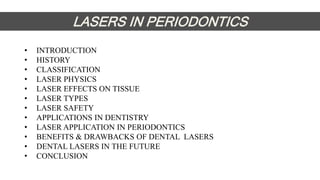
- 1. • INTRODUCTION • HISTORY • CLASSIFICATION • LASER PHYSICS • LASER EFFECTS ON TISSUE • LASER TYPES • LASER SAFETY • APPLICATIONS IN DENTISTRY • LASER APPLICATION IN PERIODONTICS • BENEFITS & DRAWBACKS OF DENTAL LASERS • DENTAL LASERS IN THE FUTURE • CONCLUSION LASERS IN PERIODONTICS
- 2. INTRODUCTION • The word LASER is an acronym for “light amplification by stimulated emission of radiation. • It refers to a device that emits light that is spatially coherent and collimated. • A laser beam can remain narrow over a long distance and it can be tightly focused.
- 3. HISTORY Einstein 1960 brought forth the concept of simulated emission of light. This ultimately introduced the concept of Lasers. Charles Hard Townes, 1951 an American physicist invented the MASER (Microwave Amplification by Stimulated Emission of Radiation) Maiman 1960 introduced the first Laser using synthetic ruby rod (RUBY LASER) Goldman 1965 established the first laser medical laboratory using ruby laser. CO2 laser was 1st invented by Kumar Patel in 1964 and it was 1st applied to periodontal surgery by Pick in 1985.
- 4. CLASSIFICATION 1.According to ANSI & OHSA standards lasers are classified as Class I : Low powered lasers that are safe to use. Class IIa : Low powered visible lasers that are hazardous only when viewed directly for longer than 1000 seconds. Class IIb : Low powered visible lasers that are hazardous when viewed for more than 0.25 seconds.
- 5. Class IIIa : Medium powered lasers & hazardous if viewed for less than 0.25 seconds without magnifying optics. Class IIIb : Medium powered lasers, hazardous when viewed directly. Class IV : High powered lasers, that produce ocular skin and fire hazards.
- 6. 3.Based on the penetration power of the beam: • Hard tissue lasers: Erbium lasers. • Soft tissue lasers: Diode, Nd:YAG,CO2 laser. 4.Based on the emission mode: • Continuous wave mode • Gated pulse mode • Free running pulsed mode
- 7. 5.Based on the laser material used: • Gas lasers: CO2, Argon, He-Ne lasers • Liquid lasers: Dye lasers • Solid state lasers: Ruby , Nd:YAG lasers • Semiconductors: Gallium, Arsenide (diode laser).
- 10. LASER EFFECTS ON TISSUES • Reflection • Transmission • Scattering • Absorption
- 11. LASER TYPES
- 14. LASER OPERATION PARAMETERS FOCUSED DE-FOCUSED • Laser beam hits tissue at its focal point- narrowest diameter • Beam moved away from its focal point
- 15. CONTACT NON-CONTACT • Tip is in contact with tissue • Tip is kept 0.5 to 1 mm away from tissue • Concentrated delivery of laser energy • Laser energy delivered at the surface is reduced
- 16. THEORETICAL ZONES OF TISSUE CHANGE ASSOCIATED WITH SOFT TISSUE EXPOSURE TO LASER LIGHT
- 17. BENEFITS OF LASER – TISSUE INTERACTION SOFT TISSUE: • Cut, coagulate, ablate or vaporize target tissue elements • Sealing of small blood vessels • Sealing of small lymphatic vessels • Sterilizing of tissue- Eschar • Decreased post-operative tissue shrinkage
- 18. THEORETICAL ZONE OF TISSUE CHANGE ASSOCIATED WITH HARD DENTAL TISSUE EXPOSURE TO LASER LIGHT In dental hard tissue the water component is vapourized at 100 °c and the resulting jet of steam expands and then explodes the surrounding matter into small particles. This micro-explosion of the apatite crystal is termed SPALLATION
- 19. • Photothermal • Photochemical • Photoacoustic • Biostimulation • Photodynamic • Photovaporolysis • Photoplasmolysis LASER EFFECTS ARE DUE TO:
- 20. PHOTOTHERMAL 1. Transformed into heat 2. Primary photothermal laser – tissue reactions are Incision/Excision Ablation/Vaporization Hemostasis/Coagulation 3. All these interactions are based upon the Spot Size
- 22. • The photoacoustic effect is a conversion between light and acoustic waves due to absorption and localized thermal excitation. • When rapid pulses of light are incident on a sample of matter, they can be absorbed and the resulting energy will then be radiated as heat. • This heat causes detectable sound waves due to pressure variation in the surrounding medium. PHOTOACOUSTIC
- 23. PHOTOVAPOROLYSIS • Ascendent heat levels-phase transfer from liquid to vapor PHOTOPLASMOLYSIS • Tissue removed by formation of electrically charged ions and particles in a semi-gaseous high energy state.
- 24. PHOTOCHEMICAL • Absorption by chromophores • Tissue response in terms of change of covalent structure BIOSTIMULATION • Believed to work towards healing by stimulation of factors and processes • Below surgical threshold • Useful for pain relief, increased collagen growth and anti- inflammatory activity
- 25. WHAT DOES THE OPERATOR CONTROL?
- 26. ARGON LASER LASER CHARACTERISTICS WAVELENGTH 488 – 514 nm ACTIVE MEDIUM Argon gas DELIVERY SYSTEM Optical fiber FIBER DIAMETER 300 microns MODE OF OPERATION Continuous wave
- 27. DIODE LASER LASER CHARACTERISTICS WAVELENGTH 810 – 980 nm ACTIVE MEDIUM Semi-conductor diode DELIVERY SYSTEM Optical fiber- quartz or silica FIBER DIAMETER 300 microns MODE OF OPERATION Continuous wave, gated pulse mode
- 28. • The chief advantage of the diode lasers is one of a smaller size, portable instrument. • Hot tip effect heat accumulation at tip thick coagulating layer • DIODENT - visible red diode 655 nm • Less tissue penetration, deeper coagulation
- 29. ND:YAG LASER LASER CHARACTERISTICS WAVELENGTH 1064 nm ACTIVE MEDIUM Neodymium in YAG crystal DELIVERY SYSTEM Optical fiber FIBER DIAMETER 300 microns MODE OF OPERATION Continuous wave, pulsed wave
- 30. CO2 LASER LASER CHARACTERISTICS WAVELENGTH 9300, 9600, 10600 nm ACTIVE MEDIUM Carbon dioxide gas DELIVERY SYSTEM Articulated arm FIBER DIAMETER Periotip aperture- 0.5mm MODE OF OPERATION Continuous wave, gated pulsed mode. Used in focused and de-focused modes
- 31. • Use limited to soft tissue procedures as it produces severe thermal damage, like cracking, melting and carbonization of the adjacent root cementum and dentin. Spencer (1996), Israel et al(1997), Barone et al (2002) • Highly absorbed by main mineral component of hard tissue, especially phosphate ions leading to Carbonization of organic components and melting of inorganic ones CARBONIZATION
- 32. Er YAG- 2940 nm: Zharikov et al 1975. Er Cr YSGG- 2780 nm: Zharikov et al 1984 and Moulton et al 1988. 1988: Phagdiwala: Er YAG laser: ability to ablate the dentinal hard tissue. 1989: Pulsed Erbium laser: Keller and Hibst - enamel , dentin and bone. 1995: Commercially available. 1997: Introduced for use in dentistry. ERBIUM FAMILY OF LASERS
- 33. APPLICATIONS IN DENTISTRY • Treatment of aphthous ulcer(photo dynamic therapy) • Dentin desensitization • Soft tissue biopsy • Vestibuloplasty • Modification of root canal walls • Sterilization of root canals • Apicoectomy • Bleaching • Tooth preparation • Cavity preparation • removal of impacted tooth
- 34. LASER APPLICATION IN PERIODONTICS • Frenectomy • Frenotomy • Gingivectomy • Gingivoplasty • Exposing implants in second stage surgery • Depigmentation of gingiva • Crown lengthening • Gingival curettage • Peri-implantitis • Osseous surgery
- 35. C. In addition, the bacteriocidal effects of FR pulsed Nd YAG laser plus intraoperative use of topical antibiotics are designed for the reduction of microbiotic pathogens (antisepsis)within the periodontal sulcus and surrounding tissues. A second pass with the 635µ/ sec “long pulse” laser finishes debriding the pocket D. Gingival tissue is compressed against the root surface to close the pocket and aid with formation and stabilization of a fibrin clot E. The wound is stabilised, the teeth are splinted and occlusal trauma is minimized to promote healing F. Oral hygiene is stressed and continued periodontal maintenance is scheduled. No probing is performed for at least 6 months APPLICATIONS OF LASER (LANAP) A. The primary endpoint of LANAP is debridement of inflamed and infected connective tissue within the periodontal sulcus B. B. Removal of calcified plaque and calculus adherent to the root surface
- 36. Conventional method- tactile feel. Latest: Er YAG laser with fluorescent feedback system for calculus detection. Rationale: Difference in the fluorescence emission properties of calculus and dental hard tissue when subjected to irradiation with 655 nm diode laser. Commercially available as Key Laser III, Ka Vo, Germany SUBGINGIVAL CALCULUS DETECTION- UNIQUE APPLICATION FOR LASER
- 37. SUB- GINGIVAL CALCULUS REMOVAL AUTHOR AND YEAR LASER STUDY DESIGN OBSERVATION PERIOD FINDINGS Cobb et al 1992 Nd YAG EXP (Laser, laser +RP, RP +LASER) Control Immediately after treatment Low effectiveness of calculus removal Decrease in no. of bacteria Scharwz F et al 2003 Diode Exp (laser) Control(SRP) Immediately after treatment Not effective for calculus removal. Thermal damage to root
- 38. ROOT SURFACE ALTERATIONS DIODE LASER Nd YAG LASER • Dry or saline moistened root surface – no detectable alterations • Morlock BJ et al 1992 : surface pitting, craters, melting, carbonization of root surface
- 39. ROOT SURFACE ALTERATIONS CO2 LASER ERBIUM LASER • Spencer P, Cobb CM et al 1996 • Carbonised layer on root surface • Cyanamide, cyanate ions- detected on carbonised layer – FTIS method • Aoki et al 2000: Er YAG with coolant: • Micro irregular surface • No thermal effects as cracking, fissuring
- 42. • Helium – neon laser • Gallium – aluminum – arsenide diode laser • Gallium – arsenide diode laser • Argon ion laser • Defocused Co2 laser • Defocused Nd:YAG laser Biostimulation effects of low level laser LOW LEVEL LASER THERAPY
- 43. • Reduction of discomfort / pain (Kreisler MB et al 2004). • Promotion of wound healing (Qadri t et al 2005). • Bone regeneration (Merli LA et al 2005). • Suppression of inflammatory process. (Qadri T et al 2005). • Activation of gingival and periodontal ligament fibroblast (Kreisler M et al 2003), growth factor release (Saygun I et al 2007). • Alteration of gene expression of inflammatory cytokines (Safavi SM et al 2007). • Photo biostimulation (Garcia et al 2012) BIOSTIMULATION OF LOW LEVEL LASERS
- 44. LASERS USED: CO2 AND ERBIUM FAMILY Involves use of lasers for calculus removal, osseous surgery, de-toxification of the root surface and bone, granulation tissue removal Advantage of Laser: Better access in furcation areas, hemostasis, less postoperative discomfort, faster healing. SURGICAL POCKET THERAPY - LASERS
- 45. MANAGEMENT OPTIONS- Conventional- plastic curettes and antibiotics. New option- Laser Rationale: Disinfection and de-contamination of implant surface. Granulation tissue removal. LASERS USED: DIODE, CO2, ERBIUM FAMILY. LASERS CONTRA-INDICATED: ND:YAG (IMPLANT DAMAGE). IMPLANT THERAPY- MANAGEMENT OF PERIIMPLANTITIS
- 46. • Reports - laser created wounds heal more quickly and produce less scar tissue than conventional scalpel surgery. • Contrary evidence from studies in pigs, rats and dogs indicate that the healing of laser wounds is delayed, that more initial tissue damage may result, and that wounds have less tensile strength during the early phase of healing. (Pick et al 1990) HEALING AFTER LASER THERAPY
- 47. • Abergel et al (1984) experiment with cultured human skin fibroblasts showed that collagen production and DNA synthesis were delayed when the fibroblasts were exposed to Nd: YAG laser radiation. • Iliria et al (2003) analyzed the biocompatibility of root surfaces treated by Er: YAG laser and concluded that laser irradiation promoted faster fibroblast adhesion and growth than surfaces treated with root planing. • Garcia et al (2012) LLLT enhanced healing biostimulation
- 48. ADVANTAGES OF LASER IN SURGICAL PERIODONTICS • Minimum collateral effects result in decreased tissue damage and thus enhance healing • Patient comfort can be enhanced • Hemostasis and coagulation are possible, making the laser essential for medically compromised patients • Some procedures can be performed with topical anesthesia only • The concept of minimally invasive dentistry (MID) can be achieved • Lasers are safe if the user adheres to protocols
- 49. Diode and Nd YAG Effective for cutting and reshaping of soft tissue Good hemostasis Greater thermal effects Thicker coagulated layer CO2 laser Rapid ablation of soft tissue Good hemostasis Effective even for thick tissue Risk of charring- thermal damage GINGIVAL SOFT TISSUE PROCEDURES
- 50. Er YAG Fine cutting can be done Less hemostasis as compared to other lasers Very less thermal damage: use with irrigation Width of thermally affected layer: 5-20 microns (Aoki et al 2005) Safer even in thin tissues Useful to remove melanin and metal tattoos
- 51. LASER SAFETY • Protective eye wear must be worn by the patient and the operator. • Surgical environment must have a warning sign posted with limited access to the treatment room. • High volume suction must be used to evacuate the laser plume formed by tissue ablation. • Normal infection control must be followed. • The laser itself must be in good order. • Mask must be of appropriate filtering capacity (0.1micron filtration mask) to prevent inhalation of laser plume which may be infectious or carcinogenic.
- 52. EYE DAMAGE PART OF EYE DAMAGED LASER TYPE • Corneal damage • Er Cr YSGG, Ho YAG, Er YAG, CO2 • Lens damage • Diode, Nd YAG, Ho YAG, Er Cr YSGG, Er • Aqueous damage • Ho YAG, Er Cr YSGG, Er YAG
- 53. LASER BENEFITS • Less pain, • Less need for anesthetics, • No risk of bacteremia, • Excellent wound healing with no scar tissue formation, • Hemostasis, • Usually no need for sutures, • Ability to remove both hard and soft tissue, • Laser can be used in combination with scalpels.
- 54. LASER DRAWBACKS • Relatively high cost of the device • A need for additional education( especially basic physics) • Every wavelength has different properties and should be used based on that knowledge • The need for implementation of safety measures.
- 55. LASER IN THE FUTURE • Optical coherence tomography using laser to create a 3 dimensional image will be tremendous advance for dental diagnosis. • Laser doppler instruments will be able to measure blood flow rates to assess inflammation. • Selective ablation of calculus and bacteria and enamel hardening for caries resistance are some new procedures which are under development.
- 56. CONCLUSION • Lasers are becoming more commonplace and even routine, either as an adjunctive treatment methodology or as stand-lone additions to the dental armamentarium. • Researchers continue to investigate new laser wavelength and clinical applications as they apply to dentistry. • The growing number of dental practitioners, will continue to advance the application of Einstein’s “splendid light” in their operatories, to the benefit of patients.
- 57. REFERENCES • Cobb et al : Lasers and the treatment of periodontitis: the essence and the noise. Periodontology 2000, Vol. 75, 2017, 205–295 • Journal of Indian Society of Periodontology;vol:19;(July-August 2015) • Robert A. Convissar: Principles and Practice of LASER DENTISTRY • Carranza: Clinical Periodontology; 10th edition. • Dental clinics of North America “ Lasers in Clinical dentistry”. Oct 2004. Vol 48. Issue 4