

Recommended
More Related Content
What's hot
What's hot (20)
Similar to 09 Acute inflamation
Similar to 09 Acute inflamation (20)
More from Dr UAK
More from Dr UAK (20)
Recently uploaded
Recently uploaded (20)
09 Acute inflamation
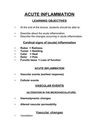
- 1. ACUTE INFLAMMATION LEARNING OBJECTIVES • At the end of the lecture, students should be able to: • Describe about the acute inflammation. • Describe the changes occurring in acute inflammation. Cardinal signs of (acute) inflammation • Rubor = Redness • Tumor = Swelling • Calor = Heat • Dolor = Pain • Functio laesa = Loss of function ACUTE INFLAMMATION • Vascular events (earliest response) • Cellular events VASCULAR EVENTS (ALTERATION IN THE MICROVASCULATURE) • Haemodynamic changes • Altered vascular permeability Vascular changes • Vasodilation
- 2. – increased permeability of vessels due to widened intercell. junctions and contraction of endothelial cells (histamin, VEGF, bradykinin) • Protein poor transudate (edema) • Protein rich exsudate • Leukocyte-dependent endothelial injury – proteolysis – protein leakage • → Platelet adhesion → thrombosis Cellular events • Leukocytes margination → rolling → adhesion → transmigration • Emigration of: – neutrophils (1-2 days) – monocytes (2-3 days) • Chemotaxis – endogenous signaling molecules - lymphokines – exogenous - toxins • Phagocytosis - lysosomal enzymes, free radicals, oxidative burst • Passive emigration of RBC - no active role in inflamm. - hemorrhagic inflammation REVIEW OF NORMAL CAPILLARY EXCHANGE • NOT ALL CAPILLARIES ARE OPEN IN 1 BED • Difference in concentration of substance in blood and isf promote diffusion • Plasma colloid osmotic pressure is more than the osmotic pressure in isf, so it retains the fluid inside the lumen
- 3. HAEMODYNAMIC CHANGES • Persistent progressive vasodilation -- rapid: histamine, serotonin; -- slower: nitric oxide, kallikerin, pge2, pgi2, pgd2 (mast cells) • Opening of precapillary arteriole sphincters resulting into opening of microvascular bed • Venules relax as well • Vessels are congested (hyperemia) and pooling of blood in post capillary venules HAEMODYNAMIC CHANGES • Elevate local h.s pressure results in transudation of fluid into extra vascular tissue HAEMODYNAMIC CHANGES • Slowing or stasis of microvasc ulation • Results in inc conc. of red cells • Raised blood viscosity • Leukocyte margination along the vascular endothelium. ALTERERED VASCULAR PERMEABILITY • Leading to a marked outflow of fluid & its accumulation in the interstitial tissue (oedema)
- 4. • Contraction of endothelial cells • Retraction of endothelial cells • Direct injury to endothelial cells • Endothelial injury mediated by leukocytes • Other mechanisms CONTRACTION OF ENDOTHELIAL CELLS – Caused by histamines, bradykinins and leukotrienes within few seconds – Lasts for 15 – 30 min – Phosphorylation of contractile & cytoskeletal proteins (myosin) – Widens intercellular gaps of venules temporary (not arterioles, capillaries) RETRACTION OF ENDOTHELIAL CELLS – Caused by cytokine mediators tnf, il-1, ifn-γ – Through structural reorganization of cytoskeleton of endothelial cells – Takes 2 – 4 hrs after injury – Lasting 24 hrs or more
- 5. DIRECT INJURY TO ENDOTHELIAL CELLS – May cause delayed damage as in mild to moderate thermal or uv injury, or some bacterial toxins (late appearing sunburn) – Begins after a delay of 2-12 hrs & lasting for several hrs- days ENDOTHELIAL INJURY MEDIATED BY LEUKOCYTES – Endothelial cell-adherent leukocytes may be activated – Release proteolytic enzymes & toxic oxygen species – Damage the endothelium – Making the vessel leaky (late response) – Seen in pulmonary venules and capillaries. OTHER MECHANISMS – Certain mediators (vegf) may cause inc.No. Of vesiculovacuolar organelle causing increased transcytosis to the endothelial cell surface – Newly formed capillaries during the repair are excessively leaky until endothelial cells mature. CELLULAR EVENTS • EXTRAVASATION • PHAGOCYTOSIS
- 6. EXTRAVASATION – Margination – Rolling and adhesion – Transmigration – Chemotaxis and activation • They are then free to participate in: – Phagocytosis and degranulation – Leukocyte-induced tissue injury. CELLULAR EVENTS • MARGINATION • ROLLING • ADHESSION • EMIGRATION • CHEMOTAXIS MARGINATION • With increased vascular permeability, fluid leaves the vessel causing leukocytes to settle-out of the central flow column and “marginate” along the endothelial surface
- 7. ROLLING & ADHESION • Tumbling slowly and adhere transiently (rolling) • Coming to rest & adhere firmly(adh) • Brought about by 4 types of distinct adhesssion molecules selectins mucin-like glycoproteins integrins immunoglobulin superfamily adhesion molecule (glycam-1, psgl-1, esl-1, cd34) SELECTINS • Take part in rolling of pmns over endothelial surface Consists of • P- selectins (on endothelium & platelets) • E- selectins (conf to endothelium) • L- selectins (on most leukocytes) INTEGRINS (GLYCOPROTEINS) • Expressed on many cell types • Made up of alpha & beta chains • Consisting of beta-1 & beta-2 mol • Beta-2 (lfa-1, mac-1) bind to icam-1 • Beta-1 (vla mol) bind vcam-1 • Bring about firm adhesion b/w leukocytes & endothelium IMMUNOGLOBILIN SUPERFAMILY ADHESION MOLECULE
- 8. • 2 endo adhesion mols (icam-1,vcam-i) • Expressed on endothelial surface by the action of tnf & il-1 • Serve as ligands for integrins found on leukocytes • Help in localizing leucocytes to the site of injury. MUCIN-LIKE GLYCOPROTEINS • Glycam-1, psgl-1, esl-1, cd34 • Found in extra cellular matrix and on the cell surfaces • Serve as a ligand for other leukocyte adhesion molecules • Regulate the localization of sub population of leukocytes ROLLING AND ADHESION • Stick and release causing the leukocyte to roll along the endothelium like a tumbleweed • Stop as mutual adhesion reaches a peak (activated) EMIGRATIONEMIGRATION • Cross The Basement Membrane By Damaging It Locally With Secreted Collegenases. DIAPEDESIS
- 9. Neutrophils Monocytes Sugars L-Selectin Integrins Endothelium E-Selectin P-Selectin CAMS ICAM-1,2 VCAM-1 Platelets P-Selectin • Diapedesis gives haemorrhagic apearrance to inflammatory exudate. CHEMOTAXIS • Transmigration of leucocytes to reach the interstitial tissues , • The chemotactic factor –mediated • Crossing several barriers (endothelium, basement membrane, perivascular myofibroblasts and matrix) REFERENCES • Robbins and Cotran – Pathologic basis of disease.
- 10. Neutrophils Monocytes Sugars L-Selectin Integrins Endothelium E-Selectin P-Selectin CAMS ICAM-1,2 VCAM-1 Platelets P-Selectin • Diapedesis gives haemorrhagic apearrance to inflammatory exudate. CHEMOTAXIS • Transmigration of leucocytes to reach the interstitial tissues , • The chemotactic factor –mediated • Crossing several barriers (endothelium, basement membrane, perivascular myofibroblasts and matrix) REFERENCES • Robbins and Cotran – Pathologic basis of disease.