
Recommended
More Related Content
What's hot
What's hot (20)
Similar to Chronic pancreatitis
Similar to Chronic pancreatitis (20)
More from Suhas U
Recently uploaded
Recently uploaded (20)
Chronic pancreatitis
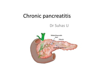
- 2. • Chronic pancreatitis is a relapsing inflammatory process that results in a variable degree of parenchymal destruction and irreversible fibrotic change in the pancreas with consequent clinical manifestations typically including characteristic abdominal pain, exocrine and endocrine insufficiency.
- 3. Types Chronic obstructive pancreatitis • is characterized by exocrine atrophy and is associated with duct stenosis caused by tumors, pseudocyst, or scarring from prior acute pancreatitis. Chronic calcifying pancreatitis • is characterized by intraductal calcifications and protein plugs, and is often associated with atrophy, stenotic ducts, and areas of acute inflammation or pseudocyst. Chronic inflammatory pancreatitis • consists of dense infiltration of mononuclear inflammatory cells.
- 4. Risk factors • Alcohol 70% • Idiopathic 20% • Other 10% Hereditary Hyperparathyroidism Hypertryglyceridemia Autoimmune pancreatitis Obstruction Trauma Pancreatic divisum
- 5. TIGAR O system by Whitmore
- 6. Toxic / metabolic Alcohol • Prolonged alcohol abuse • 3-7% 1. Increased protein concentration • Lithostathine secretion by acinar cells • Increased glycoprotein 2 secretion • Protein precipitation( protein plugs and stone) • Obstruction – acinar cells not able to secrete enzymes – autodigestion 2. Abnormal secretion of enzymes • Metabolites of alcohol – fatty acid ethyl esters and reactive oxygen species
- 7. 3. Direct acinar injury – acetaldehyde 4. Activation of pancreatic stellate cells- extensive fibrosis Smoking • Increases the risk of alcohol induced chronic pancreatitis • Increased risk for pancreatic calcifications and diabetes • Younger age
- 8. Idiopathic • 20% of cases – no obvious risk factor • Unidentified genetic or molecular derangements
- 9. Genetic mutations • Cationic tryosinogen gene(PRSS1) Mutation Cationic trypsinogen gene(PRSS1) •Secreted by acinar cells •Cleaved by duodenal enteropeptidase to trypsin •Trypsin Hydrolyse dietary proteins Activate other pancreatic zymogens Subsequent proteolytic inactivation Trypsin Resists autolytic inactivation Premature and extended activation CFTR gene Chloride ion channel Water , chloride and bicarbonate secretion from epithelial cells Viscous low volume low bicarbonate secretions Enzyme hyperconcentrations Intraglandular enzyme activation Duct sludge SPINK 1 From acinar cells Regulates premature activation of enzymes 1-2 % of normal individuals Lowers the threshold for chronic pancreatitis.
- 10. Autoimmune Lymphoplasmocytic sclerosing pancreatitis • Duct origin antigen • Infiltration of CD4/CD8 lymophocytes and IgG4 postive plasma cells o Diffuse glandular enlargement o Diffuse ductal narrowing
- 11. Recurrent or severe acute pancreatitis • Prior episodes of pancreatitis with 1. Prior episodes of necrosis 2. Postpancreatic ductal scarring 3. Persistent activation of pancreatic stellate cells
- 12. Obstructive • Post traumatic duct strictures • Cystic neoplasms / pancreatic adenocarcinoma • Anamolous anatomic variations of duct
- 13. Clinical presentation Pain abdomen • Left upper quadrant/epigastrium • Radiating to back • Variable pain pattern • Recurrent attacks of moderate or severe pain with periods of quisence • Persistent pain with significant incapacitation Weight loss and malnutrtion • Decreased intake and exocrine insufficiency with protein and fat malabsorption
- 14. Exocrine insufficiency – steatorrhoea Endocrine deficiency • Late presentation - Diabetes • >90% parenchyma fibrosed Fat soluble vitamin deficiency • Bleeding, osteopenia, osteoporosis Extrapancreatitic • Pseudocyst – intestinal or biliary obstruction • GI haemorrhage
- 15. Diagnosis Appropriate history and imaging Lab investigations – limited value • Acute exacerbation – increased amylase or lipase • Nomal with acinar cell destruction • Increased bilirubin with ALP – biliary obstruction
- 16. CT abdomen • Morphologic, duration and complications CT findings • Dilated pancreatic duct(68%) • Parenchymal atrophy(54%) • Pancreatic calcifications(50%) • Peripancretic fluid • Focal pancreatic enlargement • Biliary duct obstruction • Irregular parenchymal contour Assessing complications • Pancreatic duct disruption • Pseudocyst • Portal and splenic vein thrombosis • Splenic or pancreaticoduodenal artery aneurysms
- 19. Toxic/ Idiopathic/ Genetic Autoimmune Obstructive Focal or scattered calcifications Focal enlarged lesions in the head – inflammatory head mass Segmental or diffuse pancreatic dilatation Extrapancreatic complications Calcifications are absent Diffusely enlarged pancreas Dilated duct upstream to stenosis
- 20. MRI • Reliable alternative • Changes in pancreatic parenchyma Pancreatic atrophy Irregularities in contour • Secretin MRCP to evaluate Enhanced duct visualisation Assess pancreatic exocrine function
- 21. ERCP • Historically the gold standard investigation • Indications – when MRI/CT have inconclusive findings Main role as therapeutic procedure • Pancreatic duct complications like stones/strictures/pseudocysts / biliary stenosis
- 23. EUS • Evaluation of pancreatic mass and cystic lesions • More sensitive for early parenchymal changes • EUS directed biopsy of pancreatic mass to rule out malignancy
- 24. Functional assessment 1. Fecal elastase level Using monoclonal / polyclonal elastase antibodies >200mcg/g feces – normal 100-200mcg/g – mild to moderate exocrine deficiency <100mcg/g – severe 2. Steatorrhea Fecal fat weight After intake of 100g fat for 3days >7g/day – Steatorrhea
- 25. Treatment PAIN management • Main symptom • Initial – NSAID’s and escalated accordingly (Step up approach) • Intractable severe pain – started with narcotics ( top down approach) • Infusion pumps for intrathecal delivery of analgesics
- 27. Habit modification • Cessation of alcohol/ smoking • Avoidance of fatty food Neurolysis • Failed medical management • Persistent pain • Radiologic/ endoscopic approach • 100% alcohol injection into the celiac ganglion • Mixed results – transient improvement for not >6months
- 28. Endoscopic procedures • Pancreatic duct stenting • Proximal pancreatic duct stenosis • Decompression of pancreatic duct leak • Drainage of pseudocysts • Endoscopic stone removal • ESWL for stones
- 29. Results are • <50% effective improvement in pain / frequency of attacks • Multiple procedures • Recurrent stones/ strictures
- 30. Surgery • Indications 1) Pain intractable to pharmaco/ endoscopic therapy – 90% 2) Complications of pancreatitis – Relieve biliary/ gastrointestinal obstruction – Drain symptomatic pseudocyst – Vascular complications of chronic pancreatitis
- 31. Types Drainage procedures • Puestow/ Modified Puestow Parenchymal resection • Beger procedure • Whipples procedure • Berni procedure Combination • Frey’s Also depends on the anatomical morphology and diameter of the duct
- 32. I - Large duct disease • Enlarged duct with diameter >7-8mm • Puestow’s Longitudinal unroofing of dilated pancreatic duct in the body and neck Resection of pancreatic tail Longitudinal pancreaticojejunostomy • Modified Puestow / Partington Rochelle procedure Eliminated distal pancreatectomy
- 37. Outcomes • 75-80% pain releif over 5-10years with diffusely dilated duct and no mass • Perioperative mortality low • Endocrine and exocrine levels generally preserved at preoperative levels
- 38. Chronic pancreatitis with dominat head mass • Pure resection / resection and drainage procedures • 4 procedures 1) Pancreaticoduodenectomy 2) Beger 3) Berne 4) Frey Duodenum preserving
- 39. Whipple’s Indication – Chronic pancreaatitis with head mass with • Biliary obstruction • Imaging characteristics suspicious of malignancy Can be classical / pylorus preserving pancreaticoduodenectomy
- 40. More radical procedure for benign lesions
- 41. Beger Procedure Dominant head mass without main duct dilatation / biliary obstruction This involves • Resection of the head of pancreas • A rim of head attached to the duodenum preserved • Common bile duct left intact in the rim • Pancreaticoenteric drainage through two sided Roux en Y pancreaticojejunostomy
- 44. Frey’s Disadvantage with Beger • Does not address the disease that may coexist in body and tail of pancreas • Dominant head mass with dilated pancreatic duct • Local resection of head + lateral pancreaticojejunostomy
- 46. Berni’ modification of Beger • No transaction at neck of pancreas • Anterior surface of mass palpated and cored out using electrocautery • Roux limb is sewn to the remainder rim of pancreas
- 47. Small duct disease or diffuse sclerosis • Total pancreatectomy with islet cell transplantation • Izbicki procedure • Long term analgesic support
- 48. Total pancreatectomy • More commonly done as staged procedure with left pancreatectomy followed by head resection • Allowing initial processing of islet cells from body and tail • Isolation process relies on enzymatic and mechanical steps to dissociate islet cells from acinar cells
- 49. • Depending on proximity of islet isolation facilities and efficiency of the process • Infusion of islet cells into the portal circulation be performed in same anesthesia or postoperatively(radiological guidance)
- 50. Outcomes of islet cell transplantation • Pain relief achieved in 50-60% but recurrence of pain after 1year • Insulin independence in 40-50%, but steady decline of islet function at 10year followup. • The indications for islet autotransplantation are controversial and the overall safety and efficacy of the procedure have not been fully validated outside a handful of centers.
- 51. • Currently, the strongest arguments in favor of total pancreatectomy and islet autotransplantation • limited subset of patients with hereditary pancreatitis, who otherwise carry a significant long-term risk of developing pancreatic cancer
- 52. Izbicki procedure • V shaped longitudinal pancreatic resection • Entire pancreas is excavated along the trajectory of the main pancreatic duct from the pancreatic head (as with the Frey procedure) across the body and tail of the organ. • Pancreaticoenteric drainage is established by Roux- en-Y lateral pancreaticojejunostomy
- 53. Pancreatic enzyme supplements • Various formulations with varying composition of lipase and protease • Classical vs enteric coated enzymes • Dosing schedule is before meals • Uses For pain relief For steatorrhoea
- 54. Classical Enteric coated Pain relief ( suppressing CCK induced stimulation of pancreas) Steatorrhoea H2 blockers or PPI to suppress acid secretion Not necessary