Cerebellar haematoma
•Download as PPTX, PDF•
15 likes•2,813 views

Posterior fossa is a shallow space accommodating brainstem and cerebellum. Bleed in the cerebellum can cost life as it leads to rapid deterioration by hydrocephalus and upward herniation.

Recommended
More Related Content
What's hot
What's hot (20)
Similar to Cerebellar haematoma
Similar to Cerebellar haematoma (20)
More from suresh Bishokarma
More from suresh Bishokarma (20)
Recently uploaded
Recently uploaded (20)
Cerebellar haematoma
- 1. cka SURESH BISHOKARMA, MS MCH RESIDENT, NEUROSURGERY NINAS CEREBELLAR HAEMATOMA
- 3. Volumes of the Posterior Cranial Fossa, Cerebellum Vurdem ÜE, Acer N, Ertekin T, Savranlar A, İnci MF. Analysis of the Volumes of the Posterior Cranial Fossa, Cerebellum, and Herniated Tonsils Using the Stereological Methods in Patients with Chiari Type I Malformation. The Scientific World Journal. 2012;2012:616934
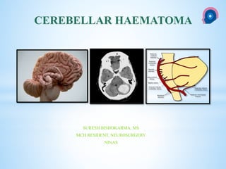
- 5. ARTERIAL SUPPLY OF CEREBELLUM
- 9. *10–15% of all ICH *High mortality: 48 hours
- 10. *Hypertension: 60-80% *AVM and Cavernoma *Anticoagulants and blood dyscrasis *Trauma *Neoplasm *Aneurysm *Amyloid: rare *Remote cerebellar hemorrhage Aetiology
- 11. In hypertensive patients : Rupture of cerebellar microaneurysms. Pathophysiology Charcot and Bouchard
- 12. Blood flow current Dentate nucleus Cerebellar hemisphere and cross midline or Cerebellar peduncle or rupture into the fourth ventricle. Brain stem compressive
- 13. Early * Vomiting, headache, ataxia Clinical feature Dentate nucleus Cerebellar hemisphere and cross midline or Cerebellar peduncle or rupture into the fourth ventricle. Brain stem compressive Early Intermediate Late
- 14. *Hydrocephalus Confused, agitate or drowsy. *VI nerve palsy: Dorsal portion of brainstem. Intermediate stage Dentate nucleus Cerebellar hemisphere and cross midline or Cerebellar peduncle or rupture into the fourth ventricle. Brain stem compressive
- 15. *Ipsilateral gaze paresis: Horizontal gaze centres. *Facial paresis: Facial colliculus *Horner’s syndrome: Sympathetic pathway running from the hypothalamus through the dorsal brain stem. *Hemiparesis: Pyramid *Stupur coma and decerebrate *Pinpoint pupil®:Descending sympathetic pathways from hypothalamus to cervical cord is affected. Parasympathetic control of pupil in midbrain is preserved *Medullary involvement: Cardiovascualar instability and ataxic respiration or apnoea . Late Dentate nucleus Cerebellar hemisphere and cross midline or Cerebellar peduncle or rupture into the fourth ventricle. Brain stem compressive
- 16. *Hydrocephalus, *Direct brainstem compression by the hematoma and surrounding swelling, or both. Depressed GCS: Dentate nucleus Cerebellar hemisphere and cross midline or Cerebellar peduncle or rupture into the fourth ventricle. Brain stem compressive
- 17. Brainstem compression Upwards herniation through the tentorial incisura or Downward tonsillar herniation through the foramen magnum. Death Dentate nucleus Cerebellar hemisphere and cross midline or Cerebellar peduncle or rupture into the fourth ventricle. Brain stem compressive
- 18. WORK UP
- 19. *Critical care: BP control and respiratory support *Investigation *Imaging Management include
- 20. CT scan head CTA MRI with MRA DSA IMAGING • Location of hemorrhage ( vermian, hemispheric or both) • Size of hemorrhage • IVE • Invasion into brain stem • Presence of hydrocephalus • Sign of brain stem impairment • Presence and extent of perilesional edema • Evidence of tight posterior fossa (TPF): Weisberg
- 21. EVIDENCE OF TIGHT POSTERIOR FOSSA (TPF) WEISBERG 1986 14 patients WEISBERG 1986;14 PATIENTS
- 22. TREATMENT
- 23. *GCS *Cerebellar atrophy *Size and Volume of hematoma *Hydrocephalus *Degree of basal cisternal compression *Brain stem sign *Location of the hematoma *Anatomy of posterior fossa DECISION
- 24. *Controversial *Surgical evacuation of the hematoma *External ventricular drainage *Conservative treatment.
- 25. *Outcomes after nonsurgical management were variable, with mortality rates between 9 and 75%.
- 26. *Posterior fossa craniectomy and evacuation of the hematoma are not without risks. *Postoperative recurrent hemorrhage can be fatal
- 27. *In one series, the presence of hydrocephalus invariably resulted from brainstem compression and it was suggested that the presence of hydrocephalus necessitated a posterior fossa craniectomy and evacuation of the hematoma. Mathew P, Teasdale G, Bannan A, Oluoch-Olunya D: Neurosurgical management of cerebellar haematoma and infarct. J Neurol Neurosurg Psychiatry 59:287–292, 1995.
- 28. *Ventricular drainage alone was observed to be ineffective in some cases. *Outcomes after nonsurgical management were variable, with mortality rates between 9 and 75%. affirmed
- 29. *When surgery is indicated *controversy exists regarding whether ventricular drainage only, evacuation of the hematoma, or both procedures should be performed. *Some surgeons recommend drainage of hydrocephalus as the only or initial procedure in all cases. *Others recommend evacuation of the hematoma whenever surgery is indicated. Current management
- 30. Size and volume Cisterns Hydrocephalus Brain stem size Consciousness Surgical evacuation: Indication
- 31. *GCS 14 or 15 and < 3 cm : Conservatively *GCS scores of 13 or less and ≥ 3 cm: surgery *Clot size between 2-3cms, if level of consciousness has altered, should be considered. Size of hematoma Kobayashi et al; 52 patients
- 32. 3 to 4 cm or a volume of more than 15 ml : Surgical evacuation of the hematoma. Kobayashi et. Al. Treatment of hypertensive cerebellar hemorrhage: Surgical or conservative management? Neurosurgery 34:246–251, 1994.
- 33. *Size threshold – 3 cm vs. 4 cm *Radiographic evidence of brainstem compression *Accounts for edema *Clinical examination
- 34. *Patient with hematoma size of >70cm3 did not respond to any treatment and died within 48hrs.
- 35. Timing of Surgical Intervention “Prophylactic” vs. at time of deterioration ISSUES TO CONSIDER
- 36. *Quadrigeminal cistern into 3 groups: *Grade I (normal), *Grade II (compressed), *Grade III (absent). *Good outcomes: *Grade I: 88%, *Grade II: 69%, *Grade III: 0% CISTERNS Taneda et al. 75 Patients
- 37. *The appearance of the fourth ventricle was divided into 3 groups: *Grade I (normal size and configuration), *Grade II (partially compressed and shifted) *Grade III (completely obliterated). 4th ventricle Kirollos et al. 50 patients
- 38. *Studied in 25 patients with cerebellar bleed. *Stable Grade I and II: Conservatively. *Grade I or II compression : only ventricular drainage *15 (60%) Grade I or II compression did not require clot evacuation. *Acute deterioration to comatose state occurred in 6 (43%) of the 14 patients with Grade III compression who were conscious at presentation; none of them experienced good outcomes. Kirollos et al
- 39. BEST *Glasgow coma scale score of 14 or greater *Small hemorrhage (< 30 mm ) *Without hydrocephalus *Without basal cistern effacement CRITERIA FOR MEDICAL CONSERVATIVE TREATMENT WORST *Comatose *Flaccid *Without brainstem reflexes *Large midline hemorrhage
- 40. *Stereotactic aspiration, *Endoscopic bur hole evacuation, *Local infusion of a thrombolytic agent : TPA Other modalities
- 41. 1. Low GCS at admission 2. Obliteration of 4th ventricle and peri-mesenchephalic cistern. 3. Hydrocephalus 4. T2W MRI: high signal intensity in brain stem PROGNOSIS
- 42. MANAGEMENT ALGORITHM Kirollos RW et al. Management of spontaneous cerebellar hematomas: a prospective treatment protocol. Neurosurgery.2001;49(6):1378-86. 4th Ventricle Kirollos’s grade I GCS <13 CONSERVATIVE CSF- D II GCS >13 CONSERVATIVE GCS <13 HYDROCEPHALUS YES CSF-D IMPROVEMENT NO IMPROVEMENT EVACUATE CLOT NO EVACUATE III ANY GCS EVACUATE CLOT + CSF D CSF-D: EVD or VPshunt.
Editor's Notes
- PICA: posterior part of cerebellar hemisphere b. inferior vermis c. central nuclei of cerebellum AICA: the anterior inferior quarter of the cerebellum. b. the middle cerebellar peduncle, SCA: most of the cerebellar cortex, b. the cerebellar nuclei, and c. The superior cerebellar peduncles
- PICA: posterior part of cerebellar hemisphere b. inferior vermis c. central nuclei of cerebellum AICA: the anterior inferior quarter of the cerebellum. b. the middle cerebellar peduncle, SCA: most of the cerebellar cortex, b. the cerebellar nuclei, and c. The superior cerebellar peduncles
- Spontaneous cerebellar hematomas represent approximately 10%–15% of all ICH High mortality
- Charcot–Bouchard aneurysms (also known as miliary aneurysms or microaneurysms) are aneurysms of the brain vasculature which occur in small blood vessels (less than 300 micrometre diameter).
- The symptoms produced by cerebellar hemorrhage are related to destruction and compression of the cerebellum itself or to the subarachnoid hemorrhage resulting from rupture of the hematoma into the subarachnoid space.
- Hydrocephalus result from the compression of the 4th ventricle or rupture of hemorrhage into ventricle.
- Ipsilateral gaze paresis : Horizontal gaze centres. Facial paresis: Facial colliculus Horner’s syndrome: Sympathetic pathway running from the hypothalamus through the dorsal brain stem. Hemiparesis: Pyramid Pupil: Constrict ® : Descending sympathetic pathways from hypothalamus to cervical cord is affected. Parasympathetic control of pupil in midbrain is preserved until late Late: Stupur coma and decerebrate. Medullary involvement: Cardiovascualar instability and ataxic respiration or apnoea .
- Furthermore, estimation of the size of the hematoma on the basis of CT scans could be both difficult and inaccurate, because of the nebulous margins at the interface between the hematoma and the surrounding cerebellum. The mass effect produced by areas of surrounding edema could be underestimated. MRI is not superior to CT to delinate Haematoma. But it may be superior to analyse the other brainstem pathology. TPF: CT: Effacement of basal cisterns of posterior fossa and ventricular enlargement consistent with obstructive hydrocephalus. LOCATION OF HEMORRHAGE ( VERMIAN, HEMISPHERIC OR BOTH) SIZE OF HEMORRHAGE IVE INVASION INTO BRAIN STEM PRESENCE OF HYDROCEPHALUS SIGN OF BRAIN STEM IMPAIRMENT PRESENCE AND EXTENT OF PERILESIONAL EDEMA EVIDENCE OF TIGHT POSTERIOR FOSSA (TPF)
- WEINGBERG 14 PATIENTS: EFFACEMENT OF BASALCISTERN AND OBSTRUCTIVE HYDROCEPHALUS: PATIENT WITH TPF SHOWED RAPID DETERIORATION.
- The degree of cerebellar atrophy (considering the advanced age of most patients with cerebellar hemorrhage) should be evaluated. A large hematoma may not exert significant brainstem compression in the presence of cerebellar atrophy.
- Outcomes after nonsurgical management were variable, with mortality rates between 9 and 75%, because surgery was not attempted for patients considered to be at high risk in many series, whereas primarily patients in good neurological condition were included in other series.
- Indication are based on level of consciousness, clinical course, size of hematoma. typically 3 or 4 cm, above which they recommend surgical evacuation of the hemorrhage regardless of clinical status
- INDICATION: 3.5X 2.5CM VERMIAN HEMATOMA OR 4X3 CM HEMISPHERIC. BECAUSE VERMIAN LIES CLOSER TO THE BRAIN STEM AND CSF PATHWAY.
- Tissue plasminogen activator