
Recommended
More Related Content
What's hot
What's hot (20)
Similar to Gastrointestinal system embryology
Similar to Gastrointestinal system embryology (20)
Recently uploaded
Recently uploaded (20)
Gastrointestinal system embryology
- 2. Gastrulation: Epiblast cells migrate through the primitive streak. Definitive (embryonic) endoderm cells displace the hypoblast. Mesoderm spreads between endoderm and ectoderm. Langman’s fig 5.3
- 7. The developing endoderm (yellow) is initially open to the yolk sac (the cardiac region is initially most anterior)… Cranio-caudal folding at both ends of the embryo and lateral folding at the sides of the embryo bring the endoderm inside and form the gut tube. Endoderm Carlson fig 6-20
- 8. Folding creates the anterior and posterior intestinal portals (foregut and hindgut, respectively) The cardiac region is brought to the ventral side of the developing gut tube. cloacal membrane Juxtaposition of ectoderm and endoderm at: Oropharyngeal (buccopharyngeal) membrane - future mouth Cloacal membrane - future anus Note: there actually isn’t much mesoderm in these membranes, which is important for their breakdown later in development to form the oral and anal orifices. Carlson fig 6-20
- 9. Gut-associated organs begin to form as buds from the endoderm: (e.g., thyroid, lung, liver, pancreas) Midgut opening to the yolk sac progressively narrows Carlson fig 6-20
- 10. By the end of the first month: The stomach bulge is visible, Dorsal pancreas has begun to bud Connection of the midgut to the yolk sac is reduced to a yolk stalk and then a very thin vitelline duct Carlson fig 6-20
- 11. With lateral folding, mesoderm is recruited to gut wall • Lateral folding of the embryo completes the gut tube • Mesodermal layer of the gut tube is called splanchnic (visceral) mesoderm - derived from lateral plate mesoderm • Somatic mesoderm lines body cavity Langman’s fig 6-18 Carlson fig 6-20
- 18. Early mesodermal patterning: (buccopharyngeal membrane) Specific regions of the epiblast migrate through the streak at different levels and assume different positions within the embryo: Cranial to caudal: Notochord (n) Paraxial mesoderm (pm) Intermediate mesoderm (im) *Lateral plate mesoderm (lpm) Extraembryonic mesoderm (eem) Langman’s fig 5-07
- 19. 4th week 5th week Celiac artery supplies the foregut Superior mesenteric artery supplies the midgut Inferior mesenteric artery supplies the hindgut Langman’s fig 14-14 Langman’s fig 14-4 The figure on the right also shows the mesenteries; note that the liver and stomach have dorsal and ventral mesenteries whereas the rest of the gut has only a dorsal mesentery.
- 20. Foregut: pharynx thyroid esophagus parathyroid glands stomach tympanic cavity proximal duodenum trachea, bronchi, lungs liver, gallbladder pancreas Midgut: proximal duodenum to right half of transverse colon Hindgut: left half of urinary bladder transverse colon to anus Gut tube proper Derivatives of gut tube (These three regions are defined by their blood supply)
- 21. Gut = bilayered tube (endoderm surrounded by mesoderm) Regional gut tube patterning and organogenesis require bi-directional endoderm-mesoderm cross-talk and inductive signals from other nearby structures Regional patterning of the gut tube
- 22. Regional patterning of the gut tube - the Hox code Hox genes are evolutionarily conserved transcription factors that are used in regional patterning (flies to mammals). The gut has an cranial-caudal Hox gene expression pattern (code) similar to that seen in neural tissue. Some Hox genes are expressed in mesoderm, in overlapping patterns; some are expressed in endoderm. Hox gene expression boundaries correspond to morphologically recognizable elements in the GI tract. Hox gene expression is important for formation of major sphincters (red circles) Carlson fig 15-01
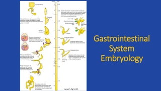
- 23. Hedgehog signaling is important for RADIAL (concentric) patterning of the entire gut tube Fetus • High hedgehog concentration directly inhibits smooth muscle differentiation (via repression of Smooth Muscle Activating Protein, or Smap) • Low Hedgehog concentration is permissive of muscle differentiation in the outer wall of the gut • High Hedgehog concentration also induces high BMP which inhibits neuron formation, thus limiting neurogenesis initially to the outer muscular wall of the gut (later in development, SHH goes away allowing development of the smooth muscle of the musularis mucosae and neurons of the submucosal plexus) Morphogen: induces different cell fates at different concentrations of signal Adult Esophagus Larsen’s fig 14-27 Wheater’s fig 14-5
- 24. Enteric Nervous System • Collection of neurons in the GI tract. • Controls motility, exocrine and endocrine secretion and microcirculation. • Regulates immune and inflammatory process. • Functions independent of CNS. Image from: Young. Gut 2000
- 25. Development of Enteric Nervous System • Primarily derived from the vagal segment of neural crest cells. • Cells initially migrate to the cranial section and then caudally • Hindgut ganglia receive contributions of cells from the cranial and sacral segments of the neural crest cells • Interstitial cells of Cajal arise from the local gut mesenchyme Image from: http://www.landesbioscience.com/curie/chapter/2823/
- 26. Development of the Enteric Nervous System • Nerve cell bodies are grouped into ganglia • Ganglia are connected to bundles of nerves forming two plexus • Myenteric (Auerbach’s) • Submucosal (Meissner’s) http://en.wikipedia.org/wiki/Enteric_nervous_system
- 27. Enteric Nervous System • Myenteric plexus • Lies between the circular and longitudinal muscles • Regulates • Motility • Secretomotor function to mucosa • Connections to • gallbladder and pancreas • sympathetic ganglia • esophageal striated muscle
- 28. Enteric Nervous System • Submucosal plexus • Lies between circular muscle layer and the muscularis mucosa • Regulates: • Glandular secretions • Electrolyte and water transport • Blood flow • Similar structure found in gallbladder, cystic duct, common bile duct and the pancreas
- 29. Enteric Nervous System • Clinical Correlations: • Motility • Achalasia • Psuedo-obstruction • Hirschsprung’s disease • Secretions • Cholera • E. Coli
- 30. Hirschsprung’s Disease • Congenital disorder • 1:5000 live births • Failure of neural crest cells to colonize the entire gut resulting in an aganglionic zone • Tonic constriction of aganglionic section • Long (20%) and Short Segment (80%) • Short segment 4:1 male:female • Isolated anomaly in 70% of cases • Multiple genes and modifier genes identified • Not mendelian
- 31. Genetics of Hirschsprung’s Disease • Associated genes encode members of the glial cell neurotrophic factor family • involved in signaling pathways • transcription factors • Genes identified • GDNF • Ret • EDNRB • Sox10
- 32. Genetics of Hirschsprung’s Disease • Glial Cell-Derived Neurotrophic Factor (GDNF) • Member of TGF-β superfamily • Binds to and activates receptor tyrosine kinase (Ret) • Defects on GDNF/Ret signaling account for • 50% familial cases • 30% of sporadic cases
- 33. Genetics of Hirschsprung’s Disease • Endothelin 3 (Et-3) is a secreted protein expressed by gut mesenchyme. • Et-3 signals via Endothelin receptor B (Ednrb) • Ednrb is expressed on migrating enteric neural crest cells • Mutations in Et-3 and Ednrb account for 5% of cases
- 34. Genetics of Hirschsprung’s Disease • Sex determining region Y – box 10 (Sox10) is a high mobility group transcription factor. • Expressed on migrating enteric neural crest cells • Mutations of Sox10 account for 5% of cases
- 35. Genetics of Hirschsprung’s Disease • Gene Interactions have been identified in isolated Mennonite populations and mouse models. • Ret and Ednrb • Ret and Et-3 • Sox10 and Et-3/Ednrb • Mechanisms are unknown – • ?Downstream signaling
- 36. Genetics of Hirschsprung’s Disease • Modifier Genes = Mutated gene that must be coupled with another mutation to result in or enhance the effect. • Neuregulin 1 (NRG1) - associates with Ret • NRG1 signals receptors to regulate neural crest cell development. The receptor is also associated with Sox10 • Modifiers have also been identified for Sox10 and Et-3 and Ednrb
- 37. Genes in Gastrointestinal Embryology • Homeobox-containing transcription factors (Hox genes) – play a role in gut regionalization • Sonic Hedgehog (Shh) – transcription factor controls endodermal-mesenchymal interactions • Defects associated with TEF and Anorectal malformations • Possible role in IBD and Malignancy