
Recommended
More Related Content
Similar to _ FEMALE BREAST ANATOMY.pdf
Similar to _ FEMALE BREAST ANATOMY.pdf (20)
More from ssuser50ebc6
Recently uploaded
Recently uploaded (20)
_ FEMALE BREAST ANATOMY.pdf
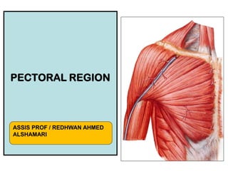
- 1. PECTORAL REGION ASSIS PROF / REDHWAN AHMED ALSHAMARI
- 2. OBJECTIVES • By the end of the lecture the students should be able to : • Identify and describe the muscles of the pectoral region. Pectoralis major. Pectoralis minor. Subclavius. Serratus anterior. • Describe and demonstrate the boundaries and contents of the axilla. • Describe the formation of the brachial plexus and its branches.
- 3. PECTORAL REGION • Skin • Superficial fascia • Deep fascia • Muscles • Vessels, nerves • Applied anatomy
- 4. Superficial fascia • Subcutaneous muscles- Platysma • Cutaneous nerves • Cutaneous vessels • Lymphatics • Adipose tissue/fat • Suspensory ligaments of coopers • Breast/ Mammary glands
- 5. Deep fascia • Pectoral fascia • Extensions and Tracings of deep fascia
- 6. Muscles/ Skeletal framework of pectoral region • Pectoral major/minor • Subclavius • Serratus anterior • Ribs • Clavicle, sternum • Humerus upper part • Clavipectoral fascia
- 7. Pectoralis Major • Origin : 2 heads; • Clavicular head: From; • Medial ½ of the front of the clavicle. • Sternocostal head: From; • Sternum. • Upper 6 costal cartilages. • Aponeurosis of the external oblique muscle. • Insertion : • Lateral lip of bicipital groove. • Nerve supply : • Medial & lateral pectoral nerves. • Action : • Adduction and medial rotation of the arm. • Clavicular head helps in flexion of arm (shoulder).
- 8. Pectoralis Minor • Origin: • From 3rd ,4th, & 5th ribs close to their costal cartilages. • Insertion: • Coracoid process. • Nerve supply: • Medial pectoral nerve. • Action: • Depression of the shoulder. • Draw the ribs upward and outwards during deep inspiration. 3 4 5
- 9. Subclavius • Origin: • From 1st rib at its costal cartilage. • Insertion: • Subclavian groove in the middle 1/3 of the inferior surface of clavicle. • Nerve supply: • Nerve to subclavius from upper trunk of brachial plexus. • Action: • Fixes the clavicle during movement of shoulder joint.
- 10. Clavipectoral Fascia • It is a thickened membrane of deep fascia between the subclavius and pectoralis minor. • It is pierced by : Lateral pectoral nerve. Thoraco- acromial artery Cephalic vein. Few lymph vessels.
- 11. Origin: • Upper eight ribs. • Insertion: • anterior aspect of the medial border and inferior angle of scapula. • Nerve supply: • Long thoracic nerve. • Action: • Draws the scapula forward in boxing, (protrusion). • Rotates scapula outwards in raising the arm above 90 degree. Serratus anterior
- 12. BREAST ANAT0MY ASSIS PROF / REDHWAN AHMED ALSHAMARI
- 13. OBJECTIVES • By the end of the lecture, the student should be able to: • Describe the shape and position of the female breast. • Describe the structure of the mammary gland. • List the blood supply of the female breast. • Describe the lymphatic drainage of the female breast. • Describe the applied anatomy in the female breast.
- 15. Mammary ridge • Mammary ridge extends from the axilla to the inguinal region. • In human, the ridge disappears EXCEPT for a small part in the pectoral region. • In animals, several mammary glands are formed along this ridge.
- 18. Parts, Shape & position of the Gland • It is conical in shape. • It lies in superficial fascia of the front of chest. • It has a base, apex and tail. • Its base extends from 2nd to 6th ribs. • It extends from the sternum to the midaxillary line laterally. • It has no capsule.
- 19. SHAPE AND POSITION OF FEMALE BREAST • 2/3 of its base lies on the pectoralis major muscle, while its inferolateral 1/3 lies on: • Serratus anterior & • External oblique muscles. • Its superolateral part sends a process into the axilla called the axillary tail or axillary process.
- 20. • Nipple: • It is a conical eminence that projects forwards from the anterior surface of the breast. • The nipple lies opposite 4th intercostal space. • It carries 15-20 narrow pores of the lactiferous ducts. • Areola : • It is a dark pink brownish circular area of skin that surrounds the nipple. • The subcutaneous tissues of nipple & areola are devoid of fat. SHAPE AND POSITION OF FEMALE BREAST
- 21. STRUCTURE OF MAMMARY GLAND • It is non capsulated gland. • It consists of lobes and lobules which are embedded in the subcutaneous fatty tissue of superficial fascia. • It has fibrous strands (ligaments of cooper) which connect the skin with deep fascia of pectoralis major. • It is separated from the deep fascia covering the underlying muscles by a layer of loose areolar tissue which forms the retromammary space. What is its Importance? (allows the breast to move freely).
- 22. STRUCTURE OF MAMMARY GLAND • It is formed of 15-20 lobes. • Each lobe is formed of a number of lobules. • The lobes and lobules are separated by interlobar and interlobular fibrous & fatty tissue, called ligaments of Cooper. (Importance)?These ligaments give the breasts support by connecting the skin of the breasts to the pectoralis muscles below them. • It has from 15-20 lactiferous ducts which open by the same number of openings on the summit of the nipple.
- 23. ARTERIAL SUPPLY • 1. Perforating branches of internal thoracic (internal mammary) artery. • 2. Mammary branches of lateral thoracic artery. • 3. Mammary branches of Intercostal arteries.
- 25. VENOUS DRAINAGE • Veins are corresponding to the arteries. • Circular venous plexus are found at the base of nipple. • Finally, veins of this plexus drain into axillary & internal thoracic veins.
- 26. AXILLARY LYMPH NODES • They are arranged into 5 groups which lie in axillary fat : • Pectoral (Anterior) group : which lies on the pectoralis minor along lateral thoracic vessels. • Subscapular (Posterior) group : which lies on posterior wall of axilla on lower border of subscapularis along subscapular vessels. • Brachial (Lateral) group : lies on lateral wall of axilla along 3rd part of axillary vessels. • Central group : lies in axillary fat at the base of axilla. • Apical group : lies at apex of axilla. • Subclavian lymph trunk: • it is formed by union of efferent lymph vessels of apical group. It usually opens in subclavian vein. On the left side it usually opens into thoracic duct.
- 27. LYMPHATIC DRAINAGE • Subareolar lymphatic plexus : • Lies beneath the areola. • Deep lymphatic plexus: • Lies on the deep fascia covering pectoralis major. • Both plexuses radiate in many directions and drain into different lymph nodes.
- 28. LYMPHATIC DRAINAGE • Central & lateral parts of the gland (75%) drain into pectoral group of axillary lymph nodes. • Upper part of the gland drains into apical group of axillary lymph nodes. • Medial part drains into internal thoracic (parasternal) lymph nodes, forming a chain along the internal thoracic vessels. • Some lymphatics from the medial part of the gland pass across the front of sternum to anastomose with that of opposite side. • Lymphatics from the inferomedial part anastomose with lymphatics of rectus sheath & linea alba, and some vessels pass deeply to anastomose with the sub diaphragmatic lymphatics.
- 31. APPLIED ANATOMY- CANCER BREAST • It is a common surgical condition. • 60% of carcinomas of breast occur in the upper lateral quadrant. • 75% of lymph from the breast drains into the axillary lymph nodes. • In case of carcinoma of one breast, the other breast and the opposite axillary lymph nodes are affected because of the anastomosing lymphatics between both breasts. • In patients with localized cancer breast, a simple mastectomy, followed by radiotherapy to the axillary lymph nodes is the treatment of choice.
- 32. Applied Anatomy • The lactiferous ducts are radially arranged from the nipple, so incision of the gland should be made in a radial direction to avoid cutting through the ducts. • Infiltration of the ligaments of Cooper by breast cancer leads to its shortening giving peau de’orange appearance of the breast.
- 33. THANK YOU
- 34. Which is correct regarding the mammary gland ? It extends from the 2nd to 8th ribs. Its base lies on the pectoralis major muscle. It has 4-8 lactiferous ducts. Its most lymph drains into the parasternal lymph nodes. The lymphatics from upper part of mammary gland drain into : The parasternal lymph nodes. Subdiaphragmatic lymph nodes. Apical group of axillary lymph nodes. Pectoral group of axillary lymph nodes. The lactiferous ducts of mammary gland are : Less than 10. From 10-15. From 15-20. More than 20.