Journal club about silver nanoparticles in PROSTHODONTICS
•Download as PPTX, PDF•
3 likes•412 views

This document describes an in vitro study that evaluated the antimicrobial activity and properties of a tissue conditioner containing silver nanoparticles. The study tested the antimicrobial effects of the tissue conditioner with 0-3% silver nanoparticles against common oral microbes like C. albicans, S. mutans, and S. aureus. The results found that 0.1% silver nanoparticles had a minimal bactericidal effect against S. aureus and S. mutans, while 0.5% had an effect against C. albicans. Control samples showed no inhibition. The study concluded the material could be an effective antimicrobial dental material, but further mechanical and toxicity testing is required.

Recommended
Recommended
More Related Content
What's hot
What's hot (20)
Similar to Journal club about silver nanoparticles in PROSTHODONTICS
Similar to Journal club about silver nanoparticles in PROSTHODONTICS (20)
More from NAMITHA ANAND
More from NAMITHA ANAND (20)
Recently uploaded
Recently uploaded (20)
Journal club about silver nanoparticles in PROSTHODONTICS
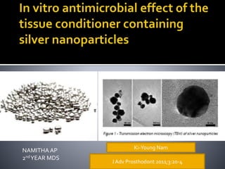
- 1. NAMITHA AP 2ndYEAR MDS J Adv Prosthodont 2011;3:20-4 Ki-Young Nam
- 2. INTRODUCTION AIM MATERIALSAND METHODS RESULTS DISCUSSION RELATED ARTICLES CONCLUSION REFERENCES
- 3. Silver nano particles Antimicrobial low toxicity profile Rapid and Broad spectrum activity well tolerated tissue response Maintenance of tissue conditioner Mechanical and chemical methods Systemic or local antibiotic agents Microbial resistance!!
- 4. Silver containing materials Central venous catheter Wound dressing Vascular graft Bone cements Endotracheal tubes Coating in dental implants • Sustained silver cation release • More effective than microsized
- 6. identify in vitro antimicrobial activity of the tissue conditioner containing silver nanoparticles on microbial strains • fungal species associated with denture stomatitisCandida albicans • oral endogenous bacteriaStreptococcus mutans • nosocomial respiratory infection-causing bacteria Staphylococcus aureus
- 7. Aqueous silver sol was prepared with 10.0 mM of analytical grade AgNO3 in distilled water • 2.0% PVP (Polyvinyl Pyrrolidon) was used as stabilizer. All solutions were deaerated by bubbling with argon gas for 1 hour then they were irradiated in the field of 20 KGy 60Co Gamma-ray sources
- 8. Samples were placed on separate culture plate dish containing 0.1 - 3.0% (vol)silver nanoparticles 0 0.1 0.5 1 2 3 Experimental disc samples (20.0×3.0 mm) of tissue conditioner (GC Soft-Liner, GC cooperation,Tokyo, Japan)
- 10. antimicrobial effects of samples were evaluated as a percentage of viable cells in withdrawn suspension (100 μL) Microbial growth was verified At 24 hours At 72 hours microbial suspensions (100 μL) of tested strains were inoculated then incubated at 37℃.
- 11. • grown in brainheart infusion (BHI) (Difco, Franklin Lakes, NJ, USA) broth and onto Mueller Hinton agar plates at 37℃ BACTERIAL STRAINS • grown in Schaedler broth (Difco, Franklin Lakes, NJ ,USA) and onto Sabouraud agar plates (Difco, Franklin Lakes, NJ, USA) at 30℃. FUNGAL STRAINS
- 12. After incubating microbial cells at 37℃ overnight, optical density (OD) of the suspension at 600 nm was adjusted to 1.0 using a spectrophotometer (Milton Roy spectrophotometer 20D+, Milton Roy, Ivyland, USA). The suspension was diluted with phosphate-buffered saline (pH 7.4) to 1:100 and suspended to final concentration of 1.0 × 10⁷ cells/mL.14,21
- 13. Each disc sample of Ag�-tissue conditioner and control were placed on the flat bottom of the separate 12-well cell culture plate(Costa�,Corning, NewYork, USA) of 22.1 mm well diameter 100 μL of initial microbial suspensions in 1.0 ml of Sabouraud broth were inoculated to each well and incubated at 37℃. After incubation for 24 hrs and 72 hrs for extended contact period, suspension (100 μL) was withdrawn
- 14. borderline of the antimicrobial effect was determined at 0.1% viable cells.
- 15. • 0.1% silver nanoparticles combined to tissue conditioner minimal bactericidal effect against Staphylococcus aureus and Streptococcus mutans strains • 0.5%silver nanoparticles combined to tissue conditioner Minimal bactericidal effect against candida albicans • Control group No microbial inhibitory effect
- 16. Optimal concentration of silver? Toxic effects of silver? Effect of addition of nanoparticles on other properties of material In vivo studies?
- 17. Within the limitation of this in vitro study, the results suggest that the tissue conditioner containing silver nanoparticles could be an antimicrobial dental material in denture plaque control. Further mechanical stability and toxicity studies are still required.
- 18. KishoreGinjupalli TusharShaw ChaitanyaTellapragada RamakrishnaAlla LokendraGupta RELATEDARTICLES 1 J Dent Mater 2018
- 19. to evaluate the antimicrobial activity and properties of irreversible hydrocolloid impression material incorporated with silver nanoparticles of varying size at different concentrations 80-100 nm 50-80 nm 30-50 nm 10-20 nm .5 1 2 5 Wt % E coli Candida albicans Staphylococcus aureus Strains used
- 20. • Kirby Baurs disc diffusion method Measurement of anti microbial activity • Allowed to contact a polished PMMA rod • Loss of adherence noted Measurement of gelation time • Loaded to disposable syringe injected onto glass slab within 60s • Another glass plate put on mix and a weight of 14.7 N was placed on top glass plate for 5s • Measure min and max diameter of disc formed and expressed in mm Measurement of flow • Stressing the specimen at 6 mins from beginning of mixing using universal testing machine Measurement of gel strength • Subjected to 10% strain for 15 s in universal testing machine • Load removed and allowed to recover for 30s Measurement of permanent deformation
- 21. Prepared irreversible hydrocolloid specimens were sliced into 3 mm thickness using sterile BP blade placed in lawn cultures prepared on Muller Hinton agar plates using respective bacterial or yeast suspensions matching to turbidity of 0.5 Mc Farland’s standerd Cultureplates incubated for 24 hrs.zones of inhibition in millimeters were measured
- 23. Silver nanoparticles of 80–100 nm size have imparted superior antimicrobial activity to the irreversible hydrocolloid in a dose-dependent manner finer nanoparticle size did not exhibit any antimicrobial activity. The addition of silver nanoparticles did not alter the properties of irreversible hydrocolloid at 0.5 and 1.0 wt% whereas at higher concentrations significant differences in flow, gelation time and strength were observed.
- 24. The results of the present study indicate that silver nanoparticles of size range 80–100 nm are superior in imparting antimicrobial activity to irreversible hydrocolloid compared to finer particle size range.
- 25. Related articles 2 J Prosthet Dent 2015
- 26. aim Materials and methods The antimicrobial activity and properties of 2 commercially available irreversible hydrocolloid impression materials were evaluated after incorporating varying concen- trations of silver nanoparticles. Antimicrobial activity was determined using the disk diffusion method. The gel strength, permanent deformation, flow, and gelation time were measured according to American Dental Association specification #18. The purpose of this in vitro study was to investigate the antimicrobial activity and properties of irreversible hydrocolloid impression materials incorporated with silver nanoparticles.
- 27. a dust-free alginate (Zelgan Plus; Dentsply India Pvt Ltd) color- changing alginate (Tropicalgin; Zhermack SpA). silver nanoparticles (Nano Labs) of 80 to 100 nm in size were added to these irreversible hydrocolloids, and their properties were evaluated. .5wt% 1wt% 2wt% 5wt%
- 30. Adding silver nanoparticles to irreversible hydrocolloid impression materials resulted in superior antimicrobial activity without adversely affecting their properties. Adding silver nano- particles to Zelgan significantly increased the gel strength compared with the control group, except at 5 wt%. However, the gel strength ofTropicalgin was unaffected except at 5 wt%. An increase in the permanent deformation was found with the incorporation of silver nanoparticles in both Zelgan and Tropicalgin. The flow of Zelgan increased with the incorporation of silver nanoparticles, whereas a decrease in the flow ofTropicalgin was observed at 1 wt% and 2 wt%. An increase in the gelation time of both Zelgan and Tropicalgin was observed with the incorporation of silver nanoparticles.
- 31. GrzegorzChladek , Anna Mertas , Izabela Barszczewska- Rybarek ,Teresa Nalewajek , Jarosław Żmudzki ,Wojciech Król and Jan Łukaszczyk RELATEDARTICLES 3 Int. J. Mol. Sci. 2011
- 32. They presented a method of incorporating AgNPs into the chemically cured silicone soft liner material UfiGel SC (UG).The aim of this work was also to evaluate the antifungal efficacy of these developed composites.
- 33. The modification process was conducted by dissolving both material components (base and catalyst) in a colloidal solution of AgNPs and evaporating the solvent. 10 ppm 20 ppm 40 ppm 80 ppm 120 ppm 200 ppm
- 34. Measurements using dynamic light scattering (DLS) for colloidal solutions of 30 ppmAgNPs showed an average NP size of 22.8 nm. when the AgNP concentration increased, the color of the samples became darker. Int. J. Mol. Sci. 2011, 12
- 35. Scanning electron microscopy measurements show the presence of both individual particles (Figure 3) and large nanoparticle agglomerates in the modified soft liners. Individual particles from all specimens usually ranged from 10 to 30 nm. In the case of a higher AgNP concentration, a greater number of nanoparticle aggregations and larger sized aggregations were observed. Starting with a concentration of 80 ppm, numerous large agglomerates were noted. Most of them ranged between 100 and 300 nm, but there were also huge aggregations that exceeded 1 μm, and individual AgNPs were still observed.
- 37. For material specimens without AgNPs, the observed Candida albicans CFU/mL value increased by 23.4% compared with the positive control.The introduction of 10 ppm AgNPs to the polymer led to an antifungal efficacy (AFE) of 16.4%. Increasing theAgNP concentration in the composite to 20 ppm resulted in an additional increase in the AFE of 8%.The average AFE value for samples with 40 ppm AgNPs was 31.5%. An additional increase in theAgNP concentration resulted in a less dynamic, but still visible, AFE increase. For the highest examinedAgNP concentration (200 ppm), the maximum composite AFE value reached was 52.2%.
- 38. In this study, they presented a method ofAgNP incorporation into chemically cured silicone soft liner materials. The AFE of the achieved composites containing 10 to 200 ppm AgPNs was 16.3% to 52.5%. This level of AFE should be capable of preventing colonisation of Candida albicans on soft denture linings. Future research should examine the mechanical characteristics of the achieved composites and confirm their microbiological characteristics in in vivo studies.
- 39. Wenjuan Liu, Bin LiuˆYuanyuan Qi RELATEDARTICLES 4 International conference on biomedical engineering and biotechnology- 2012
- 40. In this study, they investigated the appropriate adding ratio of the LZB-GC nano silver- filled antibacterial agent in the alginate impression material. antibacterial agent may affect the physical properties of the impression material. PHYSICAL PROPERTIES coagulation time compressive strength compressive strain deformation restorability fluidity of the alginate impression material by adding different content of LZB-GC antibacterial agent were characterized by standards method
- 41. PROPORTIONS OF LZB GC ANTIBACTERIAL AGENT ADDED 0.25 .5 1 1.5 2 2.5 STRAINS USED Staphylococcus auresus E coli Their antibacterial activities were tested by Film adhesion method The antibacterial ratios were assessed according to the national standard "Antimicrobial plastics-Test for antimicrobial activity and efficacy"
- 42. To evaluate the physical properties measurement indicators are shown inTable 1 is used
- 43. The antibacterial ratios increased as the content of LZB-GC increased. content of LZB-GC increased from 0.125% to 0.5%, the antibacterial ratios of Escherichia coli and Staphylococcus aureus all increased when the content of LZB-GC was 0.5% or more, the antibacterial ratios could be up to 99%.
- 44. • coagulation time, deformation restorability compressive strain, compressive strength and fluidity of all the experimental groups compared with control group had no statistically significant difference content of antibacterial agent was less than 2% • only compressive strain had statistically significant differencecontent of antibacterial agent was 2% • coagulation time, compressive strain and compressive strength had statistically significant difference content of antibacterial agent was more than 2%
- 45. It was indicated that the optimal adding ratio of the LZB-GC antibacterial agent in the alginate impression material was 0.5%, which could provide the experimental evidence and theoretical basis for its clinical application. In summary, the self-disinfection method of the alginate impression material is feasible. Whether the adding of LZB-GC nano-silver antibacterial agent having effects on the biocompatibility of the alginate impression material deserves to be explored in further study
- 46. 1. Kaur P, Luthra R. Silver nanoparticles indentistry: An emerging trend. SRM J Res Dent Sci 2016;7:162-5. 2. Silver nanoparticle incorporation effect on mechanical and thermal properties of denture base acrylic resins, J Appl Oral Sci. 2016;2, 3.Ginjupalli K, et al. Does the size matter? Evaluation of effect of incorporation of silver nanoparticles of varying particle size on the antimicrobial activity and properties of irreversible hydrocolloid impression material. Dent Mater (2018) 4.Ki-Young Nam1, Cheong-Hee Lee2 and Chul-Jae Lee3.Antifungal and physical characteristics of modified denture base acrylic incorporated with silver nanoparticles
- 47. 5.M. Samiei, M. Aghazadeh, M. Lotfi, S. Shakoei, Z. Aghazadeh,and S. M.V. Pakdel, “Antimicrobial efficacy of mineral trioxide aggregate with and without silver nanoparticles,” Iranian Endodontic Journal, vol. 8, no. 4, pp. 166–170, 2013. 6.M. Lotfi, S.Vosoughhosseini, B. Ranjkesh, S. Khani, M. Saghiri, andV. Zand, “Antimicrobial efficacy of nanosilver, sodiumhypochlorite and chlorhexidine gluconate against Enterococcus faecalis,” African Journal of Biotechnology, vol. 10, no. 35, pp.6799– 6803, 2011. 7.K.-Y. Nam, “In vitro antimicrobial effect of the tissue conditioner containing silver nanoparticles,” Journal of Advanced Prosthodontics, vol. 3, no. 1, pp. 20–24, 2011. 8.Wenjuan Liu, Bin LiuYuanyuan Qi :Study on the properties of the antibacterial alginate impression m aterial 2012 International Conference on Biomedical Engineering and Biotechnology
Editor's Notes
- sessile bacteria and fungi which colonize on plastic surface
- S. aureus, a pathogen causing respiratory infections, has often been isolated from dentures and the oral cavity4,25 and dentures have recently been reported to be a carriage of this pathogen.26 S. mutans has been associated closely with the pathogenesis of dental caries, which is of limited clinical significance for denture wearers.27 However, extensive plaque formation on denture might also contribute to the decay of residual natural teeth and to the inflammation of gingival tissue adjacent to the denture.27 C. albicans can be regularly isolated, suggesting a pathogenic association between bacteria and fungi related with denture stomatitis.
- The tissue conditioner selected in this study was GC Soft-Liner (GC cooperation, Tokyo, Japan) supplied as powder and liquid. Doses of Ag�added to the conditioner liquid are shown in Table 1. Colloidal Ag�was preliminary combined and homogenized with the conditioner liquid in a sterile glass beaker at the concentration ranging from 0 (control), 0.1, 0.5, 1.0, 2.0 to 3.0% (vol/vol %: Colloidal Ag�/conditioner liquid) respectively. Immediately afterwards, the conditioner powder was added and mixed for 30 seconds at designated powder/liquid ratio as manufacturer's instruction. In order to fabricate samples into uniform shape with regular surface, the mixed paste of conditioner was poured onto a custom-made brass mould with the hole (20 mm diameter × 3.0 mm depth). The mixed paste was sandwiched between glass-slides until it was solidified under humid condition. The total 162 samples were prepared and they were divided into six groups (n = 27) according to the concentration of Ag�incorporated. Then, within a group, nine samples were assigned to each strain. Before microbial assay, all samples were sterilized with ethylene oxide gas for 24 hrs to ensure the initial sterility of samples.
- viable cells (CFU: Colony Forming Unit) in the suspension were determined by using the spread plate method at a level of detection with in 500 CFU per plate through the serial dilution. Assays were independently performed with three repetitive tests and data were recorded as means and standard deviations. According to conventional standards,22,23 the borderline of antimicrobial effect was determined at 0.1% viable cells; 99.9% reduction of CFU as the minimum bactericidal concentration (MBC) of antibiotics. Data were analyzed by oneway ANOVA and Student t-test at a 0.05 probability level.
- there were no statistical difference between 24 hrs and extended 72 hrs incubation time (P> .05).
- Ompared with control group