
Recommended
Recommended
More Related Content
Similar to chapter-46-circulatory-and-respiratory-systems.pdf
Similar to chapter-46-circulatory-and-respiratory-systems.pdf (20)
More from ssuser1f6492
More from ssuser1f6492 (9)
Recently uploaded
Recently uploaded (20)
chapter-46-circulatory-and-respiratory-systems.pdf
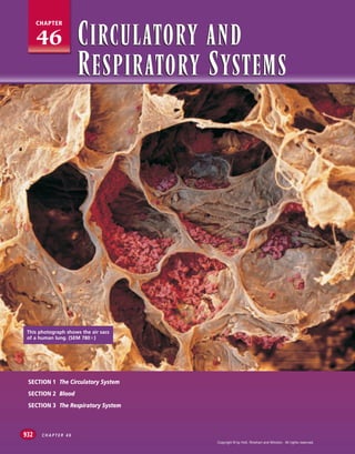
- 1. This photograph shows the air sacs of a human lung. (SEM 780!) SECTION 1 The Circulatory System SECTION 2 Blood SECTION 3 The Respiratory System C H A P T E R 4 6 932 46 CHAPTER CIRCULATORY AND RESPIRATORY SYSTEMS CIRCULATORY AND RESPIRATORY SYSTEMS Copyright © by Holt, Rinehart and Winston. All rights reserved.
- 2. 933 C I R C U L AT O R Y A N D R E S P I R AT O R Y S Y S T E M S THE CIRCULATORY SYSTEM Most of the cells in the human body are not in direct contact with the external environment. The circulatory system acts as a transport service for these cells. Two fluids move through the circulatory system: blood and lymph. THE HEART The blood, heart, and blood vessels form the cardiovascular system. The lymph, lymph nodes, and lymph vessels form the lymphatic system. The cardiovascular system and lymphatic sys- tem collectively make up the circulatory system. The circulatory system transports nutrients, hormones, and gases; gets rid of wastes; and helps maintain a constant body temperature. The central organ of the cardiovascular system is the heart, the muscular organ that pumps blood through a network of blood ves- sels. The heart beats more than 2.5 billion times in an average life span. Yet this organ is slightly larger than a fist. The heart lies within the thoracic (chest) cavity, behind the sternum (breast- bone) and between the two lungs. A tough, saclike membrane called the pericardium surrounds the heart and secretes a fluid that reduces friction as the heart beats. Notice in Figure 46-1 that a septum (wall) vertically divides the heart into two sides. The right side pumps blood to the lungs, and the left side pumps blood to the other parts of the body. Each side of the heart is divided into an upper and lower chamber. Each upper cham- ber is called an atrium, and each lower chamber is called a ventricle. SECTION 1 O B J E C T I V E S ● Describe the structure and function of the human heart. ● Trace the flow of blood through the heart and body. ● Distinguish between arteries, veins, and capillaries in terms of their structure and function. ● Distinguish between pulmonary circulation and systemic circulation. ● Summarize the functions of the lymphatic system. VO C A B U L A R Y cardiovascular system lymphatic system atrium ventricle valve aorta sinoatrial node atrioventricular node pulse artery blood pressure hypertension capillary vein pulmonary circulation systemic circulation atherosclerosis lymph The septum prevents mixing of blood from the two sides of the heart, and the valves ensure that blood flows in only one direction. FIGURE 46-1 Pulmonary (right semilunar) valve Right atrium Tricuspid (right atrioventricular) valve Right ventricle Aortic (left semilunar) valve Left atrium Mitral (left atrioventricular) valve Left ventricle Septum RIGHT LEFT Copyright © by Holt, Rinehart and Winston. All rights reserved.
- 3. C H A P T E R 4 6 934 Valves are flaps of tissue that open in only one direction. The atrioventricular (AY-tree-oh-ven-TRIH-kyuh-luhr) valve (AV valve) on the right side of the heart is called the tricuspid valve. The mitral valve, also called the bicuspid valve, is on the left. As the ventricles pump, blood pressure closes the AV valves to prevent blood from flowing backward into the atria. From the ventricles, blood is pumped out of the heart into large vessels. A semilunar (SEM-ee-LOON-uhr) valve (SL valve) separates the ventricles from these large vessels on each side of the heart. The SL valve on the right side is known as the pulmonary valve, and the SL valve on the left side is known as the aortic valve. The SL valves prevent blood from flowing back into the ventricles when the heart relaxes. Circulation in the Heart Refer to Figure 46-2 to trace the path of the blood as it circulates through the heart. Blood returning to the heart from parts of the body other than the lungs has a high concentration of carbon diox- ide and a low concentration of oxygen. Deoxygenated (O2-poor) blood enters the right atrium. The right atrium sends deoxygenated blood into the right ventricle. The muscles of the right ventricle contract and force the blood into the pulmonary arteries. The pulmonary artery sends the blood to the lungs. In the lungs, the carbon dioxide dif- fuses out of the blood, and oxygen diffuses into the blood. The oxygenated blood returns to the left atrium of the heart. Notice in Figure 46-2 that the flow of blood on the left side of the heart is illustrated with a red arrow representing oxygenated blood, which has a bright red color. 5 4 3 2 1 Determining Heart Rate Materials stopwatch or clock with second hand Procedure 1. Have your partner find the pulse in your wrist and count your heartbeats for 15 seconds while you are seated. Calculate your resting heart rate in beats per minute. 2. Have your partner count your heartbeats for 15 seconds while you are standing. Calculate your heart rate in beats per minute. 3. Have your partner count your heartbeats for 15 seconds after you jog or march in place for 1 minute. Calculate your heart rate in beats per minute. Analysis What causes your pulse? What causes the change in your heart rate in each situation? Quick Lab Trace the path of blood through the heart. Notice that illustrations of a heart are drawn as if the heart were in a person facing you.That is, the left side of the heart is shown on the right as you face the heart, and the right side of the heart is on the left as you face the heart. FIGURE 46-2 Superior vena cava sends deoxygenated blood from upper body to right atrium. Aorta sends oxygenated blood to the coronary arteries, the brain, and the rest of the body. Pulmonary veins return oxygenated blood to the left atrium from the lungs. Left atrium sends blood to the left ventricle. Left ventricle sends blood to the aorta. Right atrium sends blood to the right ventricle. Right ventricle sends blood to the pulmonary artery. Left lung Right lung Inferior vena cava sends deoxygenated blood from lower body to right atrium. Pulmonary arteries send blood to the lungs. 1 1 2 8 4 5 6 7 3 Blood from aorta to body Copyright © by Holt, Rinehart and Winston. All rights reserved.
- 4. 935 C I R C U L AT O R Y A N D R E S P I R AT O R Y S Y S T E M S The oxygenated blood is then pumped into the left ventricle. Contraction of the muscular walls of the left ventricle forces the blood into a large blood vessel called the aorta. From the aorta, blood is transported to all parts of the body. The left ventricle is the thickest chamber of the heart because it has to do the most work to pump blood to all parts of the body. Deoxygenated blood is commonly represented with the color blue. However, it is a misconception that deoxygenated blood is blue. When oxygen is attached to hemoglobin, the blood is bright red. Without oxygen, blood is dark red. The dark red blood in veins appears blue when it shows through the vein walls and skin. Control of the Heartbeat The heart consists of muscle cells that contract in waves. When the first group of cells are stimulated, they in turn stimulate neigh- boring cells. Those cells then stimulate more cells. This chain reac- tion continues until all the cells contract. The wave of activity spreads in such a way that the atria and the ventricles contract in a steady rhythm. The first group of heart-muscle cells that get stim- ulated lie in an area of the heart known as the sinoatrial node, shown in Figure 46-3. The sinoatrial (SIEN-oh-AY-tree-uhl) (SA) node is a group of spe- cialized heart-muscle cells located in the right atrium. These mus- cle cells spontaneously initiate their own electrical impulse and contract. The SA node is often called the pacemaker because it reg- ulates the rate of contraction of the entire heart. The electrical impulse initiated by the SA node subsequently reaches another special area of the heart, known as the atrioventricular (AV ) node. The AV node is located in the septum between the atria, as shown in Figure 46-3. The AV node relays the electrical impulse to the mus- cle cells that make up the ventricles. As a result, the ventricles con- tract a fraction of a second after the atria, completing one full heartbeat. In an average adult at rest, the heart beats about 70 times each minute. A heartbeat has two phases. Phase one, called systole (SIS-tohl), occurs when the ventricles contract, closing the AV valves and opening the SL valves to pump blood into the two major vessels that exit the heart. Phase two, called diastole (DIE-a-stohl), occurs when the ventricles relax, allowing the back pressure of the blood to close the SL valves and opening the AV valves. The closing of these two heart valves results in the characteristic lub dup sound we call a heartbeat. If one of the valves fails to close properly, some blood may flow backward, creating a different sound, which is known as a heart murmur. A person’s pulse is a series of pressure waves within an artery caused by the contractions of the left ventricle. When the ventricle contracts, blood surges through the arteries, and the elastic walls in the vessels expand and stretch. The most common site for taking a pulse is at a radial artery, on the thumb side of each wrist. The average pulse rate ranges from 70 to 90 beats per minute for adults. 8 7 6 Sinoatrial node Atrioventricular node Two areas of specialized tissue, known as nodes, control the heartbeat.A person whose SA node is defective can have an operation to implant an artificial pacemaker.An artificial pacemaker can also help a defective AV node. FIGURE 46-3 Copyright © by Holt, Rinehart and Winston. All rights reserved.
- 5. C H A P T E R 4 6 936 BLOOD VESSELS The circulatory system is known as a closed system because the blood is contained within either the heart or the blood vessels at all times. This type of system differs from an open system, in which blood leaves the vessels and circulates within tissues throughout the organism’s body. The blood vessels that are part of the closed circulatory system of humans form a vast network to help keep the blood flowing in one direction. Arteries and Blood Pressure The large, muscular vessels that carry blood away from the heart are called arteries. As shown in Figure 46-4, the thick walls of the arteries have three layers: an inner endothelial layer, a middle layer of smooth muscle, and an outer layer of connective tissue. This structure gives arteries a combination of strength and elastic- ity, which allows them to stretch as pressurized blood enters from the heart. You can feel this stretching of arteries—it is your pulse. Contraction of the heart moves the blood through the arteries with great force. The force that blood exerts against the inside walls of a blood vessel is known as blood pressure. Blood pressure is highest in the two main arteries that leave the heart. It is usually measured in the artery that supplies blood to the arm. To measure blood pressure, a trained person inflates a cuff that is placed around a patient’s arm, temporarily stopping the flow of blood through the artery. Connected to the cuff is a gauge containing a column of mercury (Hg) that rises as the pressure in the cuff increases. The trained person then releases the air in the cuff slowly while listening to the artery with a stethoscope and watch- ing the column of mercury. The first sounds of blood passing through the artery indicates the systolic pressure, or the pressure of the blood when the ventricles contract. In a normal adult, the sys- tolic pressure is about 120 mm of Hg for males and 110 mm of Hg for females. Continuing to release the air in the cuff, the trained person next listens for the disappearance of sound, which indicates a steady flow of blood through the artery in the arm. This indicates the diastolic pressure. In a normal adult, the diastolic pressure is about 80 mm of Hg for males and 70 mm of Hg for females. High blood pressure, or hypertension, is a leading cause of death in many countries. Blood pressure that is higher than normal places a strain on the walls of the arteries and increases the chance that a vessel will burst. Capillaries and Veins Recall that when the left ventricle contracts, it forces blood into the aorta, the body’s largest artery. From the aorta, blood travels through a network of smaller arteries, which in turn divide and form even smaller vessels, called arterioles. The arterioles branch into a network of tiny vessels, called capillaries. A capillary is shown in Figure 46-5. Endothelium Smooth muscle Connective tissue Artery (carries blood away from the heart) Notice the thick muscular layer of an artery.The layers of the artery wall are separated by elastic tissue.This tissue provides strength, preventing systolic pressure from bursting the artery. FIGURE 46-4 The diameter of a capillary is so small that red blood cells must move single file through these vessels, as shown in this photograph (1,200!).All exchange of nutrients and waste between blood and cells occurs across the thin walls of the capillaries. FIGURE 46-5 Copyright © by Holt, Rinehart and Winston. All rights reserved.
- 6. 937 C I R C U L AT O R Y A N D R E S P I R AT O R Y S Y S T E M S The network formed by capillaries is so extensive that all of the approximately 100 trillion cells in the body lie within about 125 µm of a capillary. This close association between capillaries and cells allows for rapid exchange of materials. Capillary walls are only one cell thick; gases and nutrients can diffuse through these thin walls. Wherever the concentration of oxygen or nutrients is higher in the blood than in the surrounding cells, the substance diffuses from the blood into the cells. Wherever the concentrations of carbon dioxide and wastes are higher in the cells than in the blood, these substances diffuse from the cells into the blood. Blood flows through capillaries that merge to form larger ves- sels called venules (VEN-yoolz). Several venules in turn unite to form a vein, a large blood vessel that carries blood to the heart. Veins returning deoxygenated blood from the lower parts of the body merge to form the inferior vena cava. Veins returning deoxygenated blood from the upper parts of the body merge to form the superior vena cava. Refer back to Figure 46-2, and locate the inferior vena cava and the superior vena cava. As you can see in Figure 46-6, although the walls of the veins are composed of three layers, like those of the arteries, they are thin- ner and less muscular. By the time blood reaches the veins, it is under much less pressure than it was in the arteries. With less pres- sure being exerted in the veins, the blood could flow backward and disrupt the pattern of circulation. To prevent that, valves in the veins help keep the blood flowing in one direction. Many veins pass through skeletal muscle. When these muscles contract, they are able to squeeze the blood through the veins. When these muscles relax, the valves can close, thus preventing the blood from flowing backward. Figure 46-6 shows the structure of a valve in a vein. PATTERNS OF CIRCULATION The English scientist William Harvey (1578–1657) first showed that the heart and the blood vessels form one continuous, closed system of circulation, as shown in Figure 46-7. He also reasoned that this system consists of two primary subsystems: pulmonary circulation, in which the blood travels between the heart and lungs, and systemic circulation, in which the blood travels between the heart and all other body tissues. Pulmonary Circulation Deoxygenated blood returning from all parts of the body except the lungs enters the right atrium, where it is then pumped into the right ventricle. When the right ventricle contracts, the deoxy- genated blood is sent through the pulmonary artery to the lungs. The pulmonary artery is the only artery that carries deoxygenated blood. The pulmonary artery branches into two smaller arteries, with one artery going to each lung. These arteries branch into arte- rioles and then into capillaries in the lungs. Vein (returns blood to the heart) Endothelium Smooth muscle Connective tissue The cardiovascular system transports materials throughout the body and distributes heat. FIGURE 46-7 Like an artery, a vein has three layers: the outer layer of connective tissue, the middle layer of smooth muscle, and the inner layer of endothelial tissue. FIGURE 46-6 Copyright © by Holt, Rinehart and Winston. All rights reserved.
- 7. C H A P T E R 4 6 938 From head To head From body To body Pulmonary veins Pulmonary veins Pulmonary arteries Right atrium Inferior vena cava Superior vena cava Left ventricle Right ventricle Left atrium Heart Aorta Capillaries of intestines Capillaries of legs Capillaries of liver Coronary circulation Inferior vena cava Capillaries of kidneys Hepatic portal circulation Renal circulation In the lungs, carbon dioxide diffuses out of the capillaries and oxygen diffuses into the capillaries. The oxygenated blood then flows into venules, which merge into the pulmonary veins that lead to the left atrium of the heart. From the left atrium, blood is pumped into the left ventricle and then to the body through the aorta. In Figure 46-8, trace the path blood takes as it passes through pulmonary circulation. Systemic Circulation Systemic circulation is the movement of blood between the heart and all parts of the body except the lungs. Trace the path blood fol- lows in systemic circulation in Figure 46-9. Notice that oxygenated blood is pumped out of the left ventricle and into the aorta. From the aorta, blood flows into other subsystems of systemic circulation. Coronary circulation is the subsystem of systemic circulation that supplies blood to the heart itself. If blood flow in the coronary arteries, which supply blood to the heart, is reduced or cut off, muscle cells will die. This can happen when an artery is blocked by a blood clot or by atherosclerosis (ATH-uhr-oh-skler-OH-sis), a dis- ease characterized by the buildup of fatty materials on the interior walls of the coronary arteries. Either type of blockage can lead to a heart attack. Hepatic portal circulation is a subsystem of systemic circulation. Nutrients are picked up by capillaries in the small intestine and are transported by the blood to the liver. Excess nutrients are stored in the liver for future needs. The liver receives oxygenated blood from a large artery that branches from the aorta. Renal circulation, another subsystem of systemic circulation, supplies blood to the kidneys. Nearly one-fourth of the blood that is pumped into the aorta by the left ventricle flows to the kidneys. The kidneys filter waste from the blood. The pulmonary circulation between the heart and the lungs involves the pulmonary arteries and the pulmonary veins. Deoxygenated blood flows from the right side of the heart to the lungs. Oxygenated blood is returned to the left side of the heart from the lungs.This is the opposite of systemic and coronary blood flow, in which oxygen-rich blood flows from the heart and oxygen-poor blood is returned to the heart. FIGURE 46-8 Notice three subsystems of systemic circulation. Other subsystems transport blood between the heart and the head, arms, and other organs. FIGURE 46-9 Copyright © by Holt, Rinehart and Winston. All rights reserved.
- 8. 939 C I R C U L AT O R Y A N D R E S P I R AT O R Y S Y S T E M S LYMPHATIC SYSTEM In addition to the cardiovascular system, the circulatory system also includes the lymphatic system. One function of the lymphatic sys- tem is to return fluids that have collected in the tissues to the blood- stream. Fluids diffuse through the capillary walls just as oxygen and nutrients do. Some of these fluids pass into cells, some return to the capillaries, and some remain in the intercellular spaces. Excess fluid in the tissues moves into the tiny vessels of the lym- phatic system; this fluid is called lymph. Lymph vessels merge to form larger vessels. The lymph vessels are similar in structure to capillaries, and the larger lymph vessels are similar in structure to veins. However, an important difference exists between blood ves- sels and lymph vessels. Blood vessels form a complete circuit so that blood passes from the heart to all parts of the body and then back again to the heart. In contrast, lymph vessels form a one-way system that returns fluids collected in the tissues back to the bloodstream. In addition, the lymphatic system has no pump. Like the blood in veins, lymph must be moved through the vessels by the squeezing of skeletal muscles. Like veins, the larger lymph ves- sels have valves to prevent the fluid from moving backward. Notice in Figure 46-10 that lymph vessels form a vast network that extends throughout the body. The lymph that travels in these vessels is a transparent yellowish fluid, much like the liquid part of the blood. As the lymph travels through these vessels on its way to the heart, it passes through small organs known as lymph nodes. Notice in Figure 46-10 that lymph nodes are like beads on a string. These nodes filter the lymph as it passes, trapping foreign parti- cles, microorganisms, and other tissue debris. Lymph nodes also store lymphocytes, white blood cells that are specialized to fight disease. When a person has an infection, the nodes may become inflamed, swollen, and tender because of the increased number of lymphocytes. 1. Describe the structure of the heart. 2. Outline the path that blood follows through the heart and body, starting at the superior vena cava. 3. Describe the process by which the heartbeat is regulated. 4. How are the structures of arteries, veins, and capillaries related to their function? 5. Compare oxygenation levels in pulmonary circulation and systemic circulation. 6. Explain how the lymphatic system works with the cardiovascular system. CRITICAL THINKING 7. Analyzing Information Some babies are born with a hole in the septum between the two atria. Based on what you know about blood flow through the heart, explain why this condition would be harmful to the baby. 8. Forming Reasoned Opinions In which blood vessels would you expect to find the lowest average blood pressure? Explain your answer. 9. Applying Information A man’s arm is cut by a piece of glass. Blood comes out of the wound in rapid spurts. Which type of vessel was cut? SECTION 1 REVIEW Like the cardiovascular system, the lymphatic system forms a vast network. Concentrated in certain regions of this network are lymph nodes that contain some of the disease-fighting cells of the immune system. FIGURE 46-10 Tonsils Thymus Lymph node Spleen Lymphatic vessel Bone marrow Copyright © by Holt, Rinehart and Winston. All rights reserved.
- 9. C H A P T E R 4 6 940 BLOOD Blood is a liquid connective tissue. The two main functions of the blood are to transport nutrients and oxygen to the cells and to carry carbon dioxide and other waste materials away from the cells. Blood also transfers heat to the body surface and plays a major role in defending the body against disease. COMPOSITION OF BLOOD Blood is composed of a liquid medium and blood solids (formed elements). Formed elements include red blood cells, white blood cells, and platelets. The liquid makes up about 55 percent of the blood, and formed elements make up the remaining 45 percent. A healthy adult has about 4 to 5 L of blood in his or her body. Plasma Plasma, the liquid medium, is a sticky, straw-colored fluid that is about 90 percent water and includes metabolites, nutrients, wastes, salts, and proteins. Cells receive nourishment from dis- solved substances carried in the plasma. These substances, which may include vitamins, minerals, amino acids, and glucose, are absorbed from the digestive system and transported to the cells. Plasma also carries hormones and brings wastes from the cells to the kidneys or the lungs to be removed from the body. A variety of proteins are carried in the plasma. The plasma pro- teins have various functions. Some of the proteins in the plasma are essential for the formation of blood clots. Another protein, called albumin, plays an important role in the regulation of osmotic pressure between plasma and blood cells and between plasma and tissues. Other proteins, called anti- bodies, help the body fight disease. Red Blood Cells Red blood cells, or erythrocytes (uh-RITH-ruh-siets), shown in Figure 46-11, transport oxygen to cells in all parts of the body. Red blood cells are formed in the red marrow of bones. Immature red blood cells synthesize large amounts of an iron-containing protein called hemoglobin. Hemoglo- bin is the molecule that actually transports oxygen and, to a lesser degree, carbon dioxide. During the formation of a red blood cell, its cell nucleus and organelles disintegrate. The mature red blood cell is little more than a membrane containing hemoglobin. SECTION 2 O B J E C T I V E S ● List the components of blood. ● Distinguish between red blood cells, white blood cells, and platelets in terms of their structure and function. ● Summarize the process of blood clotting. ● Explain what determines the compatibility of blood types for transfusion. VO C A B U L A R Y plasma red blood cell (erythrocyte) hemoglobin white blood cell (leukocyte) phagocyte antibody platelet fibrin blood type antigen Rh factor Notice that a mature red blood cell (RBC) is disk-shaped and is concave on both sides.A red blood cell is little more than a cell membrane filled with hemoglobin. How is this structure related to its function? FIGURE 46-11 Copyright © by Holt, Rinehart and Winston. All rights reserved.
- 10. 941 C I R C U L AT O R Y A N D R E S P I R AT O R Y S Y S T E M S Because red blood cells lack nuclei, they cannot divide and they have a limited survival period, usually 120 to 130 days. Of the more than 30 trillion red blood cells circulating throughout the body at one time, 2 million disintegrate every second. To replace them, new ones form at the same rate in the red marrow of bones. Some parts of the disintegrated red blood cells are recycled. For exam- ple, the iron portion of the hemoglobin molecule is carried in the blood to the marrow, where it is reused in new red blood cells. White Blood Cells White blood cells, or leukocytes (LOO-kuh-siets), help defend the body against disease. They are formed in the red marrow, but some must travel to lymph nodes, tonsils, the thymus, or the spleen to mature. White blood cells are larger than red blood cells and sig- nificantly less plentiful. Each cubic millimeter of blood normally contains about 4 million red blood cells and 7,000 white blood cells. White blood cells can squeeze their way through openings in the walls of blood vessels and into the intercellular fluid. In that way, white blood cells can reach the site of infection and help destroy invading microorganisms. Notice in Figure 46-12 that a white blood cell has a very different structure from that of a red blood cell. For instance, a white blood cell may be irregularly shaped and may have a rough outer surface. There are other differences between red blood cells and white blood cells as well. In contrast with the short-lived red blood cells, white blood cells may function for years. And while there is only one type of red blood cell, there are several types of white blood cells. The white blood cell shown in Figure 46-12 is the type of white blood cell known as a phagocyte (FA-guh-siet). Phagocytes are cells that engulf invading microorganisms. Locate the microorganisms that are being engulfed by the phagocyte in Figure 46-12. Another type of white blood cell produces antibodies. Antibodies are pro- teins that help destroy substances, such as bacteria and viruses, that enter the body and can cause disease. When a person has an infection, the number of white blood cells can double. leukocyte from the Greek leuco, meaning “white,” and cyte, meaning “cell” Word Roots and Origins Some white blood cells, like the phagocyte shown in blue, engulf and destroy invading microorganisms. FIGURE 46-12 Copyright © by Holt, Rinehart and Winston. All rights reserved.
- 11. C H A P T E R 4 6 942 (a) (b) Inactive platelets, such as the yellow object shown in (a), derive their name from the fact that they look like little plates. Platelets are colorless and contain chemicals that are involved in the clotting process. (b) The platelets change shape during the clotting process.When activated, the platelets settle and spread on the substrate. FIGURE 46-13 Platelets release clotting protein (enzyme) Fibrin net forms, trapping blood cells and platelets Clotting reaction occurs Blood vessel damage Stimulus Blood clot Result The release of enzymes from platelets at the site of a damaged blood vessel initiates a “clotting cascade.” FIGURE 46-14 Platelets Platelets are essential to the formation of a blood clot. A blood clot is a mass of interwoven fibers and blood cells that prevents excess loss of blood from a wound. Platelets are not whole cells. They are fragments of very large cells that were formed in the bone marrow. As you can see in Figure 46-13a, platelets get their name from their platelike structure. Platelets lack a nucleus and have a life span of 7 to 12 days. A cubic micrometer of blood may contain as many as half a million platelets. When a blood vessel tears or rips, platelets congregate at the damaged site, sticking together and forming a small plug. The ves- sel constricts, slowing blood flow to the area. Then, special clotting factors are released from the platelets and the damaged tissue. These factors begin a series of chemical reactions that occur at the site of the bleeding. The last step in this series brings about the production of a protein called fibrin. Fibrin molecules consist of long, sticky chains. As you can see in Figure 46-14, these chains form a net that traps red blood cells, and the mass of fibrin and red blood cells hardens into a clot, or scab. Hemophilia is a disorder caused by the absence of one or more of the proteins required for blood clotting. When a person with hemophilia is injured, bleeding continues for much longer than it would in a person without hemophilia. Large cuts or internal injuries can be life threatening. Today, people with hemophilia are treated with injections of the missing clotting factors. Connection Connection Eco Eco Eco Vampire Bats Help Save Stroke Victims Vampire bats have an anticoagu- lant in their saliva that prevents blood clotting when it flows from a wound. In 1995, this enzyme was isolated and named Draculin. Researchers have developed a clot-dissolving agent called Desmodus rotundus salivary plas- minogen activator (DSPA), which is based on the salivary enzyme Draculin. DSPA targets and destroys fibrin. The current treatment must be given within three hours of the onset of a stroke (a sudden loss of consciousness or paralysis that occurs when the blood flow to the brain is interrupted). Otherwise, brain-cell death and brain damage may occur. According to research, DSPA could be a safe treatment for longer periods of time and appears to have no detrimental effect on brain cells. Copyright © by Holt, Rinehart and Winston. All rights reserved.
- 12. 943 C I R C U L AT O R Y A N D R E S P I R AT O R Y S Y S T E M S BLOOD TYPES Blood type is determined by the type of antigen present on the sur- face of the red blood cells. An antigen is a substance that stimulates an immune response. Antigens that are normally present in a per- son’s body provoke no response. However, when foreign antigens enter the body, cells respond by producing antibodies. In fact, the word antigen is an abbreviation for “antibody-generating substance.” In the early 1900s, Karl Landsteiner used blood taken from his laboratory workers and made observations similar to those you see in Figure 46-15. He noticed that mixing blood samples from two people sometimes resulted in the cells clumping together, or agglutinating. When samples of two different blood types are mixed together, reactions occur between the antigens on the red blood cells and the antibodies in the plasma, causing the cells to aggluti- nate. When samples of the same blood type are mixed, no reaction occurs, and the blood cells do not agglutinate. Landsteiner’s observations led to the classification of human blood by blood types. Three of the most important human antigens are called A, B, and Rh. The A-B-O system of blood typing, described below, is based on the A and B antigens. A-B-O System The A-B-O system is a means of classifying blood by the antigens located on the surface of the red blood cells and the antibodies cir- culating in the plasma. As shown in Table 46-1, an individual’s red blood cells may carry an A antigen, a B antigen, both A and B anti- gens, or no antigen at all. These antigen patterns are called blood types A, B, AB, and O, respectively. Notice in Table 46-1 that an individual with type A blood also has anti-B antibodies against type B blood. If type B blood is given to a recipient with type A blood, the recipient’s anti-B antibodies will react with the B antigens on the donated red blood cells and the blood will agglutinate. In addition, the donor’s type B blood has anti- A antibodies. Their presence will compound the antigen-antibody reaction. The result will be agglutinated blood that will block the flow of blood through the vessels. For this reason, transfusion recip- ients must receive blood that is compatible with their own. (a) (b) Notice that there is no agglutination of red blood cells in the slide in (a), where blood samples from two people with the same blood type were mixed. Compare this with the slide in (b), where blood samples from two people with different blood types were mixed. FIGURE 46-15 TABLE 46-1 Blood Types, Antigens, and Antibodies Antigen on the Antibodies in the Blood types red blood cells plasma Can get blood from Can give blood to A A anti-B O, A A, AB B B anti-A O, B B, AB AB A and B none A, B, AB, O AB O none anti-A, anti-B O A, B, AB, O Copyright © by Holt, Rinehart and Winston. All rights reserved.
- 13. C H A P T E R 4 6 944 People who have type AB blood are universal recipients. They can receive A, B, AB, or O blood because they do not have anti-A or anti-B antibodies. People who have type O blood are universal donors. They can donate blood to people who have A, B, AB, or O blood because the blood cells of people who have type O blood do not have A or B antigens. Rh System An antigen that is sometimes present on the surface of red blood cells is the Rh factor, named after the rhesus monkey in which it was first discovered. Eighty-five percent of the United States’ popu- lation is Rh-positive (Rh!), meaning that Rh antigens are present. People who do not have Rh antigens are called Rh-negative (Rh"). If an Rh" person receives a transfusion of blood that has Rh! antigens, antibodies may react with the antigen and agglutination will occur. The most serious problem with Rh incompatibility occurs during pregnancy. If the mother is Rh" and the father is Rh!, the child may inherit the dominant Rh! allele from the father. During delivery, a small amount of the fetus’s Rh! blood may reach the mother’s bloodstream. If this happens, the mother will develop antibodies to the Rh factor. If a second Rh! child is conceived later, the mother’s antibodies can cross the placenta and attack the blood of the fetus. This condition is called erythroblastosis fetalis. The fetus may die as a result of this condition, or if the child is born alive, he or she may need an immediate transfusion of Rh! blood. To prevent this condition, an Rh" mother of an Rh! child can be given antibodies to destroy any Rh! cells that have entered her bloodstream from the fetus. The mother is, in effect, immunized against the Rh antigen before her immune system has a chance to develop its own antibodies. The antibody treatment prevents Rh sensitization in Rh" women only if their bodies have not already produced Rh antibodies. If an Rh" mother has not yet been sensi- tized, she receives the antibody treatment in the 28th week of preg- nancy and again immediately after delivery. 1. Identify the four main components of blood. 2. Explain how the structure of red blood cells, white blood cells, and platelets relates to the function of these cells. 3. Identify the stages and structures involved in the clotting process. 4. What factors determine the compatibility of blood types for transfusion? 5. Which blood types, in terms of the A-B-O and Rh antigens, can be donated to somebody with type AB– blood? CRITICAL THINKING 6. Analyzing Information Hemophilia is a disor- der in which there is a failure in one of the steps of clot formation. What might be some advan- tages and disadvantages of this disorder? 7. Evaluating Results A patient’s blood has an elevated count of leukocytes. What does this most likely indicate? 8. Relating Concepts Explain why a pregnant woman should know her blood type and the blood type of her baby’s father. SECTION 2 REVIEW www.scilinks.org Topic: Blood Types Keyword: HM60179 Copyright © by Holt, Rinehart and Winston. All rights reserved.
- 14. MILESTONES Blood Transfusions IN In 1665, Richard Lower, an English physician, transferred blood between two dogs. Jean-Baptiste Denis, a French physician and astrologer, transfused blood from a lamb to a human in 1667. The first successful human-to-human blood trans- fusions were performed in 1818 by James Blundell, a British physician, but these were followed by many failures. In 1901, Austrian-born American phys- iologist Karl Landsteiner determined the first three blood types—A, B, and O—and that incompatible types will clot. This dis- covery explained why the outcome of transfusions had been so unpredictable in the past. For his work, Landsteiner received the Nobel Prize in medicine or physiology in 1930. During World War I, scientists began to study how to preserve and transport blood for wounded soldiers. They found that the addition of sodium citrate prevented clot- ting, making the storage of blood possible for the first time. In 1937, Bernard Fantus, a physician, established the first nonprofit blood bank at Cook County Hospital in Chicago. The next year, Charles Drew, a medical doctor, found that blood plasma (the liquid com- ponent of blood) could substitute for whole blood in emergency situations. Germany’s attack on France in 1940 created a great need for blood. Based on Drew’s findings, the United States began to provide liquid plasma and whole blood for France.Working with the National Research Council and the American Red Cross, Drew set up blood collection centers. Early in the 1980s, some transfused blood was found to carry the human immunodeficiency virus (HIV), the virus that causes acquired immune deficiency syndrome (AIDS). Since 1985, careful screening for HIV, hepatitis, and other dis- eases has almost entirely removed the risk of receiving contaminated blood. Today, many people bank their own blood for later use in surgery. Blood can also be collected during surgery and returned to the patient later. The AIDS epi- demic has triggered a race to create arti- ficial blood. Several companies have begun testing blood substitutes from chemically treated animal blood and out- dated human blood. 1. What type of patients can benefit from blood transfusions? 2. Critical Thinking How might safe blood transfusion techniques affect you? 3. Critical Thinking Use the Web site below to research the latest progress on creating artificial blood. Write a short report to describe your findings. Every three seconds someone needs a blood transfusion—blood, plasma, or saline is introduced into the body. Blood and blood products are used to treat accident and burn victims, cancer patients, and patients undergoing surgeries and medical treatments. The development of safe blood transfusion techniques was an important achievement in modern medicine. Review www.scilinks.org Topic: Blood Donations Keyword: HM60178 945 Timeline 1818 First human-to- human blood transfusion. 1901 Karl Landsteiner determined three of four blood types (A, B, and O). 1914 Blood is stored for the first time. 1937 Bernard Fantus formed the first blood bank. 1939 Charles Drew set up collection centers for blood to fill World War II needs. 1950 Carl Walter and W.P. Murphy, Jr. introduced the plastic bag to collect blood. 1985 Blood is screened for HIV and other diseases. 1628 William Harvey describes the cir- culation of blood. 1667 Jean-Baptiste Denis successfully transfuses blood from a lamb to a human. Copyright © by Holt, Rinehart and Winston. All rights reserved.
- 15. C H A P T E R 4 6 946 THE RESPIRATORY SYSTEM The blood transports oxygen from the lungs to cells and carries carbon dioxide from the cells to the lungs. It is the function of the respiratory system to exchange gases with the cardiovascular system. RESPIRATION The respiratory system involves both external respiration and internal respiration. External respiration is the exchange of gases between the atmosphere and the blood. Internal respiration is the exchange of gases between the blood and the cells of the body. Once oxygen is in the cells, the cells use it to break down glucose and make ATP by the process of aerobic respiration. Without oxy- gen, the body could not obtain enough energy from food to sur- vive. Excess carbon dioxide produced as a waste product of aerobic respiration is toxic to cells and is removed from the cells by internal respiration. THE LUNGS The lungs are the site of gas exchange between the atmosphere and the blood. Notice in Figure 46-16 that the right lung has three divi- sions, or lobes. It is slightly heavier than the two-lobed left lung. The lungs are located inside the thoracic cavity, bounded by the rib cage and the diaphragm. Lining the entire cavity and covering the lungs are pleura, membranes that secrete a slippery fluid that decreases friction from the movement of the lungs during breathing. The Path of Air Refer to Figure 46-16 to trace the path air follows from the atmos- phere to the capillaries in the lungs. External respiration begins at the mouth and at the nose. Air filters through the small hairs of the nose and passes into the nasal cavity, located above the roof of the mouth. In the nasal cavity, mucous membranes warm and moisten the air, which helps prevent damage to the delicate tissues that form the respiratory system. The walls of the nasal cavity are also lined with cilia. These cilia trap particles that are inhaled and are eventually swept into the throat, where they are swallowed. SECTION 3 O B J E C T I V E S ● Differentiate external respiration from internal respiration. ● Trace the path of air from the atmosphere to the bloodstream. ● Describe how gases are exchanged in the lungs and transported in the bloodstream. ● Summarize the skeletal and muscular changes that occur during breathing. ● Describe how the rate of breathing is controlled. VO C A B U L A R Y respiratory system external respiration internal respiration lung pharynx epiglottis trachea larynx bronchus bronchiole alveolus inspiration diaphragm expiration www.scilinks.org Topic: Respiratory System Keyword: HM61307 Copyright © by Holt, Rinehart and Winston. All rights reserved.
- 16. 947 C I R C U L AT O R Y A N D R E S P I R AT O R Y S Y S T E M S The moistened, filtered air then moves into the throat, or pharynx (FER-inks), a tube at the back of the nasal cavities and the mouth. The pharynx contains passageways for both food and air. When food is swallowed, a flap of cartilage, called the epiglottis, presses down and covers the opening to the air passage. When air is being taken in, the epiglottis is in an upright position, allowing air to pass into a cartilaginous tube called the windpipe, or trachea (TRAY-kee-uh). The trachea is about 10 to 12 cm long and has walls lined with ciliated cells that trap inhaled particles. The cilia sweep the particles and mucus away from the lungs toward the throat. At the upper end of the trachea is the voicebox, or larynx (LER-inks). Sounds are produced when air is forced past two ligaments—the vocal cords—that stretch across the larynx. The pitch and volume of the sound produced varies with the amount of tension on the vocal cords and on the amount of air being forced past them. The trachea then branches into two bronchi (BRAHN-kie) (singular, bronchus), each of which leads to a lung. The walls of the bronchi consist of smooth muscle and cartilage and are lined with cilia and mucus. Within the lungs, the bronchi branch into smaller and smaller tubes. The smallest of these tubes are known as bronchioles, which are also lined with cilia and mucus. Eventually the bronchioles end in clusters of tiny air sacs called alveoli (al-VEE-oh-LIE) (singular, alveo- lus). A network of capillaries surrounds each alveolus, as you can see in the detailed view shown in Figure 46-16. All exchange of gases in the lungs occurs in the alveoli. To facilitate this exchange, the surface area of the lungs is enormous. A healthy lung contains nearly 300 mil- lion alveoli and has a total surface area of about 70 m2—about 40 times the surface area of the skin. Nasal cavity Pharynx Epiglottis Larynx Trachea Right lung Bronchus Bronchiole Heart Alveoli Diaphragm Left lung Capillaries Bronchiole Alveoli Trace the passage of air from the atmosphere to the lungs. Oxygen in the air finally reaches the alveoli, the functional units of the respiratory system.All exchange of gases between the respiratory system and the cardiovascular system occurs in the alveoli. FIGURE 46-16 Copyright © by Holt, Rinehart and Winston. All rights reserved.
- 17. C H A P T E R 4 6 948 Alveolus O2 molecule CO2 molecule Capillary Because of concentration gradients, oxygen and carbon dioxide diffuse across the alveoli and capillary walls. FIGURE 46-17 GAS EXCHANGE AND TRANSPORT In the lungs, gases are exchanged between the alveoli and the blood in the capillaries. Oxygen (O2) to be transported throughout the body moves into the bloodstream, and carbon dioxide (CO2) to be eliminated from the body moves into the alveoli. Gas Exchange in the Lungs Figure 46-17 illustrates the direction in which oxygen and carbon dioxide move in the alveoli. When air moves into the lungs, the oxygen in the air crosses the thin alveolar membranes as well as the capillary walls and dissolves in the blood. Carbon dioxide moves in the opposite direction, crossing the capillary walls and thin alveolar membranes and entering the alveoli. Air moving into the alveoli is rich in oxygen and contains little carbon dioxide. In contrast, blood in the capillaries surrounding the alveoli is low in oxygen and contains high levels of carbon diox- ide. Substances diffuse from an area of higher concentration to an area of lower concentration. Consequently, oxygen diffuses from the alveoli into the blood, and carbon dioxide diffuses from the blood into the alveoli. The enormous surface area of the alveoli increases the rate of diffusion of these two gases. Transport of Oxygen When oxygen diffuses into the blood, only a small amount remains dissolved in the plasma. Most of the oxygen—95 to 98 percent— moves into the red blood cells, where it combines with hemoglo- bin, an iron-containing protein. Each hemoglobin molecule contains four iron atoms. Each iron atom can bind to one oxygen molecule. Thus, one hemoglobin molecule can carry up to four molecules of oxygen. There are about 250 million hemoglobin mol- ecules in each red blood cell. When oxygenated blood reaches body tissues, the oxygen concentration is higher in the blood than in the body tissues. Thus, oxygen is released from hemoglobin and diffuses out of the capillaries and into surrounding cells. Copyright © by Holt, Rinehart and Winston. All rights reserved.
- 18. 949 C I R C U L AT O R Y A N D R E S P I R AT O R Y S Y S T E M S (a) INSPIRATION (b) EXPIRATION Rib muscles contract (rib cage expands) Rib muscles relax (rib cage contracts) Ribs Diaphragm Diaphragm relaxes (diaphragm moves up) Diaphragm contracts (diaphragm moves down) The diaphragm, a large skeletal muscle that separates the thoracic cavity from the abdominal cavity, and the muscles between the ribs control the movement of the thoracic cavity during breathing. If these muscles were paralyzed, then inspiration and expiration would not occur. FIGURE 46-18 Transport of Carbon Dioxide Because the concentration of carbon dioxide (CO2) is higher in the cells, it diffuses out of the cells and into the blood. Only about 7 percent of the carbon dioxide dissolves in the plasma. Approximately 23 percent binds to hemoglobin. The remaining 70 percent is carried in the blood as bicarbonate ions (HCO3 !). As shown in the equation below, CO2 reacts with water in the plasma to form carbonic acid (H2CO3 ). In turn, the carbonic acid disasso- ciates into bicarbonate ions and hydrogen ions (H+): H2O " CO2 ∏ H2CO3 ∏ HCO3 ! " H+ Thus, most of the CO2 travels in the blood as bicarbonate ions. When the blood reaches the lungs, the reactions are reversed: HCO3 ! " H+ ∏ H2CO3 ∏ H2O " CO2 Bicarbonate ions combine with hydrogen ions to form carbonic acid, which in turn forms carbon dioxide and water. The carbon dioxide diffuses out of the capillaries into the alveoli and is exhaled into the atmosphere. MECHANISM OF BREATHING Breathing is the process of moving air into and out of the lungs. Inspiration, shown in Figure 46-18, is the process of taking air into the lungs. When you take a deep breath, your chest expands as muscles contract to move the ribs up and outward. At the same time, your diaphragm, a large skeletal muscle that separates the thoracic cavity from the abdominal cavity, flattens and pushes down on the abdomen. Muscles in the abdominal wall in turn relax. This action provides room for the flattened diaphragm. Copyright © by Holt, Rinehart and Winston. All rights reserved.
- 19. C H A P T E R 4 6 950 When the diaphragm flattens and the ribs are lifted up and out, the volume of the lungs increases. An increased volume reduces the air pressure within the lungs. At this point, the air pressure inside the lungs is lower than the air pressure outside the body. As a result, air from the atmosphere moves into the lungs. During expiration, the process of releasing air from the lungs, the reverse movements take place, as shown in Figure 46-18. As the diaphragm and rib muscles relax, the elastic tissues of the lungs recoil, deflating the lungs. The volume of the lungs decreases. Because the volume is smaller, the air pressure inside the cavity becomes greater than the air pressure outside the body. This pres- sure difference forces air out of the lungs until the pressures are again equal. Regulation of Breathing The rate at which oxygen is used depends on the activity of the cells. The greater their activity, the more oxygen they need and the faster the body needs to breathe. The slower their activity, the slower the body needs to breathe. Both rate and depth of breathing change in order to provide oxygen and eliminate car- bon dioxide. The rate of breathing is controlled by the brain and brain stem, which monitors the concentration of carbon dioxide in the blood. As activity increases, high levels of carbon dioxide in the blood stimulate nerve cells in the brain. The brain stem in turn stimulates the diaphragm to increase the breathing rate and depth. When the carbon dioxide concentration in the blood returns to lower levels, the sensors in the brain send a message to the respiratory muscles to return to a slower breathing rate. All this is controlled subcon- sciously by control centers in the brain. However, a person can temporarily override the respiratory control system at any time, holding his or her breath until losing consciousness. Then the brain stem takes control, and normal breathing resumes. This mechanism allows humans to swim underwater for short periods and to sleep without concern for breathing. 1. How does internal respiration differ from exter- nal respiration? 2. Outline the path of oxygen from the atmosphere to the bloodstream. 3. Explain the process of gas exchange in the lungs. 4. Differentiate between oxygen transport and carbon dioxide transport in the bloodstream. 5. Sequence the skeletal and muscular changes that take place when a person inhales. 6. What factors regulate the rate of breathing? CRITICAL THINKING 7. Relating Concepts Predict the effect that increasing altitude would have on blood-oxygen saturation. 8. Applying Information Why does a single-celled organism not need a respiratory system? 9. Predicting Results Normally, arterial blood is about 98 percent saturated with oxygen. What are two conditions that could result in lower oxygen saturation? SECTION 3 REVIEW expiration from the Latin expir, which means “to breathe out” Word Roots and Origins Copyright © by Holt, Rinehart and Winston. All rights reserved.
- 20. C I R C U L AT O R Y A N D R E S P I R AT O R Y S Y S T E M S CHAPTER HIGHLIGHTS plasma (p. 940) red blood cell (erythrocyte) (p. 940) hemoglobin (p. 940) white blood cell (leukocyte) (p. 941) phagocyte (p. 941) antibody (p. 941) platelet (p. 942) fibrin (p. 942) blood type (p. 943) antigen (p. 943) Rh factor (p. 943) Vocabulary cardiovascular system (p. 933) lymphatic system (p. 933) atrium (p. 933) ventricle (p. 933) valve (p. 934) aorta (p. 935) sinoatrial node (p. 935) atrioventricular node (p. 935) pulse (p. 935) artery (p. 936) blood pressure (p. 936) hypertension (p. 936) capillary (p. 936) vein (p. 937) pulmonary circulation (p. 937) systemic circulation (p. 937) atherosclerosis (p. 938) lymph (p. 939) Vocabulary The Circulatory System SECTION 1 ● The human circulatory system is made up of the cardiovascular system and the lymphatic system. ● The heart is a muscular organ that pumps blood through an intricate network of blood vessels. ● Blood flows from the body into the heart, which then pumps blood to the lungs. After oxygenation, blood returns to the heart, which pumps blood to the rest of the body. ● Arteries carry blood away from the heart. Materials are exchanged at the capillaries. Veins contain valves and carry blood back to the heart. ● In pulmonary circulation, blood travels between the heart and lungs. In systemic circulation, blood travels between the heart and all other body tissues. ● The lymphatic system returns lymph, fluid that has collected in the tissues, to the bloodstream. Blood SECTION 2 ● Blood is composed of plasma (water, metabolites, wastes, salts, and proteins), red blood cells, white blood cells, and platelets. ● Red blood cells transport oxygen. White blood cells help defend the body against disease. Platelets are essential to the formation of a blood clot. ● Blood clotting occurs when platelets release a clotting protein, which causes a clotting reaction to occur. A fibrin net forms, trapping blood cells and platelets. ● Human blood can be grouped into four types—A, B, AB, and O—based on proteins on the surface of red blood cells. Another antigen called Rh factor, is sometimes present on red blood cells. respiratory system (p. 946) external respiration (p. 946) internal respiration (p. 946) lung (p. 946) pharynx (p. 947) epiglottis (p. 947) trachea (p. 947) larynx (p. 947) bronchus (p. 947) bronchiole (p. 947) alveolus (p. 947) inspiration (p. 949) diaphragm (p. 949) expiration (p. 950) Vocabulary The Respiratory System SECTION 3 ● External respiration is the exchange of gases between the atmosphere and the blood. Internal respiration is the exchange of gases between the blood and the cells of the body. ● The lungs are the site of gas exchange between the atmosphere and the blood. ● Air enters through the mouth or nose, passes through the pharynx, larynx, trachea, bronchi and bronchioles and into alveoli. A network of capillaries surrounds each alveolus. All exchange of gases in the lungs occurs at the alveoli. ● Most oxygen is carried attached to hemoglobin. Some carbon dioxide is carried bound to hemoglobin. A small amount is dissolved in plasma. Most carbon dioxide is carried as bicarbonate ions. ● During inspiration, the diaphragm and rib muscles contract, the thoracic cavity expands, and air is pulled into the lungs. During expiration, the diaphragm and rib muscles relax, the thoracic cavity contracts, and air is forced out of the lungs. ● The rate of breathing is controlled by nerve centers in the brain that monitor the level of carbon dioxide in the blood. 951 Copyright © by Holt, Rinehart and Winston. All rights reserved.
- 21. CHAPTER REVIEW C H A P T E R 4 6 952 USING VOCABULARY 1. Distinguish between systolic pressure and dias- tolic pressure. 2. Choose the term that does not belong in the following group: erythrocyte, hemoglobin, leuko- cyte, and platelet. Explain why it does not belong. 3. For each pair of terms, explain the relationship between the terms. a. atrioventricular valve and semilunar valve b. artery and vein c. expiration and inspiration 4. Word Roots and Origins The word phagocyte is derived from the Greek word phagein, which means “to eat.” The suffix cyte means “cell.” Using this information, explain why the term phagocyte is a good name for the biological process that the term describes. UNDERSTANDING KEY CONCEPTS 5. Identify the parts of the human heart, and describe the function of each part. 6. Outline the route that blood takes through the heart, lungs, and body. 7. Relate the structure of arteries, veins, and capil- laries to the function of each. 8. Compare the pulmonary arteries and the aorta. 9. Compare the pulmonary veins and the inferior vena cava. 10. Summarize the roles of the lymphatic system. 11. Discuss the function of each of the components of blood. 12. Identify the structure that red blood cells lack that limits their life span. 13. Describe three differences between white blood cells and red blood cells. 14. Sequence the process of blood-clot formation that occurs after a vessel is injured. 15. Explain the A-B-O blood-typing system. 16. Identify the role of the Rh factor in determining blood compatibility for transfusion. 17. Compare external respiration with internal respiration. 18. Sequence the path oxygen travels from the envi- ronment into the blood. 19. Compare the transport and exchange of oxygen and carbon dioxide. 20. Describe the movement of the diaphragm and the rib muscles during inspiration and expiration. 21. Name the factor that stimulates the brain stem to increase the breathing rate. 22. CONCEPT MAPPING Use the following terms to create a concept map that shows the relationship between the cardiovascu- lar, lymphatic, and respiratory systems: artery, capillary, vein, lymphatic system, pulmonary circulation, systemic circulation, atrium, ventricle, aorta, and vena cava. CRITICAL THINKING 23. Inferring Relationships A person with anemia can have too few red blood cells or low hemoglobin levels. The most common symptom is a lack of energy. Why would anemia cause this symptom? 24. Applying Information Explain how the lymphatic system moves lymph through the body without the aid of a pumping organ like that of the cardio- vascular system. 25. Analyzing Concepts One function of the cardio- vascular system is to help maintain a uniform body temperature. Explain how the constant cir- culation of blood throughout the body can accomplish this task. 26. Interpreting Graphics Copy the blood-type table below on a sheet of paper. Fill in the missing information for each type. 27. Calculating Data Calculate the number of times a person’s heart will beat if the person lives 75 years. Assume that the average heart beats 70 times per minute. 28. Recognizing Relationships Assuming that the heart of an overweight person beats an addi- tional 10 times per minute, explain why being overweight can put additional strain on the heart. TABLE 1 Blood Type Can Antigen receive Can Blood on the red Antibodies blood donate type blood cell in plasma from blood to A B O, A A, AB B B O, B B, AB AB A, B Neither O, A, A nor B B, AB O Neither A, B O, A, A nor B B, AB Copyright © by Holt, Rinehart and Winston. All rights reserved.
- 22. 953 C I R C U L AT O R Y A N D R E S P I R AT O R Y S Y S T E M S Standardized Test Preparation DIRECTIONS: Choose the letter of the answer choice that best answers the question. 1. In what direction does blood move during ventricular systole? A. from the atria to the veins B. from the ventricles to the atria C. from the atria to the ventricles D. from the ventricles to the arteries 2. What is the function of the lymphatic system? F. It opens two-way vessels. G. It helps the body fight infections. H. It interacts with the respiratory system. J. It transports intercellular fluid away from the heart. 3. Fibrin is a protein that does which of the following? A. transports oxygen B. helps form a blood clot C. destroys invading microorganisms D. stimulates the production of antibodies INTERPRETING GRAPHICS: The graph below shows how systolic pressure is affected by salt intake. Use the graph to answer the question that follows. 4. What is the relationship between salt intake and blood pressure? F. As salt intake increases, blood pressure increases. G. As salt intake increases, blood pressure decreases. H. Salt intake of 20 g per day results in stable blood pressure. J. Salt intake of 30 g per day results in stable blood pressure. DIRECTIONS: Complete the following analogy. 5. superior vena cava : deoxygenated blood :: pulmonary veins : A. type A blood B. type B blood C. oxygenated blood D. deoxygenated blood INTERPRETING GRAPHICS: The model below shows a cross section of the heart. Use the model to answer the question that follows. 6. Which numbers point to vessels that bring blood into the heart? F. 1, 4, and 7 G. 1, 5, and 6 H. 4, 5, and 6 J. 5 and 6 only SHORT RESPONSE Even a small increase or decrease in blood volume has an effect on blood pressure. When an accident victim suffers significant blood loss, the person is transfused with plasma rather than whole blood. Why is plasma effective in meeting the immediate threat to life? EXTENDED RESPONSE Polio is a disease that paralyzes muscles by affecting the nerves that make the muscles move. Part A List muscles involved in breathing. Part B Explain how polio might affect breathing. Slow, deep breathing helps a person relax. If you suffer from test anxiety, focus on your breathing in order to calm down. Systolic pressure (mm Hg) Daily salt intake (g) 110 0 0 10 20 30 120 130 140 150 160 170 1 2 3 4 9 8 7 6 5 Right lung Left lung Copyright © by Holt, Rinehart and Winston. All rights reserved.
- 23. C H A P T E R 4 6 954 Measuring Lung Volumes and CO2 Production ■ Use indirect measurement to determine lung capacity. ■ Determine the effect of exercise on breathing rate and CO2 production. ■ measuring ■ analyzing data ■ hypothesizing ■ experimenting ■ collecting data ■ safety goggles ■ lab apron ■ disposable gloves ■ 1 L bromothymol indicator solution ■ drinking straws ■ 100 mL Erlenmeyer flasks, 2 per group ■ 100 mL graduated cylinders ■ marker ■ plastic wrap ■ spirometer ■ stopwatch or clock with second hand Background 1. A spirometer is an instrument used to measure the volume of air a person can breathe. 2. Compare the diagram of a spirometer on the right with the spirometer you will be using to complete this investigation. The marking pen creates a line that can be compared with the scale on the left side to measure liters of air. 3. Tidal volume is the volume of air inhaled or exhaled during a normal breath. 4. Lung capacity is the total volume of air that the lungs can hold. Total lung capacity is 5 to 6 L. What factors might increase or reduce lung capacity? 5. Expiratory reserve volume is the amount of air that can be forcefully exhaled after a normal exhalation. 6. Vital capacity is the maximum amount of air that can be inhaled or exhaled. 7. Carbon dioxide is soluble in water. You can deter- mine the relative amount of CO2 in your breath by using an indicator to react with the CO2. Higher CO2 levels will react with the indicator solution faster. Tidal Volume, Expiratory Volume, and Vital Capacity 1. Make a data table in your notebook like the one shown on the next page (Part A Lung Volumes). 2. Place a clean mouthpiece in the end of the spirometer. CAUTION Many diseases are spread by body fluids, such as saliva. Do NOT share a mouthpiece with anyone. Inhale a normal breath. Hold your nose, then exhale a normal breath into the spirometer. Record your data in the table. 3. Measure your expiratory reserve volume by first breathing a normal breath and exhaling normally. Then put the spirometer tube to your mouth as you forcefully exhale whatever air is left in your lungs. Be sure to force out as much air as possible. Record your data in the table. PART A MATERIALS PROCESS SKILLS OBJECTIVES INQUIRY LAB 1 2 3 4 5 6 7 SPIROMETER Total lung capacity (in liters) Tidal volume Vital capacity Residual volume Paper Pen Mouthpiece Copyright © by Holt, Rinehart and Winston. All rights reserved.
- 24. 955 C I R C U L AT O R Y A N D R E S P I R AT O R Y S Y S T E M S 4. The table includes values for young adult males.The aver- age volumes for young adult females are 20–25 percent lower than those of males. Calculate the average volumes for young adult females.Athletes can have volumes that are 30–40 percent greater than the average for their gen- der. Calculate the average volumes for athletes. 5. Dispose of your mouthpiece in the designated waste container. Breathing Rate and CO2 Production 6. Discuss with your partners the use of bromothymol blue as an indicator of CO2. Develop a hypothesis that describes a relationship between air volume exhaled during rest or exercise and the volume of CO2 exhaled. 7. Make a data table in your notebook like the one on this page, titled “Part B CO2 Production”. 8. Label two flasks as 1 and 2. 9. CAUTION Wear safety goggles at all times during this procedure. If you get the indicator solution on your skin or clothing, wash it off at the sink while calling to your teacher. If you get the indicator solution in your eyes, imme- diately flush it out at the eyewash station while calling to your teacher. 10. Add 100 mL of indicator solution to each flask. Cover the mouth of each flask with plastic wrap. 11. Remove the plastic wrap from flask 1. Begin the stop- watch. Blow gently through one straw into flask 1 until the solution turns a yellowish color, exhaling slowly so that the solution does not bubble up. CAUTION Be careful not to inhale the solution or get it in your mouth. Stop the stopwatch. 12. Record in your data table the time in seconds that it took to see a color change in flask 1. 13. Exercise by jogging in place or doing jumping jacks for 2 min. Begin the stopwatch immediately. Blow gently through a new straw into flask 2 until the solution becomes the same yellowish color as the solution in flask 1. Stop the stopwatch. 14. In your data table, record the amount of time in sec- onds that it took to get the same yellow color in flask 2 as you got in flask 1. 15. Calculate the difference in the amount of time it took to see a color change in the two flasks. What can you infer about the amount of CO2 you exhaled before and after exercise? 16. Clean up your materials. Pour the solu- tions down the sink, and rinse the sink thoroughly with water. Wash your hands before leav- ing the lab. Analysis and Conclusions 1. How did your tidal volume compare with that of your classmates? 2. What are the independent and dependent variables in Part B? How did you vary the independent variable and measure changes in the dependent variable? 3. Why were the flasks covered with plastic wrap? 4. Do your data support your hypothesis from Part B? Explain your answers. 5. How do you know whether you produced more carbon dioxide before or after you exercised? 6. What were some of the possible sources of error in your experiment? Further Inquiry Design an experiment to determine whether exercise affects heart rate in the same way it affects breathing rate and tidal volume. PART B PART A Lung Volumes Average for young adult males Average for young adult females Average for athletes Your readings Tidal volume 500 mL Expiratory reserve volume 100 mL Vital capacity 4,600 mL Time for color change in flask 1 Time for color change in flask 2 Difference in time between flask 1 and flask 2 PART B CO2 Production Copyright © by Holt, Rinehart and Winston. All rights reserved.