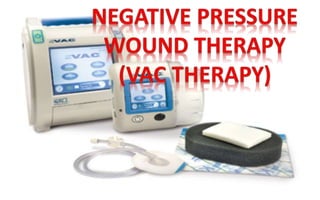
Negative pressure wound therapy
•
12 likes•951 views

negative pressure wound management by dr ,mohammad abdel ghany

Recommended
More Related Content
What's hot
What's hot (20)
Similar to Negative pressure wound therapy
Similar to Negative pressure wound therapy (20)
Recently uploaded
Recently uploaded (20)
Negative pressure wound therapy
- 1. intro
- 2. Dr. Mohammed Abdel Ghany Ismail MD plastic and reconstructive surgery El-mehala Plastic Surgeons Club
- 3. Definition • New theraputic technique using subatmospheric pressure or vacuum-assisted drainage to remove fluid from a wound surface • A sponge or foam interface is used to distribute the negative pressure allover the wound surface
- 4. History • Started 1989 USA California • Released 1994 in Europe • Published 1997;Annals of Plastic Surgery (175 casas)
- 5. Principles and Mechanism Exact unknown, but theories: 1. Fluid-based mechanism 2. Mechanical mechanism 3. Reduction of bacterial load
- 6. 1- Fluid-based mechanism Removes excess interstitial fluid • Improves microcirculation and oxygen delivery • Allows transport of nutrients • Removal of waste products &local toxins in wounds • (proteolytic enzymes, acute phase proteins, metalloproteinases,proinflammatory mediators, and cytokines)
- 7. 2- Mechanical mechanism Cellular response to mechanical tissue stress • Negative pressure causes the contact wound dressing to collapse • the force is transferred to the wound edges, drawing them closer together • pieces of tissue are drawn into contact dressing inducing mechanic alstress, stimulating angiogenesis and tissue growth.
- 8. Application • The wound is cleaned and necrotic tissue debrided • Nonadherent contact layer, Xeroform , between prepared wound bed and contact dressing • A piece of sponge is cut to fit the wound approximately • The sponge is placed within the wound and a drainage tube placed over it (if circumferential holes, another piece of sponge should be put over the top of the tube)
- 10. Application (cont.) • Adhesive transparent waterproof dressing (opsite) overlaps the surrounding skin, and a seal created with the skin and the drainage tube. • Suction tubing is attached to a cannister (through another tube). • The cannister slots into the VAC unit.
- 12. Variation • Contact dressing may be foam, sponge or gauze. • Transparent dressing is placed directly on the sponge; a hole is cut in it; and a suction tube with a flat adhesive end is stuck over the hole.
- 13. Pressure • Apply subatmospheric pressure. • Continuous or intermittent pressure application • Continuous with early wound+excess exudate • Intermittent pressure shown to produce more rapid granulation tissue deposition(5-2,5-1). • Pressure of 50 to 125 mm Hg applied. • Maximum increase in blood flow seen at 125 mm Hg.
- 15. Frequency of dressing changes • No less than three times a week for non- infected wounds • More frequently for infected wounds • Twice a week is suitable
- 16. Indications • Acute ,chronic, traumatic, and dehisced wounds. • Partial-thickness burns, ulcers (diabetic, pressure, venous stasis) • Flaps, and grafts
- 17. Acute wounds Large soft tissue injuries with compromised tissue, contaminated wounds, hematomas, gunshot wounds. 1. Debride wound of all nonviable tissue. 2. Remove foreign bodies. 3. Hemostasis. 4. Cover vital structures such as major vessels, viscera, and nerves with muscle or soft tissue. 5. If significant contamination or patient has signs of sepsis, change dressings at 24-hour intervals with repeated debridement .
- 18. Chronic wounds Pressure ulcers, long-term dehisced wounds, venous stasis ulcers, vascular and diabetic ulcers) • Debride wound of all nonviable tissue. • Converts a chronic wound into a subacute wound that responds more rapidly.
- 19. Contraindications 1. Exposed vessels, nerves, and organs 2. Malignancy in wound 3. Untreated osteomyelitis 4. Nonenteric or unexplored fistulas 5. Fresh anastomotic site 6. Necrotic tissue with eschar present TIP: Some surgeons may cover vessels, nerves, and organs temporarily using either a nonadherent contact layer or a white foam contact dressing on a case-by-case basis.
- 20. Precautions • Active bleeding • Anticoagulant • Difficult haemostasis • Near BV,organs; protect with fascia or barrier • Irradiated
- 21. Benefits 1. Protect the wound and prevent environmental contamination, and prevent wound desiccation. 2. Prepare wound bed for surgical closure or allow healing by secondary intention 3. Improve granulation tissue formation, manage infection and reduce wound size 4. Improve patient comfort: Decrease pain, decrease number of dressing changes 5. Reduce costs: Shorten time to closure or next additional surgery, minimize wound complications, and allow management of wounds as outpatient
- 22. Special Clinical Applications Extremities and Orthopedic Injuries • NPWT has become a first-line treatment for allowing definitive reconstruction to be performed in a stable, clean wound on an elective basis.(Streubel et al.,2012) Use of negative-pressure wound therapy in orthopaedic trauma
- 23. Values in Orthopaedic Trauma 1) Allows serial debridement of only nonviable soft tissue and bone, minimizing unnecessarily aggressive debridements. 2) Removes edema and increases perfusion. 3) Viable soft tissue is drawn together so that the wound does not enlarge with edema and retraction. 4) Bone is kept in a moist environment, minimizing desiccation.!! 5) Can be placed directly over hardware.!! 6) After fasciotomies, reducing edema and allowing primary closure sooner
- 24. Special Clinical Applications (cont.) Grafting Over Bone • NPWT may be used on surgically exposed cranial diploe or other bones by drilling at 1 cm intervals to bleeding level to partially remove the corex; • accelerates formation of granulation tissue for skin grafting.
- 25. Cases
- 29. 1 Vac therapy is not magic but may be a suitable solution for some difficult wounds that are not suitable or ready for reconstruction immediately
- 30. 2 Negative pressure wound dressing prepares the wounds well and changes a hard option to apply into simple one (free flap graft)
- 31. 3 It added a revolution in soft tissue rec. of foot and ankle because it has enabled surgeons to close wounds by simple technique that usually needed complex pedicled or microsrgical flap
- 32. 4 At last we need more cases of application in our own experience to judge this method efficiently