Downloaded 39 times
![DC/TMD Protocol 1: Introduction 5
1.4 How to cite this document
Citation of English language source
Cite this document as follows (with <italic text> denoting new text to be inserted):
Ohrbach R, Gonzalez Y, List T, Michelotti A, Schiffman E. Diagnostic Criteria for
Temporomandibular Disorders (DC/TMD) Clinical Examination Protocol: Version
02June2013. www.rdc-tmdinternational.org Accessed on <date>.
For example:
Ohrbach R, Gonzalez Y, List T, Michelotti A, Schiffman E. Diagnostic Criteria for
Temporomandibular Disorders (DC/TMD) Clinical Examination Protocol: Version
02June2013. www.rdc-tmdinternational.org Accessed on July 1, 2013.
Citation of translation of full document
Translated versions of this document should be cited as follows if the title is also
translated:
Ohrbach R, Gonzalez Y, List T, Michelotti A, Schiffman E. Diagnostic Criteria for
Temporomandibular Disorders (DC/TMD) Clinical Examination Protocol: Version
02June2013. [<Title in target language: <target language> Version 02June2013]
<Developer name or names>, Trans. www.rdc-tmdinternational.org Accessed
on <date>.
For example, if the instrument title was translated, the citation would look read as
follows:
Ohrbach R, Gonzalez Y, List T, Michelotti A, Schiffman E. Diagnostic Criteria for
Temporomandibular Disorders (DC/TMD) Clinical Examination Protocol: Version
02June2013 [Diagnostiska Kriterier för Temporomandibulär Dysfunktion
(DC/TMD) Kliniskt Undersöknings Protokoll: Swedish Version 02June2013]. List
T, Trans. www.rdc-tmdinternational.org Accessed on July 1, 2013.
Citation of translation of partial document
Translated versions of this document should be cited as follows if the title is not
translated:
Ohrbach R, Gonzalez Y, List T, Michelotti A, Schiffman E. Diagnostic Criteria for
Temporomandibular Disorders (DC/TMD) Clinical Examination Protocol: Version
02June2013 [in <target language>]. <Developer name or names>, Trans.
www.rdc-tmdinternational.org Accessed on <date>.
For example, if only the complete specifications were translated into Swedish while
retaining the title in English, the citation for the actual examination as performed would
read as follows:
Ohrbach R, Gonzalez Y, List T, Michelotti A, Schiffman E. DC/TMD Complete
Specifications for Examination: Version 02June2013 [in Swedish]. List T, Trans.
www.rdc-tmdinternational.org Accessed on July 1, 2013.
…and the user would likely cite the full Protocol as well since it specifies all other
aspects of the examination.](https://image.slidesharecdn.com/qwtp5n8wtzcafraw77vw-signature-d95bbf363b3219feeb75d250b5e2cf4e78e1dbfd2c7fe7186d561218c53aeafe-poli-150525021224-lva1-app6892/85/Www-unlock-pdf-com-dc-tmd-protocol-2014-06_02-5-320.jpg)

![DC/TMD Protocol 2: General Instructions 11
2. Lateral excursions are corrected by adding or subtracting the midline
discrepancy, as determined by the direction of any discrepancy between the
maxillary and mandibular midlines. If any midline discrepancy has been
negated via creation of an arbitrary reference midline, the principle remains
the same but the value that is added or subtracted is ‘0’.
3. Protrusive movements are corrected by adding the measurement of
protrusive movements to the horizontal overjet. When an anterior crossbite or
prognathic relationship occurs, the negative overjet is added to the protrusive
movement. And when a severe overjet (recorded as a positive number) is
accompanied by mandibular protrusion that remains posterior to the maxillary
teeth (recorded as a negative number), the sum of the two values correctly
indicates the extent of mandibular movement in the protrusive direction.
(d) Movement even with pain. With one exception [Pain-free opening], every time the
patient moves his/her mouth vertically or excursively, he or she is instructed to
move it as far as possible in that direction, even if it is painful.
(e) Movement without guidance. Movements for range of motion and for joint sounds
are made without any assistance by the examiner. Only for Maximum Assisted
Opening is the jaw actively pushed on by the examiner. The examiner can touch
the patient to provide non-verbal cues for the direction of intended movement.
2.6 Classification of Anatomic Structures
(a) Description of problem. When the patient points to the location where he or she
feels pain (e.g., by history or from the examination), it may be obvious as to which
structure is associated with the complaint or, if the patient points to either the
preauricular area or to the boundary of a structure – e.g., anterior area of the
masseter muscle – it may not be obvious. When pointing to the preauricular area,
the structures associated with a pain complaint may be joint, muscle, or both; when
pointing to the marginal area of a muscle, the structures associated with a pain
complaint may not be within the muscle tissue at all. The following procedures,
when consistently followed, will provide reliable identification of which structure
(muscle, joint, or other structure) is associated with the reported pain.
(b) Identify involved areas of pain. The first time that a participant points to a location
that is uncertain in terms of anatomical identity, the examiner will investigate the
possible underlying structures. Subsequent pointing by the participant to that
same location should allow the examiner to code the structure(s), based on the
initial inquiry.
1. Patient points to pain locations. When a patient indicates that a mobility test
is painful, the examiner determines the location(s) of the pain induced during
the procedure by asking the patient to point to each area of pain with one
fingertip or to show the area of involvement by “painting” the boundary with a
fingertip. The examiner then determines what area(s) the patient has
indicated and then records all of the selected locations (as available,
depending on the item): temporalis, masseter, sub-mandibular, posterior
mandibular, lateral pterygoid, temporalis tendon, and TMJ. These pain-site
locations are independent. If the patient reports the location of the pain as, for
example, “my joint”, the examiner will ask the patient to point to or show the
location.](https://image.slidesharecdn.com/qwtp5n8wtzcafraw77vw-signature-d95bbf363b3219feeb75d250b5e2cf4e78e1dbfd2c7fe7186d561218c53aeafe-poli-150525021224-lva1-app6892/85/Www-unlock-pdf-com-dc-tmd-protocol-2014-06_02-11-320.jpg)

![DC/TMD Protocol 2: General Instructions 16
For the purposes of the DC/TMD, “familiar” pain is defined as pain that is “similar”
or “like” the individual’s clinical pain, regardless of its intensity from the clinical
procedure vs the typical clinical pain, and experienced by the patient in the
reference anatomical region within the prior 30 days. Another way to define the
term, in the context of a clinical examination, is reproduction of the pain qualia in
response to clinical provocation. The examiner reminds the patient, as needed,
that the goal of the examination is to replicate (duplicate) the patient’s pain in order
to locate the source. “Familiar” pain is included as a criterion for a pain diagnosis
because replication of pain, in terms of differential diagnosis, is essential; the
necessary goal in establishing the presence of replication is for the patient to
describe the provoked pain in the same way as the pain of complaint, because it is
the same type of pain. Moreover, the “familiar” pain probe provides the examiner
an opportunity to more fully explore the patient’s pain experience by also inquiring
into the context, frequency, and location of any elicited pain symptoms in order to
determine their relevance to the chief complaint. The examiner then integrates this
information into the history in order to create a coherent set of findings that reduce
the discrepancy between reported symptoms vs signs and which supports the
diagnoses.
(b) “Familiar pain” probe. For any procedure that produces pain, the examiner asks as
a follow-up probe whether that pain is familiar to the pain that the patient may have
experienced in this region in the past 30 days. A “yes” response is followed by the
probe, “Familiar to what?” in order to elicit a description of the context in which the
pain experience occurred; one type of response points to an event associated with
the pain (e.g., chewing gum). Clinical patients who experience pain much or all of
the time will more often respond to the “familiar to what?” probe with a response
such as “the pain I am seeking care for”.
(c) Time period for pain. For DC/TMD pain diagnoses, the standard time period for a
diagnosis is pain within the prior 30 days. This means that for Familiar Pain to be
endorsed as [yes], the pain must have occurred within the prior 30 days; pains that
occurred previous to the recent 30-day period are not acceptable for Familiar Pain
at the time of the examination per the DC/TMD 30-day time period and are
recorded as [no]. Certain protocols or specific clinical situations may require that
this time period be modified, but that should be documented in the clinical record.
And, it should be carefully explained in any publication that cites the methods being
used. Note that the ICHD-2 criteria require that headache be present within the
prior 3 months in order for the disorder to be classifiable. In certain settings, the
user may way to modify the time period for headache pain accordingly.
(d) Complicating factors. There are three issues which affect the patient’s report
regarding possible familiar pain: location, temporal characteristics, and intensity.
Location and temporal characteristics are readily addressed through appropriate
probes, while the intensity characteristic is more complex.
1. Location characteristics. For unilateral pain complaints (that is, a patient is
asymptomatic on one side and symptomatic on the other side within the past
30 days), if the patient says that procedure-induced pain on the
asymptomatic side is “familiar”, the examiner should probe as follows:
i. verify side(s) and specific locations of symptoms in the past 30 days;
ii. verify that the familiar pain within a side that had been reported as
asymptomatic (E1) occurred within the past 30 days and verify that the
pain was familiar;](https://image.slidesharecdn.com/qwtp5n8wtzcafraw77vw-signature-d95bbf363b3219feeb75d250b5e2cf4e78e1dbfd2c7fe7186d561218c53aeafe-poli-150525021224-lva1-app6892/85/Www-unlock-pdf-com-dc-tmd-protocol-2014-06_02-16-320.jpg)
![DC/TMD Protocol 2: General Instructions 18
2.11 Examination-related Pain Interview
(a) Interview structure. There is a standard set of questions related to the
examination-based interview of presence of pain, whether it is familiar pain,
whether it is familiar headache, and where the pain is perceived. The inquiry into
pain starts with an examination-specific question based on the particular
procedure. This initial question is prompted after each relevant procedure in the
Specifications; the specific questions for each type of clinical procedure are listed
at the beginning of Section 5. The pain interview is comprised of a standard
hierarchy of questions. Additional questions are needed if the patient responds in
other ways, and such questions should follow the intent of the specific questions
described here.
(b) Ambiguous responses from patient. When requested, the patient must clearly
indicate “pain” or “no pain”. If the patient provides other descriptors (e.g., achiness,
tightness, pressure, uncomfortable, etc), the examiner will clarify this with “Is that
pain or not?” If headache is a primary focus of the examination, then this question
may need to be modified, such as “Is that headache pain or not?” No other
question is provided for addressing this particular ambiguity, as any other question
will tend to have leading characteristics. For other forms of ambiguity, the intent is
to clarify and not to lead the patient towards making specific responses.
(c) Repeat questioning. A simple question such as “Did you feel pain [from that
procedure]?” may be shortened after the first few uses of it, to “Pain?”. Examiners
are encouraged to develop shorthand versions of the repeated items within the
Examination-related Pain Interview for use with a given patient after the patient
understands the intent of the question. Another example is the repeat question of
“Show me where you felt pain” followed by the question “Were there any other
areas?”; the latter question is repeated until the patient says “no”. The first time
that this form of questioning occurs, such as in E1 (Pain location), is an opportunity
to review with the patient that inclusive reporting of all pain locations is requested.
(d) Efficient Completion of the Examination-related Pain Interview. After the first
several positive responses from the pain provoking procedures, the examiner can
instruct the patient to respond in an abbreviated form. For example, the patient
might be instructed to report, in response to positive palpation findings, as follows:
“yes, familiar” or “yes, not familiar”.
2.12 Examination Form
(a) Overview. The DC/TMD includes an examination form which standardizes the
examination and the recording of findings. This form, Section 9 and available on
the website as a stand-alone document, is inclusive of all procedures described in
this protocol. The form is available in versions for the US and for international
settings.
(b) Completion of all items. Unless a procedure is indicated as supplementary, the
procedure should be completed. Completion of a procedure does not mean that all
response bubbles have to be completed (see next section for further explanation).
Rather, the form is intended to be a record of what is normal as well as abnormal,
hence reporting negative findings (as [N]) is as important as reporting positive
findings. Completion of all items relevant to a given complaint also supports
reliable decision-making. Procedures noted as “supplementary” are used as
indicated by the clinical circumstance. If the examination form is used for a](https://image.slidesharecdn.com/qwtp5n8wtzcafraw77vw-signature-d95bbf363b3219feeb75d250b5e2cf4e78e1dbfd2c7fe7186d561218c53aeafe-poli-150525021224-lva1-app6892/85/Www-unlock-pdf-com-dc-tmd-protocol-2014-06_02-18-320.jpg)
![DC/TMD Protocol 2: General Instructions 19
screening examination, then only items indicated for the type of screening
examination would be completed.
(c) Form structure logic. While all items should be completed, not all response fields
need to be completed. Parts 1, 2, and 3 are typical checklists. The remaining
sections of the examination utilize a conditional reporting format.
1. If the response from the initial inquiry about presence of pain from the
procedure is reported by the patient as “no”, then record [N]. All other
response options for that item are left blank since no response is needed.
2. If initial inquiry response about presence of pain is “yes”, the examiner
records [Y] to Pain. Then the examiner will ask a series of further questions
depending on the type of examination procedure that induced the pain.
3. Familiar Pain and Familiar Headache co-exist as parallel forms of inquiry
because some individuals will report temporalis area “pain” as different from
temporalis “headache” with respect to an examination procedure provoking
that particular pain or headache experience. This was addressed in section
2.9.
4. A response for Referred Pain is recorded if Pain is “yes” and independent of
whether Familiar Pain or Familiar Headache are “yes”.
5. Example. In Part 4B, if the patient reports pain only in the right temporalis
and right masseter during maximum unassisted opening, [Y] would be
marked in the Pain column for the right temporalis and the examiner would
proceed with the Examination-related Pain Interview, asking if the pain was
familiar, if the provoked pain (or noxious experience) was familiar to
headache, and if the pain was referred. For the masseter muscle, the
Examination-related Pain Interview would address if the pain was familiar and
if the pain was referred. For all other muscles and both TMJs, [N] is marked
in the Pain column and because the remaining columns are not applicable,
they are left blank.
(d) Description of the form structure. The conditional sections of the examination form
are as follows.
Parts 4 and 5 use the conditional structure as described above under (c), and
illustrated with the example at (c)5.
In Parts 6 and 7, mandatory response fields are listed under Examiner and Patient.
The examiner section and the patient-report section are separated by a vertical
line. The patient-report section starts the conditional response fields which are
completed only if the patient reports ‘yes’ (recorded as [Y]) to the presence of a
clicking sound; the examiner then begins the Examination-Related Pain Interview
relevant to joint sounds.
In part 8, Locking is reported as an observed event during any part of the
examination, and the response fields for Reduction are completed. If Locking is
reported as “no”, then the remaining fields for that type of locking in that joint are
left blank.
In Parts 9 and 10, if Pain to palpation is not reported, then [N] is marked and the
remainder of the row for that muscle is left blank. If Pain to palpation is reported,
then a prolonged palpation is performed and the Examination-related Pain](https://image.slidesharecdn.com/qwtp5n8wtzcafraw77vw-signature-d95bbf363b3219feeb75d250b5e2cf4e78e1dbfd2c7fe7186d561218c53aeafe-poli-150525021224-lva1-app6892/85/Www-unlock-pdf-com-dc-tmd-protocol-2014-06_02-19-320.jpg)
![DC/TMD Protocol 3: Description of Examination Procedures 26
of this particular procedure. Ask the patient about any pain produced by this
procedure.
E4_D Opening Terminated. If patient indicates to the examiner that the procedure should
be stopped, [Yes] is endorsed. If the patient indicates for the procedure to be
terminated simultaneous with the examiner’s percept that the assisted opening
has reached its maximum, [No] should be endorsed. For all other situations, [No]
should be endorsed.
E5 Lateral and Protrusive Movements
Rationale. Excursive movements complement open movements for full assessment
of jaw mobility. These measurements are supplemental and may be omitted.
The rationale for assessing lateral movements is to document the extent of the
excursive movement(s) and any resultant (induced) in pain. Moreover, in certain
settings, measurement of excursive movements serves to document if condylar
movement was limited versus normal. The examination form provides fields for
recording the extent of movement.
General. Lateral excursive measurements are made between the maxillary and
mandibular reference midlines, while protrusive excursive measurement is made
between the labial surfaces of the maxillary and mandibular reference teeth. If
the patient cannot perform a movement, indicate this on the recording form by
leaving the section blank. For the lateral excursive movements, if the patient is
confused about direction s/he should move his or her jaw, touch the ipsilateral
side of the face, lip, or even shoulder, and ask the patient to move towards the
indicated side. Lateral pressure to the jaw by the examiner, as an aid to help the
patient move in the requested direction, is very difficult to calibrate and
consequently is discouraged.
E5_A Right Lateral Excursion. Ask the patient to move mandible to the patient’s right.
Record any reported pain.
E5_B Left Lateral Excursion. Ask patient to move mandible to the patient’s left. Record
any reported pain.
E5_C Protrusion. Ask patient to move the mandible forward. Record any reported pain.
Note that if the mandibular incisors cannot be protruded beyond the maxillary
incisors, the value will be negative; the form should be marked for the negative
number. If the incisors exhibit a Class III situation in maximum closure, the
horizontal overlap is recorded as a negative value (as explained in E2), but the
protrusive movement, still measured as the distance from the labial surfaces of
the maxillary to mandibular incisors, will be recorded as a positive number
(consistent with the Class I situation where the mandibular incisor is anterior to
the maxillary incisor).
E6 TMJ Noises During Open & Close Movements
Rationale. TMJ noises are a classic sign associated with TMD. However, because
TMJ noises are often not stable over time, clinical diagnosis of intracapsular
disorders is at best fair except for displacement without reduction, with limited
opening. Because patients are often concerned about joint noises, the
assessment of TMJ noise remains part of the clinical examination.](https://image.slidesharecdn.com/qwtp5n8wtzcafraw77vw-signature-d95bbf363b3219feeb75d250b5e2cf4e78e1dbfd2c7fe7186d561218c53aeafe-poli-150525021224-lva1-app6892/85/Www-unlock-pdf-com-dc-tmd-protocol-2014-06_02-26-320.jpg)
![DC/TMD Protocol 3: Description of Examination Procedures 27
General. Since the 1992 publication of the RDC/TMD, much research regarding joint
sounds has been published, and the Validation Project attempted to improve
upon joint sound characterization and assessment. While the assessment
method has been improved with the recognition of assessing pain with the
sound, the definition of the joint sounds is nevertheless the same as in the
RDC/TMD (1992).
Instructions and Palpation. The examiner explains that the jaw joints (TMJ) will be
examined for whether they make any noise while the patient moves his or her
jaw, and the patient is asked to report any noise that was felt or heard. Palpation
is generally performed by placing one finger on the skin overlying the lateral pole
of the TMJ condyle, and light palpation pressure is used.
Examiner Detection of Noises. Using palpation, the examiner will determine if joint
noises are present during opening and closing. The patient will also report
whether they heard or felt a joint sound. It is essential that the patient bring the
posterior teeth into MICP before each open-close cycle. This maneuver insures
that the full range of opening and closing has been assessed. Joint noises can
be assessed either unilaterally or bilaterally, depending on circumstance.
Bilateral assessment is sometimes essential in order to determine if the noise
from a single click is being conducted via bone to both joints. In contrast, if the
patient report of noises is clinically important, then patients typically do better by
focusing on one joint at a time. Sometimes, tooth contact can be sufficiently
“loud” or noticeable such that the sound can be misperceived by the examiner as
a joint noise. In order to control for this, ask the patient to intentionally tap the
teeth together lightly before starting the open-close procedure in order to
establish a reference.
Definitions of noises.
(a) Click. A distinct noise, of brief and very limited duration, with a clear
beginning and end, which usually sounds like a “click”. Also referred to as
a snap or pop.
(b) Crepitus. A noise that is continuous, over a longer period of jaw movement
than a click or pop and can occur during part or the whole of the opening
and/or closing movement. The noise is not muffled, and it may be
comprised of multiple overlapping grating sounds such that it becomes
“continuous”; distinguish this from the discrete sound characteristics
associated with a click. Such joint noise is also often referred to as
crunching, grating, or grinding sounds.
(c) Eminence click. The eminence click has to include at least an opening
click and is detected when the condyle-disk complex translates around the
eminence accompanied by a bodily shift of the mandible. The examiner
observes for noise near the end-range of normal range of movement (i.e.,
at the end of normal-range opening or beginning of closing from a normal-
range maximal opening). Noise detected at the end-range of vertical jaw
movement that is limited is not likely representative of an eminence click.
An eminence click is not reported on the examination form; it is identified
only to distinguish it from the “click” that is reported.
Recording joint noises. Only clicking noises that meet the following criteria are
scored.
(a) Opening click. If from MICP to maximum opening, a click is noted on at
least one of three opening movements, record Open Click as [yes].](https://image.slidesharecdn.com/qwtp5n8wtzcafraw77vw-signature-d95bbf363b3219feeb75d250b5e2cf4e78e1dbfd2c7fe7186d561218c53aeafe-poli-150525021224-lva1-app6892/85/Www-unlock-pdf-com-dc-tmd-protocol-2014-06_02-27-320.jpg)
![DC/TMD Protocol 3: Description of Examination Procedures 28
(b) Closing click. If from maximum opening to MICP, a click is present on at
least one of three closing mandibular movements, record Close Click as
[yes].
(c) Crepitus. Crepitus can be scored in addition to a click.
(d) None. Indicates that neither click nor crepitus were present during
opening, closing, or both; this is scored as a [no] for each of Click and
Crepitus in this examination.
Patient report of joint noises with movement. Any sounds that the patient
perceives during any part of the joint noise evaluation are recorded separately for
each of right and left joints. When the examiner has completed assessment of
joint noise, the patient is asked if s/he felt or heard any joint noise. If the patient
says “yes”, the examiner follows with identification of type and side of all noises.
If the patient reports distinct sounds such as clicking, popping or snapping
sounds, these are coded as a [click] on the form. If the patient reports longer
duration sounds including crunching, grinding or gratings sounds, these are
coded as [Crepitus] on the form. Otherwise, [No] is coded for that side. If the
patient uses terms other than those listed above under “Definitions”, the
examiner then asks for a description rather than assuming that a particular term
necessarily refers to a specific sound such as “click” or “grating” and then
determines the type of sound. A sound that the patient describes need not
necessarily map to [click] or [creptitus] and such noises are most likely recorded
as [No].
Pain inquiry. The patient is asked about pain that occurs at the same time as the
clicking noise. Opening itself may cause pain, so the examiner should not
assume that a “yes” response to this inquiry necessarily indicates that the click
itself was painful.
E7 TMJ Noises During Lateral & Protrusive Movements
Rationale. This test is an extension of the evaluation of TMJ noises during opening
and closing movements.
Procedures. The examiner asks the subject to move his or her mandible to the right,
to the left, and protrude just as previously performed when measuring the extent
of these movements. Make sure the subject closes into MICP prior to and at the
end of each movement, as with opening/closing noise assessment. A minimum
of three movements is required. A noise is scored as positive any time it occurs
during the lateral or protrusive movements (out or in). After the movements are
completed, then assess for "Pain with click" and "Familiar pain" for any clicks
reported by the patient. The definitions of the noises are the same as for TMJ
Noises During Open and Close Movements.
E8 Joint Locking
Rationale. Joint locking in the clinic is uncommon but it does occur. Documenting
whether locking occurs or not is a useful function within the examination, given
the associated pain, disability, and treatment complexity that can be associated
with joint locking.
Closed lock procedures. If the patient’s mandible suddenly locks during the process
of opening, then a lock “While opening” would be recorded as [yes]. If the lock is](https://image.slidesharecdn.com/qwtp5n8wtzcafraw77vw-signature-d95bbf363b3219feeb75d250b5e2cf4e78e1dbfd2c7fe7186d561218c53aeafe-poli-150525021224-lva1-app6892/85/Www-unlock-pdf-com-dc-tmd-protocol-2014-06_02-28-320.jpg)
![DC/TMD Protocol 3: Description of Examination Procedures 29
self-reducing or the patient engages in a specific maneuver to unlock the
mandible, then the patient effected a reduction (code as [yes]), whereas if the
examiner has to reduce the lock, then the examiner should be coded as [yes]
and the patient as [no].
Open lock procedures. If the patient’s mandible becomes locked in the wide open
position, then that type of lock is recorded as [yes]. Recording of the reduction of
the lock is the same as described for the Closed lock procedures (described just
previously).
E9 Muscle and TMJ Pain with Palpation
Rationale. Pain induced in muscles via palpation is a classic clinical test. The intent
is to determine if the patient reports pain from palpation of a muscle or joint and
determine if any induced pain duplicates or replicates the patient’s pain
complaint. Several approaches are available, depending on the purpose of the
examination. See Part 2.12.e for description of the examination form pertinent to
temporalis and masseter palpation.
General
(a) Palpating the muscles and joint capsules for pain requires that the examiner
press on a specific site using the spade-like pad of one finger (the second or
third digits; or index finger or middle finger) with standardized pressure. The
“spade-like” area of the finger is the space between the tip (just adjacent to the
edge of finger nail) and the finger-print area. One finger is used and finger
placement is as shown in the illustrations, Section 7.
(b) Within each group of muscle sites as indicated on the examination form, apply
pressure to the right side and then to the left side. When applying pressure,
support the head or mandible by placing the other hand on the opposite side of
the head in order to provide stability. During palpation, the patient's mandible
should be in a comfortable position, without the teeth touching.
(c) Each major muscle (temporalis, masseter) is divided into 3 zones for purposes
of insuring that the muscle is examined in a consistent manner. Responses
may be recorded for each zone or for the muscle as a whole; alternate
examination forms exist for each approach. Within each zone, 3-5 areas should
be palpated, depending on the size of the muscle and the intent of the
examination.
(d) Due to the inherent difficulty in an examiner exactly calibrating his/her finger
pressure when going from measurement scale to actual palpation, the examiner
should target no less than the indicated value. For example, a target of 1 kg
should be at least as great as 1.0 kg but not less. Target “pressures” are 1 kg
(as measured by a force meter) to the temporalis and masseter muscles and
around the lateral pole of the TMJ and 0.5 kg to the lateral condylar pole of the
TMJ and any supplemental muscle areas
(e) If the examiner is unable to palpate at the specified amount of pressure due to
the patient’s physical withdrawal or the patient’s request for the palpation to be
more gentle, then such information is recorded in the Examination Comments
section, and the palpation should be modified accordingly.
(f) In a given setting, diagnosis of myalgia may be the sufficient end-point and
further investigation for presence of referred pain may not be needed. If so, the](https://image.slidesharecdn.com/qwtp5n8wtzcafraw77vw-signature-d95bbf363b3219feeb75d250b5e2cf4e78e1dbfd2c7fe7186d561218c53aeafe-poli-150525021224-lva1-app6892/85/Www-unlock-pdf-com-dc-tmd-protocol-2014-06_02-29-320.jpg)
![DC/TMD Protocol 3: Description of Examination Procedures 31
examination for referred pain was not performed. However, in practice 5-
seconds of pressure is tolerable.
(e) Method 2. Use 2 seconds duration for every palpation. If identification of
referred pain is not important in a given examination or setting, then palpation
with only 2 seconds duration will be generally sufficient to identify myalgia and
arthralgia. This approach is particularly well-suited if the clinical evaluation
serves as a gateway for referral. Instructions include: Now I am going to apply
pressure to different areas of your head and jaw, and I will ask about pain. And: I
will hold the pressure for 5 seconds [or 2 seconds], and I will then ask you about
pain; please tell me yes or no, and if so I will then ask you if the pain is familiar,
and if the pain stays under my finger or if you feel it also in different areas of your
head or jaw.
Description of Specific Extraoral Muscle Sites
(a) Temporalis Anterior – 1.0 kg. Start just posterior to the bony crest lateral to the
eyebrow and superior to the zygomatic process of the temporal bone. Request
muscle contraction via patient clenching as necessary in order to insure that
muscle tissue is beneath the finger. The area for palpation lies along a curve
parallel to the anterior extent of the muscle; lightly palpate for the bony crest
defining the anterior boundary of the temporal fossa.
(b) Temporalis Middle – 1.0 kg. Start just anterior of the ear and superior to the
zygomatic process of the temporal bone; the area for palpation is directly
superior.
(c) Temporalis Posterior – 1.0 kg. Start just above the superior tip of the ear; the
area for palpation is directly superior. Ask the patient to clench and then relax
to help identify muscle boundaries, as necessary.
(d) Origin of Masseter – 1.0 kg. Request that the patient first clench and then relax
in order to confirm (1) the location of the posterior extent of the muscle with
respect to the anterior border of the TMJ condyle and (2) the anterior border of
the masseter. Start at the posterior extent, just inferior to the zygomatic
process of the temporal bone; the area for palpation is directly anterior.
(e) Body of the Masseter – 1.0 kg. Start at the posterior boundary of the muscle,
midway between origin and insertion. The area for palpation is directly anterior.
(f) Insertion of the Masseter – 1.0 kg. Start at the posterior boundary of the
muscle, just superior to the inferior mandibular border; the area for palpation is
directly anterior.
Description of Specific Joint Palpation Sites.
(a) Lateral Pole – 0.5 kg. Place index finger just anterior to the tragus of the ear
and on the skin overlying the patient's TMJ. In order to confirm location, ask the
patient to open or protrude slightly until the examiner feels the lateral pole of the
condyle translated forward.
(b) Around the Lateral Pole – 1.0 kg. While the mandible is in the comfort position
or in a slightly protruded position, place index finger just anterior to the tragus of
the ear and dorsal to the TMJ. While the mandible is supported from the other
side, the index finger presses while orbiting around the lateral pole in a circular
fashion over the superior aspect of the condyle and then anteriorly – i.e., from
the 9:00 to the 3:00 position, and then continuing fully around the condyle. Two-](https://image.slidesharecdn.com/qwtp5n8wtzcafraw77vw-signature-d95bbf363b3219feeb75d250b5e2cf4e78e1dbfd2c7fe7186d561218c53aeafe-poli-150525021224-lva1-app6892/85/Www-unlock-pdf-com-dc-tmd-protocol-2014-06_02-31-320.jpg)
![DC/TMD Protocol 3: Description of Examination Procedures 32
five seconds duration for this procedure yields the appropriate pace of finger
movement.
Examiner calibration. The examiner calibrates the necessary fingers at the specified
palpation “pressure”, just prior to that set of palpations.
Examination sequence. A practice trial is optional and may be performed on the
deltoid muscle or frontalis muscle. Figures related to palpation are shown in Section
7. In order to maximize examiner consistency, the following sequence is used:
• Temporalis and masseter, right side and then left side (1 kg), following
order of zones as listed above.
• Lateral pole of TMJ, right side and then left side (0.5 kg)
• Around the TMJ condyle, right side and then left side (1 kg)
• Supplemental muscles (0.5 kg), as indicated.
Palpation and pain inquiry. The examiner inquires about pain from the palpation,
whether the pain is familiar, and the anatomical extent of the pain. For the latter,
the examiner asks the patient to show (with a fingertip) where the pain was felt.
Verbal responses such as “to my ear” or “deep” are accepted as evidence of
extension of the palpation-induced pain, while ambiguous responses from the
patient are interpreted as [no]. This protocol requires that “referral” denotes pain
felt in a different structure than that which is being examined via palpation.
E10 Supplemental Muscles
Rationale. Additional sites may be examined as per the protocol in E9.
E11 Examiner Comments
Rationale. The examiner records any observations that are deemed pertinent towards
understanding any findings. Any barriers that compromised any part of the
examination are also recorded here; see Section 2.4(d) for details. Any clinical
observations that may affect diagnosis should be recorded here as well.](https://image.slidesharecdn.com/qwtp5n8wtzcafraw77vw-signature-d95bbf363b3219feeb75d250b5e2cf4e78e1dbfd2c7fe7186d561218c53aeafe-poli-150525021224-lva1-app6892/85/Www-unlock-pdf-com-dc-tmd-protocol-2014-06_02-32-320.jpg)

![DC/TMD Protocol 5: Complete Specifications for Examination 42
5 Complete Specifications for DC/TMD Examination
5.1 Overview
The Complete Specifications are intended to facilitate maximal reliability in clinical
technique for the researcher and clinician.
The verbal commands and associated procedures used for each component of the
clinical examination are listed in a table format for ready reference. The reader should
refer to Section 2 (General Instructions) and Section 3 (Description of DC/TMD
Examination Procedures) for further details regarding these procedures. Figures, as
referenced below, are found in Section 7. The enumeration of the examination
procedures in this section corresponds to the enumeration in Section 3 and to the
DC/TMD Examination Form (Section 9). Section 6 contains the Examination-Related
Pain Interview, which is repeatedly referenced in the below Protocol. Section 8 lists only
the required verbal commands (See 5.1, Conventions for clarification), which serve two
purposes: facilitate learning the core component of the structured examination, and the
required commands are the only part of the DC/TMD Clinical Examination Protocol that
must be translated for use of the examination specifications in another language.
5.2 Conventions for Section 5.3
1. “Verbal Commands” as used by the examiner are of four forms:
a. Bold text identifies verbal commands that should be stated verbatim by the
examiner.
b. Non-bold text identifies verbal commands or statements for which strict
implementation is not expected. The examiner should follow the intent of the
command or statement and convey that intent to the patient.
c. [Square-bracketed text] denotes optional commands.
i. “Place your mouth in a comfortable position [with your back teeth apart]” refers
to a standard reference position by the patient which is required prior to most
examination procedures. This command is used contingently on what the
patient does. If the patient automatically returns his/her mandible to a
“comfortable position” after completion of a procedure, then nothing more
needs to be done by the examiner. Otherwise, the examiner should use the
command. It is included with each set of procedures in order to remind the
examiner that the next procedure takes as its starting point this neutral position.
ii. All other optional commands address common situations and should be used
as needed depending on the patient response during the examination
procedure.
d. <Angle-bracketed text> identifies instructions to the examiner.
2. Italicized text denotes comments and clarifications regarding verbal commands;
overlap between the comments and clarifications in this section and that in Sections
1 and 2 is intentional.
3. ALL UPPERCASE TEXT under “Verbal Commands” or “Examiner Procedures”
denotes conditional instructions.
4. |Response options| are placed between vertical bars.](https://image.slidesharecdn.com/qwtp5n8wtzcafraw77vw-signature-d95bbf363b3219feeb75d250b5e2cf4e78e1dbfd2c7fe7186d561218c53aeafe-poli-150525021224-lva1-app6892/85/Www-unlock-pdf-com-dc-tmd-protocol-2014-06_02-42-320.jpg)
![DC/TMD Protocol 5: Complete Specifications for Examination 43
5.3 Specifications
CONSTRUCT VERBAL COMMAND EXAMINER PROCEDURE
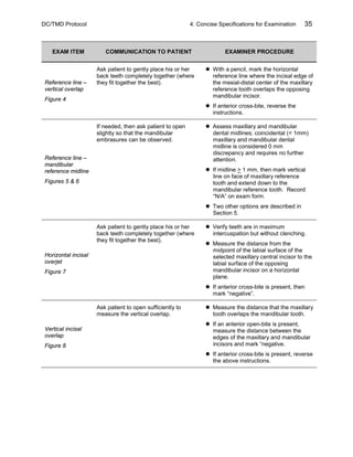
E1. Examiner Confirmation of Pain and Headache Locations
Identifying
information
<none> Examiner enters patient name, examiner
name, and date on examination form.
Instructions to the
patient
Before I start the exam, I want to
review a few things with you.
I will be asking you about pain, and
only you know if you have pain.
When I ask about pain, I want you to
say either yes or no; if you are not
sure, give me your best answer.
If you feel pain, I will also ask if that
pain is familiar. Familiar pain refers
to pain that is similar or like the pain
you may have had in that same part
of your body in the last 30 days.
If you feel pain in the temple area, I
will ask if that pain is like any
headache you may have had in the
temple area in the last 30 days.
Pain as defined here is absolute but
translation into local terms (or other
language) requires attention to cultural
standards. Intention is to clearly place
responsibility for determination of pain
on the patient, and the only response
that can be accepted is either “yes” or
“no”.
Definition of “familiar pain” may require
some elaboration when it is first asked
during the examination. Other related
words include “similar” or “feels like”.
Scope of
examination:
anatomic areas
of interest
Figure 1
For the purposes of this
examination, I am interested in pain
that you may have in these areas….
…. and also inside the mouth.
Examiner touches, bilaterally at the
same time, the following 4 areas in
sequence: temporalis, preauricular,
masseter, and
posterior/submandibular areas.
Examiner says “here” while touching
each of the above areas.
The areas are not named anatomically
as they are touched.
E1a
Location of pain:
last 30 days
Figures 2 & 3
In the last 30 days, have you had
pain in these areas [that I touched]?
IF “YES”:
Could you point with your finger to
each of the areas where you have felt
pain [in the last 30 days]?
Are there any other areas where you
have felt pain [in the last 30 days]?
IF “YES”, EXAMINER CONFIRMS:
Let me confirm where you just pointed.
IF PATIENT REPORTS NO PAIN IN THE
AREAS OF INTEREST:
Record “None” for each of right side
and left side in Q1a.
IF PATIENT REPORTS PAIN IN THE
AREAS OF INTEREST:
Examiner inquires into all locations.
Examiner touches involved areas to
confirm location with patient and
inquires “here?”
Record pain locations in Q1a.](https://image.slidesharecdn.com/qwtp5n8wtzcafraw77vw-signature-d95bbf363b3219feeb75d250b5e2cf4e78e1dbfd2c7fe7186d561218c53aeafe-poli-150525021224-lva1-app6892/85/Www-unlock-pdf-com-dc-tmd-protocol-2014-06_02-43-320.jpg)
![DC/TMD Protocol 5: Complete Specifications for Examination 44
CONSTRUCT VERBAL COMMAND EXAMINER PROCEDURE
E1b
Location of
headache in the
last 30 days.
In the last 30 days, have you had any
headaches?
IF “YES”:
Could you point with your finger to
each of the areas where you have felt
headaches [in the last 30 days]?
Are there any other areas where you
have felt headaches [in the last 30
days]?
IF “YES”, EXAMINER CONFIRMS:
Let me confirm your headache locations
where you just pointed.
IF PATIENT REPORTS NO HEADACHE
OR NO HEADACHE IN INDICATED
AREAS:
Record “None” for each of right side
and left side in Q1b.
IF PATIENT REPORTS HEADACHE:
Examiner inquires into all locations.
Examiner touches involved areas to
confirm location with patient, and
inquires “here?”.
Record pain locations in Q1b.
E2. Incisal Relationships
Select maxillary
and mandibular
reference teeth
In order to visualize the teeth
Open slightly.
I will place some pencil marks on your
teeth; I will remove them at the end of
the examination.
The potential maxillary and mandibular
reference teeth need to be visualized
at the same time as they are selected
jointly.
Choose maxillary right central incisor
(US #8; FDI #11) if the incisal edge is
horizontal, the tooth is vertically
oriented, and the tooth is not rotated;
else, select tooth US#9 (FDI #21) if it
better fits these criteria. See Section 3,
E2, for further instructions. Enter
selected tooth on the examination
form.
Note that the mesial-distal center of the
maxillary reference tooth will be the
specific maxillary reference position for
all vertical and protrusive mobility
measurements.
Select mandibular reference tooth,
which opposes the mesial-distal center
of the maxillary reference tooth.
The location of the incisal edge of the
mandibular reference tooth that is
opposite the mesial-distal center of the
maxillary reference tooth represents
the mandibular reference position for
all vertical mobility measurements.
The buccal surface of the mandibular
reference tooth that is opposite the
mesial-distal center of the maxillary
reference tooth represents the
mandibular reference position for
protrusive mobility measurements.](https://image.slidesharecdn.com/qwtp5n8wtzcafraw77vw-signature-d95bbf363b3219feeb75d250b5e2cf4e78e1dbfd2c7fe7186d561218c53aeafe-poli-150525021224-lva1-app6892/85/Www-unlock-pdf-com-dc-tmd-protocol-2014-06_02-44-320.jpg)
![DC/TMD Protocol 5: Complete Specifications for Examination 45
CONSTRUCT VERBAL COMMAND EXAMINER PROCEDURE
If anterior cross-bite or open bite, then
specific measures will also include
marking the “negative” field on the
exam form.
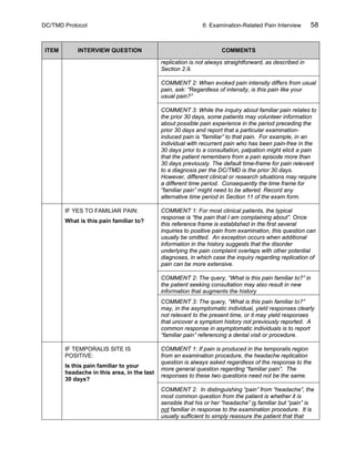
Reference line –
vertical overlap
Figure 4
Place your back teeth completely
together.
With a pencil, mark the horizontal
reference line where the central portion
of the incisal edge of the maxillary
reference tooth overlaps the opposing
mandibular incisor.
If anterior cross-bite, reverse the
instructions.
Reference line –
mandibular
reference midline
Figures 5 & 6
[Place your back teeth completely
together.]
<If needed, then ask patient to open
slightly so that the mandibular
embrasures can be observed.>
Assess maxillary and mandibular
dental midlines; if the discrepancy
between the maxillary and mandibular
dental midline is < 1mm, then record ‘0’
mm.
If the discrepancy in the midline is > 1
mm, select one of the following:
Method 1: Measure the distance of any
discrepancy (> 1mm) in the frontal
plane between the maxillary and
mandibular dental midlines and note
the direction of the discrepancy of the
mandibular midline relative to the
maxillary midline. The mandibular
dental midline is now the reference for
measuring lateral mandibular
movement.
Method 2: Draw a vertical line on the
face of the maxillary central incisor and
extend the line to the opposing
mandibular incisor. The vertical pencil
marks on the two incisors are now the
reference midlines.
Method 3: Extend, using a vertical
pencil mark, the maxillary dental
midline onto the corresponding
mandibular incisor. The vertical pencil
mark on the mandibular incisor is now
the reference midline for the mandible.
If anterior cross-bite, reverse the above
instructions with regard to maxillary
and mandibular.
If using Method 1, record the
measurement and direction.
If using Methods 2 or 3, record the
value ‘0’.](https://image.slidesharecdn.com/qwtp5n8wtzcafraw77vw-signature-d95bbf363b3219feeb75d250b5e2cf4e78e1dbfd2c7fe7186d561218c53aeafe-poli-150525021224-lva1-app6892/85/Www-unlock-pdf-com-dc-tmd-protocol-2014-06_02-45-320.jpg)
![DC/TMD Protocol 5: Complete Specifications for Examination 46
CONSTRUCT VERBAL COMMAND EXAMINER PROCEDURE
Horizontal incisal
overjet
Figure 7
[Place your back teeth completely
together.]
Verify teeth are in maximum
intercuspation.
Measure the distance from the
mesiodistal midpoint of the facial
surface of the selected maxillary
central incisor to the facial surface of
the opposing mandibular incisor on a
horizontal plane.
If anterior cross-bite is present, then
mark “negative”.
Record the measurement.
Vertical incisal
overlap
Figure 8
<Ask patient to open sufficiently to
measure the vertical overlap.>
Measure the distance that the maxillary
tooth overlaps the mandibular tooth.
If an anterior open-bite is present,
measure the distance between the
edges of the maxillary and mandibular
incisors and mark “negative.
If anterior cross-bite is present, reverse
the above instructions.
Record the measurement.
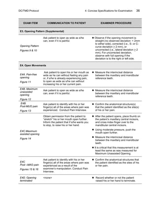
E3. Opening Pattern (Supplemental)
Opening Pattern
Figures 9 & 10
Place your back teeth completely
together.
I would like you to slowly open your
mouth as wide as you can, even if it
is painful, close, and put your back
teeth completely together again.
Repeat 2 more times.
Alternative Format:
In English, the common expression is
“open as wide as you can” but other
languages may differ; for example, “as
much as you can” is often better in
other languages.
Observe if the opening movement is:
straight (no observed deviation: < 2mm
to either side of the midline), corrected
(i.e., S- or C-curve deviation, >2mm),
or uncorrected (i.e., lateral deviation,
>2mm).
For uncorrected deviation, observe at
full opening if the deviation is to the
right or left side.
More than one option may be selected;
this allows recording of any type of
movement in case the movement is not
consistent across repeated trials.
Repeat 2 more times.
INTENTIONAL BLANK ROW](https://image.slidesharecdn.com/qwtp5n8wtzcafraw77vw-signature-d95bbf363b3219feeb75d250b5e2cf4e78e1dbfd2c7fe7186d561218c53aeafe-poli-150525021224-lva1-app6892/85/Www-unlock-pdf-com-dc-tmd-protocol-2014-06_02-46-320.jpg)
![DC/TMD Protocol 5: Complete Specifications for Examination 47
CONSTRUCT VERBAL COMMAND EXAMINER PROCEDURE
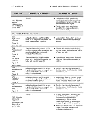
E4. Open and Close Movements
4A. PAIN FREE OPENING
Ruler position for
vertical
movement
measurements
<none> Place the ‘0’ edge of a prepared
millimeter ruler (see Section 1) at the
incisal edge of the mandibular
reference tooth.
E4A
Pain-free opening
Figure 11
I would like you to open your mouth
as wide as you can without feeling
any pain, or without increasing any
pain you may have right now.
Alternative Format:
In English, the common expression is
“open as wide as you can” but other
languages may differ; for example, “as
much as you can” is often better in
other languages. This alternative
format applies to 4B and 4C as well.
Measure the inter-incisal distance
between the maxillary and mandibular
reference teeth.
Record this measurement.
4B. MAXIMUM UNASSISTED OPENING
Starting position
<none> Maximum unassisted opening can
often be assessed immediately after
taking the measurement for pain-free
opening, without intervening closure of
the mandible by the patient.
E4B
Maximum
unassisted
opening
Figure 12
I would like you to open your mouth
as wide as you can, even if it is
painful.
Use ruler position as under 4A.
Measure the inter-incisal distance
between the maxillary and mandibular
reference teeth.
Record this measurement.
E4B
Post-MUO pain
Figure 13
Did you feel any pain with this
movement?
See PAIN INTERVIEW
6.2.1: Maneuver-induced pain
6.2.4: Familiar pain
Confirm the anatomical structures that
the patient identified as the sites of
pain.
Record this finding.
4C. MAXIMUM ASSISTED OPENING
Instructions
In a moment I will try, [if possible], to
open your mouth wider with my
fingers. If you want me to stop, raise
your hand and I will stop
immediately.
<none>](https://image.slidesharecdn.com/qwtp5n8wtzcafraw77vw-signature-d95bbf363b3219feeb75d250b5e2cf4e78e1dbfd2c7fe7186d561218c53aeafe-poli-150525021224-lva1-app6892/85/Www-unlock-pdf-com-dc-tmd-protocol-2014-06_02-47-320.jpg)
![DC/TMD Protocol 5: Complete Specifications for Examination 48
CONSTRUCT VERBAL COMMAND EXAMINER PROCEDURE
E4C
Maximum
assisted opening
Figure 14
I will place my ruler. [pause]
Now open [your mouth] as wide as
you can, even if painful, just as you
did before. [pause]
You will feel my fingers.
Please relax your jaw so that I can
help you open wider, if possible.
[pause]
Use ruler position as under 4A.
Insure that the patient initially opens to
the same extent as was measured for
Maximum Unassisted Opening.
If not, ask patient to open more.
Place thumb on the patient’s maxillary
central incisors, and cross index finger
over to the mandibular central incisors.
(Orientation of fingers is relative to
examiner standing in front of the
patient.)
Provide support to the mandible with
the fingers, before saying “Please
relax…”.
Using moderate pressure, push the
mouth open further, until either (1) you
feel resistance from the tissue, or (2)
the patient raises his/her hand. NOTE:
Use clinical judgment with respect to
avoidance of over-stretching.
Measure the inter-incisal distance
between the maxillary and mandibular
reference teeth.
Record this measurement.
E4C
Post –MAO pain
Figures 15 & 16
Did you feel any pain when I tried to
open your mouth wider with my
fingers?
See PAIN INTERVIEW
6.2.1: Maneuver-induced pain
6.2.4: Familiar pain
Confirm the anatomical structures that
the patient identified as the sites of
pain.
Record this finding.
4D. MAXIMUM ASSISTED OPENING TERMINATED
E4D
Opening
terminated
<none> Record whether or not the patient
raised his/her hand to terminate the
opening.
INTENTIONAL BLANK ROW](https://image.slidesharecdn.com/qwtp5n8wtzcafraw77vw-signature-d95bbf363b3219feeb75d250b5e2cf4e78e1dbfd2c7fe7186d561218c53aeafe-poli-150525021224-lva1-app6892/85/Www-unlock-pdf-com-dc-tmd-protocol-2014-06_02-48-320.jpg)
![DC/TMD Protocol 5: Complete Specifications for Examination 49
CONSTRUCT VERBAL COMMAND EXAMINER PROCEDURE
E5. Lateral & Protrusive Movements
5A. RIGHT LATERAL EXCURSION
E5A
Right lateral
excursion
Figure 17
Also: Figure 21
Open slightly, and move your jaw as
far as you can towards the right,
even if it is painful.
Hold your jaw in that position until I take
a measurement.
If the patient is confused about which
direction to move the jaw, say “Move
your jaw towards this hand” and touch
the patient´s jaw or shoulder on the
side of the desired movement.
Place ruler with zero end aligned with
either the maxillary reference midline or
the mandibular reference midline,
depending on type of ruler.
Measure from the maxillary reference
midline to the mandibular reference
midline.
If ruler obscures the opposing
reference point, move the ruler up or
down in order to read the numbers.
Record this measurement.
Return jaw
[Move your jaw back to a comfortable
position.]
<none>
E5A
Post excursion
pain
Figure 18
Did you feel any pain with that
movement?
See PAIN INTERVIEW
6.2.1: Maneuver-induced pain
6.2.4: Familiar pain
Confirm the anatomical structures that
the patient identified as the sites of his
or her pain.
Record this finding.
5B. LEFT LATERAL EXCURSION
E5B
Left lateral
excursion
Figure 19
Also: Figure 21
Open slightly, and move your jaw as
far as you can towards the left, even
if it is painful.
Hold your jaw in that position until I take
a measurement.
If the patient is confused about which
direction to move the jaw, say “Move
your jaw towards this hand” and touch
the patient´s jaw or shoulder on the
side of the desired movement.
Use ruler as for 5A.
Measure from the maxillary reference
midline to the mandibular reference
midline.
If ruler obscures the opposing
reference point, move the ruler up or
down in order to read the numbers.
Record this measurement.
Return jaw
[Move your jaw back to a comfortable
position.]
<none>](https://image.slidesharecdn.com/qwtp5n8wtzcafraw77vw-signature-d95bbf363b3219feeb75d250b5e2cf4e78e1dbfd2c7fe7186d561218c53aeafe-poli-150525021224-lva1-app6892/85/Www-unlock-pdf-com-dc-tmd-protocol-2014-06_02-49-320.jpg)
![DC/TMD Protocol 5: Complete Specifications for Examination 50
CONSTRUCT VERBAL COMMAND EXAMINER PROCEDURE
E5B
Post excursion
pain
Figure 18
Did you feel any pain with that
movement?
See PAIN INTERVIEW
6.2.1: Maneuver-induced pain
6.2.4: Familiar pain
Confirm the anatomical structures that
the patient identified as the sites of his
or her pain.
Record this finding.
5C. PROTRUSIVE
E5C
Protrusive
excursion
Figure 20
Open slightly and move [slide] your
jaw forward [straight out in front of you]
as far as you can, even if it is painful.
Hold your jaw in that position until I take
a measurement.
Place ruler with zero end against the
mesial-distal center of the maxillary
reference tooth and with ruler aligned
forward such that the labioincisal edge
of the opposing mandibular incisor
touches the mm markings on the ruler.
Measure from labial surface of the
maxillary reference tooth to the labial
surface of the mandibular reference
tooth.
Record this measurement.
Return jaw
[Move your jaw back to a comfortable
position.]
<none>
E5C
Post protrusion
pain
Figure 18
Did you feel any pain with that
movement?
See PAIN INTERVIEW
6.2.1: Maneuver-induced pain
6.2.4: Familiar pain
Confirm the anatomical structures that
the patient identified as the sites of his
or her pain.
Record this finding.
E6. TMJ Noises During Open & Close Movements
General
instructions to
examiner
<none> Patients may use a variety of terms for
the single-occurrence joint noise (e.g.,
“click”, “pop”, “snap”); any of these
terms denote a “click” for purposes of
the exam, and the instructions below
refer to the term “Click” on the
recording form.
Patients may use a variety of terms for
the fine, multiple-occurrence joint noise
(e.g., “grating”, “grinding”, “crepitus”);
any of these terms denotes “crepitus”
for purposes of the examination, and
the instructions below refer to the term
“Crepitus” on the recording form.](https://image.slidesharecdn.com/qwtp5n8wtzcafraw77vw-signature-d95bbf363b3219feeb75d250b5e2cf4e78e1dbfd2c7fe7186d561218c53aeafe-poli-150525021224-lva1-app6892/85/Www-unlock-pdf-com-dc-tmd-protocol-2014-06_02-50-320.jpg)
![DC/TMD Protocol 5: Complete Specifications for Examination 51
CONSTRUCT VERBAL COMMAND EXAMINER PROCEDURE
Hand position for
palpation of joint
noise
Figure 22
<none> Use of bilateral or unilateral palpation
depends on examiner preference and
circumstances.
Bilateral palpation: Place finger of each
hand over the corresponding TMJ.
This method requires that the patient
monitor noises from both TMJs at the
same time, which may be difficult.
Unilateral palpation: Use same finger
placement for a single joint, as
described for bilateral palpation,
assessing first the right TMJ and then
the left TMJ.
Repeat following instructions for each
TMJ separately.
Instructions
regarding joint
noises
I will be evaluating the jaw joints for
whether they make any noises. I
would like you to pay attention as
well, since I will ask you at the end
whether you heard or felt any noises.
[Focus on both joints.]
If doing bilateral palpation, touch both
joints and ask patient to attend to both
joints.
If doing unilateral palpation, touch the
targeted joint and ask patient to attend
to that joint.
Full closure of
mandible
Place your back teeth completely
together.
Insure that the teeth are in maximal
intercuspal position in order to insure
that the TMJ is in the closed position.
Examiner
detection of open
and closing joint
noises
Slowly open as wide as you can,
even if it is painful, and then slowly
close until your back teeth are
completely together again.
Repeat 2 more times.
Mandible should be opened and closed
slowly, allowing about 2 seconds to
open and about 2 seconds to close.
Guide patient accordingly.
At the end of closing, distinguish
noises from teeth contacting.
Record a noise as a “click” or “crepitus”
if it is present on at least 1 of the 3
movements. Multiple types of noise
can be present in a single joint.
Patient inquiry
regarding joint
noises
Did you hear or feel noises in either
jaw joint when you opened or
closed?
IF “YES”:
What type of noise?
<The patient may be prompted by
offering the words of different jaw joint
sounds: click, pop, snap, grate, grind,
crunch.>
Examiner can interview patient if
necessary in order to confirm location
(right vs left, or both) of noises.
If patient detects joint noise but is
unable to classify the noise as a click
or crepitus, then examiner may repeat
the open-close movement again in
order for the patient to reassess type of
noise.](https://image.slidesharecdn.com/qwtp5n8wtzcafraw77vw-signature-d95bbf363b3219feeb75d250b5e2cf4e78e1dbfd2c7fe7186d561218c53aeafe-poli-150525021224-lva1-app6892/85/Www-unlock-pdf-com-dc-tmd-protocol-2014-06_02-51-320.jpg)
![DC/TMD Protocol 5: Complete Specifications for Examination 53
CONSTRUCT VERBAL COMMAND EXAMINER PROCEDURE
Inquiry regarding
joint noises
Did you hear or feel any noises in
this [right, left] joint when you moved
your jaw forward or to the side?
IF “YES”:
What type of noise?
<The patient may be prompted with the
words of different jaw joint sounds:
click, pop, snap, grate, grind, crunch.>
Examiner touches the patient’s right
TMJ while asking the question.
Examiner can interview patient if
necessary in order to localize location
(right vs left) of noises.
Pain inquiry
IF PATIENT REPORTS CLICK:
Did you feel any pain when that click
occurred?
See PAIN INTERVIEW
6.2.3: Click-related pain
6.2.4: Familiar pain
Record this finding.
Repeat for left
joint
<none> Repeat all of the above for the left joint
if assessing joints separately.
E8. Joint Locking
Locking Closed
<For observed closed lock during the
examination:>
Can you “unlock” your jaw?
Inability to further open the mouth from
a partially opened position, even
momentarily, is positive.
Record if the patient or examiner
reduced the closed lock or if it could
not be reduced.
Locking Open
<For observed open lock during the
examination:>
Can you “unlock” your jaw?
Inability to close the mouth from a
wide-open position, even momentarily,
is positive.
Record if the patient or examiner
reduced the open lock or if it could not
be reduced.
INTENTIONAL BLANK ROW](https://image.slidesharecdn.com/qwtp5n8wtzcafraw77vw-signature-d95bbf363b3219feeb75d250b5e2cf4e78e1dbfd2c7fe7186d561218c53aeafe-poli-150525021224-lva1-app6892/85/Www-unlock-pdf-com-dc-tmd-protocol-2014-06_02-53-320.jpg)
![DC/TMD Protocol 5: Complete Specifications for Examination 54
CONSTRUCT VERBAL COMMAND EXAMINER PROCEDURE
E9. Muscle and TMJ Pain with Palpation
General
Figure 24
<none> Select coverage method for palpation
of the larger muscles: comprehensive
where every part of the muscle is
palpated, or sampling areas of the
muscle.
Select time duration for stimulus
application: (a) 2 seconds for efficiency
and ignoring referred pain diagnosis,
(b) 5 seconds to minimize false
negative diagnoses of referred pain
and to better identify hyperalgesia.
Instructions
Now I am going to apply pressure to
different areas of your head, face and
jaw, and I will ask you about pain,
familiar pain, and familiar headache.
In addition, I will ask whether the
pain stays only under my finger or if
you feel it also anywhere else
besides under my finger.
I will prompt you with the words “pain?”,
“familiar pain?”, “familiar headache?”,
and “only under my finger?”.
[Inquiry “go anywhere else?” can be
used instead of “only under my finger” if
the examiner prefers.]
Each time, I will apply pressure and
hold it for 5 seconds.
<none>
Examiner
calibration
Figure 25
<none> Examiner uses “finger algometer” and
calibrates the respective finger of each
of right and left hand to 1.0 kg.
Temporalis
and
masseter
muscles
Figures 24 &
26-30
[Patient can be asked to clench the
teeth together in order to identify the
borders of the muscles.]
[Please relax your jaw.]
See PAIN INTERVIEW
6.2.2: Palpation-Induced pain
6.2.4: Familiar pain
6.2.5: Referred pain
Palpate the temporalis and masseter
muscles, one side at a time.
Palpate the entire muscle. For
systematic coverage, use three vertical
zones for the temporalis and use three
horizontal bands for the masseter.
Apply 1 kg for total of 5 seconds.
Record findings.](https://image.slidesharecdn.com/qwtp5n8wtzcafraw77vw-signature-d95bbf363b3219feeb75d250b5e2cf4e78e1dbfd2c7fe7186d561218c53aeafe-poli-150525021224-lva1-app6892/85/Www-unlock-pdf-com-dc-tmd-protocol-2014-06_02-54-320.jpg)
![DC/TMD Protocol 5: Complete Specifications for Examination 55
CONSTRUCT VERBAL COMMAND EXAMINER PROCEDURE
Examiner
calibration
Figure 33
<none> Examiner uses “finger algometer” and
calibrates the respective finger of each
of right and left hand to 0.5 kg; if
unsure, use more, not less, force.
TMJ: lateral pole
Figures 31-33
Open slightly, and move [slide] your
lower jaw forward and then move
[slide] it back to its normal position
with your teeth slightly apart.
See PAIN INTERVIEW
6.2.2: Palpation-Induced pain
6.2.4: Familiar pain
6.2.5: Referred pain
Examine right side first, then examine
left side. See Figures as a guide for
the location of each individual
palpation.
Place index finger anterior to the tragus
of the ear and over the patient’s TMJ.
Apply 0.5 kg and hold for 5 seconds.
Record findings.
Examiner
calibration
Figure 36
<none> Examiner uses “finger algometer” and
calibrates the respective finger of each
of right and left hand to 1 kg.
TMJ: around
lateral pole
Figures 34-36
Open slightly, and move [slide] your
lower jaw forward a little bit and keep
it there.
See PAIN INTERVIEW
6.2.2: Palpation-Induced pain
6.2.4: Familiar pain
6.2.5: Referred pain
Examine right side first, then examine
left side. See Figures as a guide for
the location of each individual
palpation.
Place finger at posterior aspect of
lateral pole.
Mandible is protruded enough to gain
access to the dorsal aspect of the
lateral pole but also retain access to
the anterior aspect as well.
Roll finger completely around the
lateral pole of condyle. The finger
should “hug” or contact the lateral
aspect of the condylar pole while
moving in one smooth circular
movement that should take 5 seconds
to complete.
Record findings.
INTENTIONAL BLANK ROW](https://image.slidesharecdn.com/qwtp5n8wtzcafraw77vw-signature-d95bbf363b3219feeb75d250b5e2cf4e78e1dbfd2c7fe7186d561218c53aeafe-poli-150525021224-lva1-app6892/85/Www-unlock-pdf-com-dc-tmd-protocol-2014-06_02-55-320.jpg)
![DC/TMD Protocol 5: Complete Specifications for Examination 56
CONSTRUCT VERBAL COMMAND EXAMINER PROCEDURE
E10. Supplemental palpation sites
Examiner
calibration
Figure 38
<none> Examiner uses “finger algometer” and
calibrates the respective finger of each
right and left hand to 0.5 kg.
Posterior
mandibular
region
Figures 37, 39
Relax your jaw.
[Extend your head.]
See PAIN INTERVIEW
6.2.2: Palpation-Induced pain
6.2.4: Familiar pain
6.2.5: Referred pain
The target is the posterior digastric
muscle. This region is defined as that
area between the insertion of the
sternocleidomastoid muscle and the
posterior border of the mandible.
Submandibular
region
Figures 37, 39
Relax your jaw.
[Drop your chin to your chest.]
See PAIN INTERVIEW
6.2.2: Palpation-Induced pain
6.2.4: Familiar pain
6.2.5: Referred pain
The target is the medial pterygoid muscle.
This region is defined as the area 2 cm
anterior to the angle of the mandible, and
medial to the mandible.
Lateral pterygoid
area
Figure 40
Open slightly and move your jaw to
the side.
See PAIN INTERVIEW
6.2.2: Palpation-Induced pain
6.2.4: Familiar pain
6.2.5: Referred pain
The target is the lateral pterygoid muscle.
Place finger on buccal side of alveolar
ridge above the maxillary molars and
move finger distally, superiorly, and
medially and palpate.
Tendon of the
temporalis
Figure 41
Open your mouth.
See PAIN INTERVIEW
6.2.2: Palpation-Induced pain
6.2.4: Familiar pain
6.2.5: Referred pain
Place finger on anterior ridge of the
coronoid process. Palpate on the superior
aspect of the process.
E11. Examiner Comments
Examination
comments
<none> Include description of any physical
barriers to the examination as well as any
exceptions or modifying circumstances.
END OF DC/TMD EXAMINATION](https://image.slidesharecdn.com/qwtp5n8wtzcafraw77vw-signature-d95bbf363b3219feeb75d250b5e2cf4e78e1dbfd2c7fe7186d561218c53aeafe-poli-150525021224-lva1-app6892/85/Www-unlock-pdf-com-dc-tmd-protocol-2014-06_02-56-320.jpg)
![DC/TMD Protocol 6: Examination-Related Pain Interview 57
6 Examination-Related Pain Interview
6.1 Overview
Examination-related Pain Interview is a structured format for repeatedly eliciting and
clarifying the pain status with any positive examination findings. The intent is for the
examiner to probe in a neutral manner, with the patient providing the description of any
pain experienced.
6.2 Structured Pain Interview
ITEM INTERVIEW QUESTION COMMENTS
6.2.1 For range of motion maneuver-induced pain
Did you feel pain with that
movement?
The intent of “with that movement” is whether the procedure
caused pain or caused existing pain to change.
IF YES to pain:
Could you point with your finger
to each of the areas where you
felt pain?
Are there any other areas where
you felt pain with that
movement? Point [with your
finger] to those areas.
Go to FAMILIAR PAIN INQUIRY.
Refer to STRUCTURAL LOCALIZATION OF PAIN, Section
2.
6.2.2 For palpation-induced pain
Did you feel pain [in the area
where I applied pressure]?
IF YES to pain:
Go to FAMILIAR PAIN INQUIRY.
6.2.3 For click-related pain
Was that click painful? Insure that the patient distinguishes pain concurrent with the
click vs pain associated with the movement (i.e., opening,
closing, excursive movements).
IF YES to pain:
Go to FAMILIAR PAIN INQUIRY.
6.2.4 Familiar Pain Inquiry
Is this pain familiar to any pain
you have experienced in this
area in the last 30 days?
COMMENT 1: Regarding “familiar”, the examiner might
elaborate with something like “Is this pain familiar, that is,
similar or like, the pain that you have experienced in that
area in the last 30 days?” Capturing the construct of](https://image.slidesharecdn.com/qwtp5n8wtzcafraw77vw-signature-d95bbf363b3219feeb75d250b5e2cf4e78e1dbfd2c7fe7186d561218c53aeafe-poli-150525021224-lva1-app6892/85/Www-unlock-pdf-com-dc-tmd-protocol-2014-06_02-57-320.jpg)
![DC/TMD Protocol 6: Examination-Related Pain Interview 59
ITEM INTERVIEW QUESTION COMMENTS
response is fine. Alternatively, the relevant examination
procedure can be repeated: (“Would you like me to repeat
that procedure?”), and this allows the patient to reassess
his/her experience when s/he responds to the pain inquiry
questions.
COMMENT 3: When patients report “headache” in other
masticatory structures (e.g., masseter region, TMJ region),
this inquiry should also be performed for diagnostic
purposes. The standard examination form does not have
response option for this information but the finding can be
recorded in the comments section of the examination form.
COMMENT 4: A 30-day time frame is used here in order to
retain congruence with the time-frame for the masticatory
system pain. The International Classification of Headache
Disorders, version 2 (ICHD-2) criteria specify different time
periods for Infrequent, Frequent and for Chronic Tension-
Type Headaches. The user may want to alter the reference
time period in this protocol for headache in order to be
consistent with the ICHD-2 criteria. However, for Headache
Secondary to TMD, there are currently no established time
periods.
COMMENT 5: Palpation of asymptomatic areas can elicit a
positive pain response, and during the inquiry a patient
might report that the pain in such areas is “familiar”. It may
be that the clinical pain is present in that location and which
the patient believed to be asymptomatic, in which case the
area is recoded as painful in item 1 of the examination.
Alternatively, a patient may report that palpation pain in an
asymptomatic location is “familiar” because that pain
experience is similar to the pain that is experienced in
another area of the body. The response for “familiar” pain in
this example is |no| since the pain provoked by examination
must be familiar to a pain experienced in the same body
region.
Examiner verifies that reported
pain areas in Q1 of the exam are
consistent with body areas
reported as positive for familiar
pain.
6.2.5 Referred Pain Inquiry
Did you feel the pain just under
my finger, or did you also feel it
somewhere else [in different
areas of your head, face, or
jaw]?
COMMENT 1. For purposes of classifying “referred pain”,
the location of perceived pain in response to palpation
procedures needs to extend beyond the boundary of the
structure that was examined. The examiner determines
whether the pain location(s) denote a different anatomic
structure or the same structure. Only if the pain is localized
in a different anatomic structure is it termed “referred pain”.](https://image.slidesharecdn.com/qwtp5n8wtzcafraw77vw-signature-d95bbf363b3219feeb75d250b5e2cf4e78e1dbfd2c7fe7186d561218c53aeafe-poli-150525021224-lva1-app6892/85/Www-unlock-pdf-com-dc-tmd-protocol-2014-06_02-59-320.jpg)
![DC/TMD Protocol 6: Examination-Related Pain Interview 60
ITEM INTERVIEW QUESTION COMMENTS
If there is ambiguity with respect to what structure pain is felt
in when described as “deep” by the patient, ask the patient
to locate on the surface of the area of pain or, as
appropriate, to inside the mouth.
COMMENT 2. This inquiry can be productively shortened,
as indicated in Section 5.3, to “only under my finger?” for
purposes of the repeat administrations associated with an
examination that has many positive findings to palpation.
An alternative short-hand inquiry is “go anywhere else?”.
[Show me where you felt the
pain.] Point with your finger to
all of the areas where you just
felt pain [in response to my
pressure].
After the patient points, ask:
Did you feel it anywhere else?
6.3 Efficient Completion
After the first several positive responses from pain-provoking procedures and depending
on the purpose of the examination and the patient’s ability to easily distinguish the
presence of pain, familiar pain, familiar headache, and referred pain, the examiner can
instruct the patient to respond to the Pain Interview in a more abbreviated manner. For
example, the patient might be instructed to report, in response to positive palpation
findings, as follows: “yes, familiar” or “yes, not familiar” in order to eliminate asking
whether each positive pain finding is familiar or not. After establishing that positive pain
responses are familiar to the patient’s clinical pain from at least 2 positive examination
procedures, the examiner need only ask if the pain is familiar but need not ask “familiar to
what” unless an area not yet recorded in examination item 1 is reported as positive in
response to a procedure.](https://image.slidesharecdn.com/qwtp5n8wtzcafraw77vw-signature-d95bbf363b3219feeb75d250b5e2cf4e78e1dbfd2c7fe7186d561218c53aeafe-poli-150525021224-lva1-app6892/85/Www-unlock-pdf-com-dc-tmd-protocol-2014-06_02-60-320.jpg)

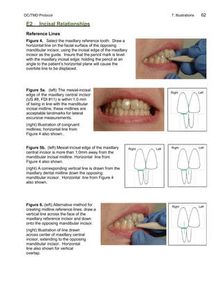
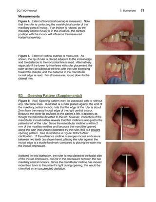
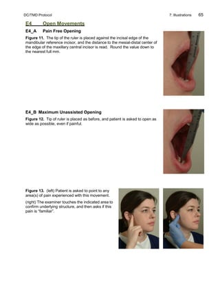
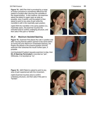
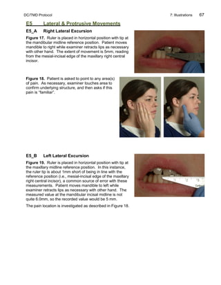
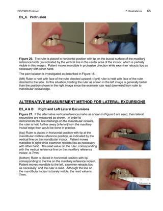
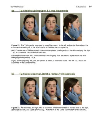
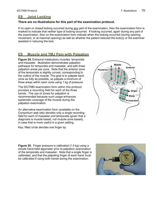
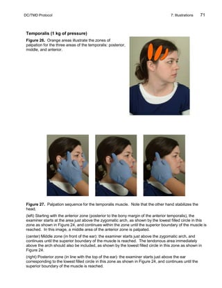
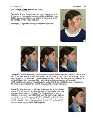
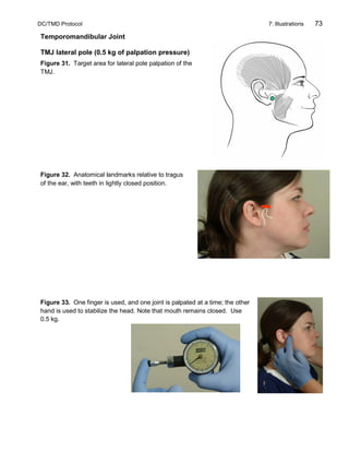
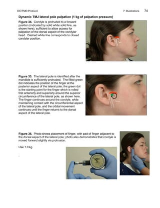
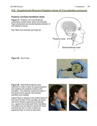
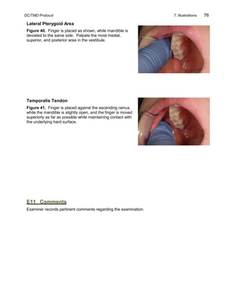
![DC/TMD Protocol 8: Required Examination Commands 77
8 Required Examination Commands
8.1 Overview
The examiner commands that are required to be used verbatim, as shown in the complete
examination sequence (Section 4) are reproduced here in order to facilitate learning and
review.
8.2 Examination-related Pain Interview
Sub-section numbering follows that of the Examination-Related Pain Interview (Section 6)
6.2.1 For range of motion maneuver-induced pain
o Did you feel pain with that movement?
o IF YES to pain:
o Could you point with your finger to each of the areas where you felt pain?
o Are there any other areas where you felt pain with that movement? Point
[with your finger] to those areas.
o Go to FAMILIAR PAIN INQUIRY.
6.2.2 For palpation-induced pain
o Did you feel pain [in the area where I applied pressure]?
o IF YES to pain: Go to FAMILIAR PAIN INQUIRY.
6.2.3 For click-related pain
o Was that click painful?
o IF YES to pain: Go to FAMILIAR PAIN INQUIRY.
6.2.4 Familiar Pain Inquiry
o Is this pain familiar to any pain you have experienced in this area in the last 30
days?
o IF YES TO FAMILIAR PAIN: What is this pain familiar to?
o IF TEMPORALIS SITE IS POSITIVE: Is this pain familiar to your headache in this
area, in the last 30 days?
o Examiner verifies that reported pain areas in the past 30 days are consistent with
positive familiar pain reports.
6.2.5 Referred Pain Inquiry
o Did you feel the pain just under my finger, or did you also feel it somewhere else
[in different areas of your head, face or jaw]?
o [Show me where you felt the pain.] Point with your finger to each of the areas
where you just felt pain [in response to my pressure].
o Did you feel it anywhere else?](https://image.slidesharecdn.com/qwtp5n8wtzcafraw77vw-signature-d95bbf363b3219feeb75d250b5e2cf4e78e1dbfd2c7fe7186d561218c53aeafe-poli-150525021224-lva1-app6892/85/Www-unlock-pdf-com-dc-tmd-protocol-2014-06_02-77-320.jpg)
![DC/TMD Protocol 8: Required Examination Commands 78
8.3 Required Components from Section 5: Complete Examiner
Commands
CONVENTIONS FOR THIS TABLE: Bold text under “Verbal Command” must be used verbatim;
procedures associated with cells containing bold text may require additional (ordinary) language for
the completion of the procedure. Blank cells under “Verbal Command” indicate that ordinary
language is sufficient for that entire procedure; non-bolded text is also used to illustrate ordinary
language use associated with particular procedures. Procedures with <none> under “Verbal
Command” indicate that there is no verbal command from the examiner.
CONSTRUCT VERBAL COMMAND
E1. Examiner Confirmation of Pain and Headache Locations
Identifying
information
<none>
Instructions to the
patient
Before I start the exam, I want to review a few things with you.
I will be asking you about pain, and only you know if you have pain. When I
ask about pain, I want you to say either yes or no; if you are not sure, give me
your best answer.
If you feel pain, I will also ask if that pain is familiar. Familiar pain refers to
pain that is similar or like the pain you may have had in that same part of your
body in the last 30 days.
If you feel pain in the temple area, I will ask if that pain is like any headache
you may have had in the temple area in the last 30 days.
Scope of
examination:
anatomic areas
of interest
For the purposes of this examination, I am interested in pain that you may
have in these areas….
…. and also inside the mouth.
E1a
Location of pain:
last 30 days
In the last 30 days, have you had pain in these areas [that I touched]?
IF “YES”:
Could you point with your finger to each of the areas where you have felt pain
[in the last 30 days]?
Are there any other areas where you have felt pain [in the last 30 days]?
E1b
Location of
headache in the
last 30 days.
In the last 30 days, have you had any headaches?
IF “YES”:
Could you point with your finger to each of the areas where you have felt
headache [in the last 30 days]?
Are there other areas where you have felt headache [in the last 30 days]?
E2. Incisal Relationships
Select maxillary
and mandibular
reference teeth
I will place some pencil marks on your teeth; I will remove them at the end of the
examination.
Reference line –
vertical overlap
Place your back teeth completely together.
Reference line –
mandibular
reference midline
[Place your back teeth completely together.]](https://image.slidesharecdn.com/qwtp5n8wtzcafraw77vw-signature-d95bbf363b3219feeb75d250b5e2cf4e78e1dbfd2c7fe7186d561218c53aeafe-poli-150525021224-lva1-app6892/85/Www-unlock-pdf-com-dc-tmd-protocol-2014-06_02-78-320.jpg)
![DC/TMD Protocol 8: Required Examination Commands 79
CONSTRUCT VERBAL COMMAND
Horizontal incisal
overjet
[Place your back teeth completely together.]
Vertical incisal
overlap
<Ask patient to open sufficiently to measure the vertical overlap.>
E3. Opening Pattern (Supplemental)
Starting Position Place your back teeth completely together.
Opening Pattern
I would like you to slowly open your mouth as wide as you can, even if it is
painful, close, and put your back teeth completely together again.
Repeat 2 more times.
E4. Open Movements
4A. PAIN FREE OPENING
Starting position [Place your mouth in a comfortable position.]
Ruler position for
vertical
movement
measurements
<none>
E4A
Pain-free opening
I would like you to open your mouth as wide as you can without feeling any
pain, or without increasing any pain you may have right now.
4B. MAXIMUM UNASSISTED OPENING
Starting position [Place your mouth in a comfortable position.]
E4B
Maximum
unassisted
opening
I would like you to open your mouth as wide as you can, even if it is painful.
E4B
Post-MUO pain
Did you feel any pain with this movement?
4C. MAXIMUM ASSISTED OPENING
Starting position [Place your mouth in a comfortable position.]
Instructions
In a moment I will try, [if possible], to open your mouth wider with my fingers. If
you want me to stop, raise your hand and I will stop immediately.
E4C
Maximum
assisted opening
I will place my ruler. [pause]
Now open [your mouth] as wide as you can, even if painful, just as you did
before. [pause]
You will feel my fingers.
Please relax your jaw so I can help you open wider, if possible. [pause]
E4C
Post –MAO pain
Did you feel any pain when I tried to open your mouth wider with my fingers?](https://image.slidesharecdn.com/qwtp5n8wtzcafraw77vw-signature-d95bbf363b3219feeb75d250b5e2cf4e78e1dbfd2c7fe7186d561218c53aeafe-poli-150525021224-lva1-app6892/85/Www-unlock-pdf-com-dc-tmd-protocol-2014-06_02-79-320.jpg)
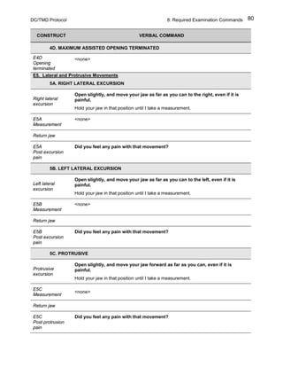
![DC/TMD Protocol 8: Required Examination Commands 81
CONSTRUCT VERBAL COMMAND
E6. TMJ Noises During Open & Close Movements
Hand position for
palpation of joint
noise
<none>
Instructions
regarding joint
noise
I will be evaluating the jaw joints for whether they make any noises. I would
like you to pay attention as well, since I will ask you at the end whether you
heard or felt any noises.
[Focus on both joints.]
Full closure of
mandible
Place your back teeth completely together.
Examiner
detection of open
and closing joint
noise
Slowly open as wide as you can, even if it is painful, and then slowly close
until your back teeth are completely together again.
Repeat 2 more times.
Patient inquiry
regarding joint
noise
Did you hear or feel noises in either jaw joint when you opened or closed?
What type of noise?
Pain inquiry
IF PATIENT REPORTS CLICK:
Did you feel any pain when that click occurred?
E7. TMJ Noises During Lateral & Protrusive Movements
General <none>
Joint sounds:
lateral and
protrusive
movements
[Perform the following set of procedures for examining each of the right joint, then
the left joint. Inform patient which joint to focus on.]
Place your back teeth completely together, open slightly, and move your jaw to
the right as far as you can, even if painful; move your jaw back and place your
back teeth completely together.
Repeat 2 more times.
Place your back teeth completely together, open slightly, and move your jaw to
the left as far as you can, even if painful; move your jaw back and place your
back teeth completely together.
Repeat 2 more times.
Place your back teeth completely together, open slightly, and move your jaw
forward as far as you can, even if painful; move your jaw back, and place your
back teeth completely together.
Repeat 2 more times.
Inquiry regarding
joint sounds
Did you hear or feel any noises in this [right, left] joint when you moved your
jaw forward or to the side?
What type of noise?](https://image.slidesharecdn.com/qwtp5n8wtzcafraw77vw-signature-d95bbf363b3219feeb75d250b5e2cf4e78e1dbfd2c7fe7186d561218c53aeafe-poli-150525021224-lva1-app6892/85/Www-unlock-pdf-com-dc-tmd-protocol-2014-06_02-81-320.jpg)
![DC/TMD Protocol 8: Required Examination Commands 82
CONSTRUCT VERBAL COMMAND
Pain inquiry
IF PATIENT REPORTS CLICK:
Did you feel any pain when that click occurred?
E8. Joint Locking
Locking Closed Can you “unlock” your jaw?
Locking Open Can you “unlock” your jaw?
E9. Muscle and TMJ Pain with Palpation
Introduction
Now I am going to apply pressure to different areas of your head, face and jaw,
and I will ask you about pain, familiar pain, and familiar headache.
In addition, I will ask whether the pain stays only under my finger or if you feel
it also anywhere else besides under my finger.
I will prompt you with the words “pain”, “familiar pain”, “familiar headache”, and “only
under my finger?”.
[Inquiry “go anywhere else?” can be used instead of “only under my finger” if the
examiner prefers.]
Each time, I will apply pressure and hold it for 5 seconds.
Calibration <none>
Temporalis
& masseter
<none>
Calibration <none>
TMJ: lateral pole
Open slightly, and move your lower jaw forward and then move it back to its
normal position with your teeth slightly apart.
Calibration <none>
TMJ: around
lateral pole
Open slightly, and move your lower jaw forward a little bit and keep it there.
E10. Supplemental Palpation Sites
Calibration <none>
Posterior
mandibular
region
Relax your jaw.
Submandibular
region
Relax your jaw.
Lateral pterygoid
area
Open slightly and move your jaw to the side.
Tendon of the
temporalis
Open your mouth.](https://image.slidesharecdn.com/qwtp5n8wtzcafraw77vw-signature-d95bbf363b3219feeb75d250b5e2cf4e78e1dbfd2c7fe7186d561218c53aeafe-poli-150525021224-lva1-app6892/85/Www-unlock-pdf-com-dc-tmd-protocol-2014-06_02-82-320.jpg)

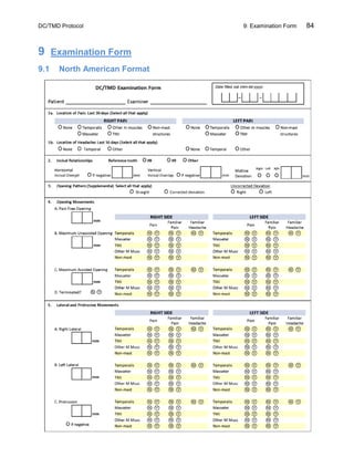
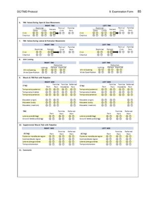
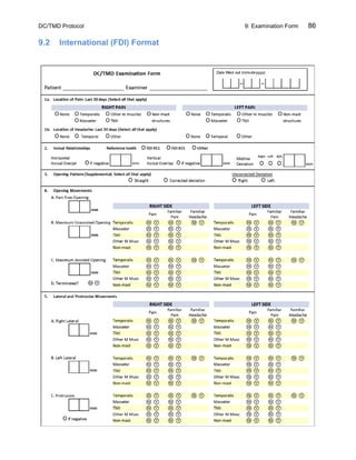
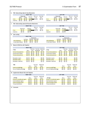

This document provides guidelines for conducting a standardized clinical examination for temporomandibular disorders (TMD) based on the Diagnostic Criteria for TMD (DC/TMD). The examination involves assessing pain location, jaw range of motion, joint sounds, and muscle tenderness. Sections provide both concise and fully operationalized procedures for clinical and research use. Calibration of examiners is emphasized to ensure reliability of findings. The examination is intended to identify clinical features for TMD diagnoses; additional testing may be needed for comprehensive assessment of orofacial pain.


