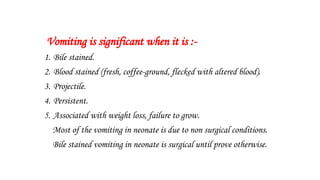
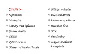
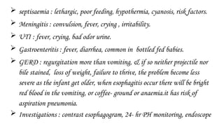
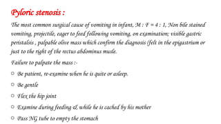
Vomiting in neonate
Causes & diagnosis
Vomiting is significant when it is :-
1. Bile stained.
2. Blood stained (fresh, coffee-ground, flecked with altered blood).
3. Projectile.
4. Persistent.
5. Associated with weight loss, failure to grow.
Most of the vomiting in neonate is due to non surgical conditions.
Bile stained vomiting in neonate is surgical until prove otherwise.
Causes :-
o Septisaemia
o Meningitis
o Urinary tract infection
o Gastroenteritis
o GERD
o Pyloric stenosis
o Obstructed inguinal hernia
o Mid-gut volvulus
o intestinal atresia
o hirschsprung's disease
o meconium ileus
o NEC
o Overfeeding
o congenital adrenal
hyperplasia
 septisaemia : lethargic, poor feeding, hypothermia, cyanosis, risk factors.
 Meningitis : convulsion, fever, crying , irritability.
 UTI : fever, crying, bad odor urine.
 Gastroenteritis : fever, diarrhea, common in bottled fed babies.
 GERD : regurgitation more than vomiting, & if so neither projectile nor
bile stained, loss of weight, failure to thrive, the problem become less
severe as the infant get older, when esophagitis occur there will be bright
red blood in the vomiting, or coffee- ground or anaemia.it has risk of
aspiration pneumonia.
 Investigations : contrast esophagogram, 24- hr PH monitoring, endoscope
Pyloric stenosis :
The most common surgical cause of vomiting in infant, M : F = 4 : 1, Non bile stained
vomiting, projectile, eager to feed following vomiting, on examination; visible gastric
peristalsis , palpable olive mass which confirm the diagnosis (felt in the epigastrium or
just to the right of the rectus abdominus musle.
Failure to palpate the mass :-
o Be patient, re-examine when he is quite or asleep.
o Be gentle
o Flex the hip joint
o Examine during feeding & while he is cached by his mother
o Pass NG tube to empty the stomach
Differential diagnosis of the mass:-
1. Pole of right kidney
2. Caudate lobe of the liver
3. Tip of NG tube
4. Body of vertebrae
Visible gastric peristalsis & projectile vomiting are supportive but not by themselves
diagnostic.
Ultrasound : length of pyloric canal & thickness if > 16 mm, 4mm.
Barium meal : string sign (long narrow pyloric canal)
Shoulder sign (impression of pylorus into the duodenal mucosa)
Double crack sign (folding of pyloric mucosa)
Obstructed inguinal hernia :-
Nearly all inguinal hernia in infant is (indirect).
M:F = 8:1 .
60 % in the right side, 25% in the left side, 15 % bilateral.
The most common condition requiring surgery in childhood.
Incidence 1-2 per 100 live births male.
30% firstly presented with strangulated hernia.
Presentation of inguinal hernia : history of intermittent swelling overlying external inguinal ring
painless or with occasional discomfort, bulge on crying or straining.
Silk glove sign : contiguous layer of peritoneum of empty sac.
Thickened spermatic cord in comparism to the other side.
Presentation of obstructed inguinal hernia : crying , vomiting, abdominal distention, irreducible
swelling in the groin which is tens & tender. With the delay in the diagnosis there will be induration
overlying the lump, redness, hotness which are signs of peritonitis & bowel ischemia.
Differential diagnosis :-
o Encysted hydrocele of the cord
o Undescended testis (empty scrotal sac on the
affected side)
o Torsion of the testis
o Lymphadenitis or local inguinal abscess.
Volvulus neonatorum :-
In Malrotation of midgut there is narrow mesentery of midgut &
labile to twist around the axis of superior mesenteric artery.
It is emergency condition requiring urgent intervention.
Presentation : healthy full term baby who is well for the first few
days of life the suddenly developing bile stained vomiting then
abdominal distention, blood per rectum, peritonitis, septisaemia.
Diagnosis : upper GIT contrast study (barium meal & follow through)
we see the abnormal position of DJ junction(to the right side of
midline & antero-inferior more than usual position).
Intestinal atresia :-
o Pyloric atresia
o Duodenal atresia
o Jejunoileal atresia
o Colonic atresia
Antenatal ultrasound show polyhydramnios
Down syndrome is common associated anomaly
Symptoms : vomiting , abdominal distention , constipation.
Diagnosis :
Plain x-ray : single bubble
Double bubble
Air fluid levels
Gasless lower abdomen
Contrast study (barium enema) : microcolon.
Hirschsprung's disease :-
Delayed or non passing meconium
Abdominal distention
Bile stained vomiting
Rectal examination with probe : explosive decompression of meconium & feces
Investigations :
Contrast enema : narrow, transitional ,& dilated segments.
Rectal biopsy : absent ganglion cells, hypertrophic nerve fibers, increase staining
with cholin esterase.
Electromanometry : absent rectosphincteric reflex.
NEC(necrotizing enterocolitis) :-
95% occur in pre mature.
Risk factors :
Presentation : lethargic, poor feeding, abdominal distention, bile stained vomiting ,
blood per rectum, then progress to peritonitis, edematous redness of the anterior
abdominal wall, palpable mass of intra abdominal abscess.
Diagnosis :
Plain x- ray : air fluid levels
Pneumatosis intestinalis
Portal venous gas
Air under diaphragm
Meconium ileus :-
15% 0f cystic fibrosis firstly presented with meconium ileus.
Genetic mutation ΔF508 in the cell membrane protein CFTR.
Cystic fibrosis causes changes in the composition of meconium (thicker, sticky,& tenacious).
Presentation : bile stained vomiting, abdominal distention, failure to pass meconium, loops
of distended gut palpable as they filled with meconium rather than gaseous distention.
Rectal examination : no normal meconium, there is pellets of mucus.
Plain x-ray : no air fluid level, ground glass appearance, soap bubble appearance.
Contrast study : microcolon with pellets in the terminal ileum.
Congenital adrenal hyperplasia :-
21- hydroxylase deficiency will cause block in the synthesis of cortisol & aldosterone &
17-hydroxyprogesterone shift to synthesis of androgen.
Increase stimulation of ACTH : adrenal hyperplasia
Decrease cortisol : hypoglycemia
Decrease aldosterone : salt losing metabolic disturbance (vomiting) which life
threatening situation.
Increase androgen : virilization of female body & ambiguous genitalia.
Overfeeding :-
In bottle fed babies
Healthy ( no weight loss)
Improper feeding habits.
Dr.Ali E. Joda
M.B.Ch.B – F.I.C.M.S.

Vomiting in neonates bilious and nonbilious

  • 1.
  • 2.
    Vomiting is significantwhen it is :- 1. Bile stained. 2. Blood stained (fresh, coffee-ground, flecked with altered blood). 3. Projectile. 4. Persistent. 5. Associated with weight loss, failure to grow. Most of the vomiting in neonate is due to non surgical conditions. Bile stained vomiting in neonate is surgical until prove otherwise.
  • 3.
    Causes :- o Septisaemia oMeningitis o Urinary tract infection o Gastroenteritis o GERD o Pyloric stenosis o Obstructed inguinal hernia o Mid-gut volvulus o intestinal atresia o hirschsprung's disease o meconium ileus o NEC o Overfeeding o congenital adrenal hyperplasia
  • 4.
     septisaemia :lethargic, poor feeding, hypothermia, cyanosis, risk factors.  Meningitis : convulsion, fever, crying , irritability.  UTI : fever, crying, bad odor urine.  Gastroenteritis : fever, diarrhea, common in bottled fed babies.  GERD : regurgitation more than vomiting, & if so neither projectile nor bile stained, loss of weight, failure to thrive, the problem become less severe as the infant get older, when esophagitis occur there will be bright red blood in the vomiting, or coffee- ground or anaemia.it has risk of aspiration pneumonia.  Investigations : contrast esophagogram, 24- hr PH monitoring, endoscope
  • 5.
    Pyloric stenosis : Themost common surgical cause of vomiting in infant, M : F = 4 : 1, Non bile stained vomiting, projectile, eager to feed following vomiting, on examination; visible gastric peristalsis , palpable olive mass which confirm the diagnosis (felt in the epigastrium or just to the right of the rectus abdominus musle. Failure to palpate the mass :- o Be patient, re-examine when he is quite or asleep. o Be gentle o Flex the hip joint o Examine during feeding & while he is cached by his mother o Pass NG tube to empty the stomach
  • 6.
    Differential diagnosis ofthe mass:- 1. Pole of right kidney 2. Caudate lobe of the liver 3. Tip of NG tube 4. Body of vertebrae Visible gastric peristalsis & projectile vomiting are supportive but not by themselves diagnostic. Ultrasound : length of pyloric canal & thickness if > 16 mm, 4mm. Barium meal : string sign (long narrow pyloric canal) Shoulder sign (impression of pylorus into the duodenal mucosa) Double crack sign (folding of pyloric mucosa)
  • 7.
    Obstructed inguinal hernia:- Nearly all inguinal hernia in infant is (indirect). M:F = 8:1 . 60 % in the right side, 25% in the left side, 15 % bilateral. The most common condition requiring surgery in childhood. Incidence 1-2 per 100 live births male. 30% firstly presented with strangulated hernia. Presentation of inguinal hernia : history of intermittent swelling overlying external inguinal ring painless or with occasional discomfort, bulge on crying or straining. Silk glove sign : contiguous layer of peritoneum of empty sac. Thickened spermatic cord in comparism to the other side. Presentation of obstructed inguinal hernia : crying , vomiting, abdominal distention, irreducible swelling in the groin which is tens & tender. With the delay in the diagnosis there will be induration overlying the lump, redness, hotness which are signs of peritonitis & bowel ischemia.
  • 8.
    Differential diagnosis :- oEncysted hydrocele of the cord o Undescended testis (empty scrotal sac on the affected side) o Torsion of the testis o Lymphadenitis or local inguinal abscess.
  • 9.
    Volvulus neonatorum :- InMalrotation of midgut there is narrow mesentery of midgut & labile to twist around the axis of superior mesenteric artery. It is emergency condition requiring urgent intervention. Presentation : healthy full term baby who is well for the first few days of life the suddenly developing bile stained vomiting then abdominal distention, blood per rectum, peritonitis, septisaemia. Diagnosis : upper GIT contrast study (barium meal & follow through) we see the abnormal position of DJ junction(to the right side of midline & antero-inferior more than usual position).
  • 11.
    Intestinal atresia :- oPyloric atresia o Duodenal atresia o Jejunoileal atresia o Colonic atresia Antenatal ultrasound show polyhydramnios Down syndrome is common associated anomaly Symptoms : vomiting , abdominal distention , constipation. Diagnosis : Plain x-ray : single bubble Double bubble Air fluid levels Gasless lower abdomen Contrast study (barium enema) : microcolon.
  • 12.
    Hirschsprung's disease :- Delayedor non passing meconium Abdominal distention Bile stained vomiting Rectal examination with probe : explosive decompression of meconium & feces Investigations : Contrast enema : narrow, transitional ,& dilated segments. Rectal biopsy : absent ganglion cells, hypertrophic nerve fibers, increase staining with cholin esterase. Electromanometry : absent rectosphincteric reflex.
  • 13.
    NEC(necrotizing enterocolitis) :- 95%occur in pre mature. Risk factors : Presentation : lethargic, poor feeding, abdominal distention, bile stained vomiting , blood per rectum, then progress to peritonitis, edematous redness of the anterior abdominal wall, palpable mass of intra abdominal abscess. Diagnosis : Plain x- ray : air fluid levels Pneumatosis intestinalis Portal venous gas Air under diaphragm
  • 14.
    Meconium ileus :- 15%0f cystic fibrosis firstly presented with meconium ileus. Genetic mutation ΔF508 in the cell membrane protein CFTR. Cystic fibrosis causes changes in the composition of meconium (thicker, sticky,& tenacious). Presentation : bile stained vomiting, abdominal distention, failure to pass meconium, loops of distended gut palpable as they filled with meconium rather than gaseous distention. Rectal examination : no normal meconium, there is pellets of mucus. Plain x-ray : no air fluid level, ground glass appearance, soap bubble appearance. Contrast study : microcolon with pellets in the terminal ileum.
  • 15.
    Congenital adrenal hyperplasia:- 21- hydroxylase deficiency will cause block in the synthesis of cortisol & aldosterone & 17-hydroxyprogesterone shift to synthesis of androgen. Increase stimulation of ACTH : adrenal hyperplasia Decrease cortisol : hypoglycemia Decrease aldosterone : salt losing metabolic disturbance (vomiting) which life threatening situation. Increase androgen : virilization of female body & ambiguous genitalia.
  • 16.
    Overfeeding :- In bottlefed babies Healthy ( no weight loss) Improper feeding habits. Dr.Ali E. Joda M.B.Ch.B – F.I.C.M.S.