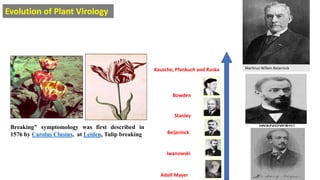
1. Viruses are submicroscopic infectious agents that are composed of nucleic acid and proteins and require a host cell to replicate. They were first observed in 1886 to cause tobacco mosaic disease.
2. In 1892, Beijerinck discovered that the infectious agent causing tobacco mosaic disease was a "contagium vivum fluidum", or "living fluid contagion", which was later shown to be tobacco mosaic virus (TMV) in 1935.
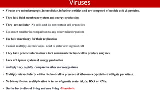
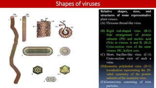
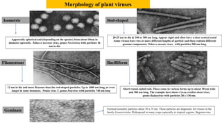
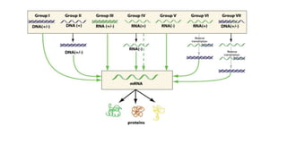
3. Viruses have a variety of shapes including rod-shaped, spherical, filamentous, and bacilliform. They contain a protein capsid that protects the viral genome, which can be DNA or RNA but not both. Viruses use