Vibriosis in marine fish
•Download as PPTX, PDF•
0 likes•426 views

Vibriosis is one of the most prevalent fish diseases caused by bacteria belonging the genus Vibrio affecting many marine and fresh water fishes. The disease characterized by septicemia, dermal ulceration, ascitis and haematopiotic necrosis.

Recommended
More Related Content
What's hot
What's hot (20)
Similar to Vibriosis in marine fish
Similar to Vibriosis in marine fish (20)
Recently uploaded
Recently uploaded (20)
Vibriosis in marine fish
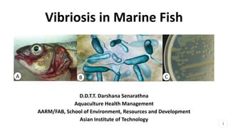
- 1. Vibriosis in Marine Fish D.D.T.T. Darshana Senarathna Aquaculture Health Management AARM/FAB, School of Environment, Resources and Development Asian Institute of Technology A B C 1
- 2. Outline Introduction Causative agent Host factors Disease pattern (Epidemiology) Pathogenesis Diagnosis Control Measures D 2
- 3. Introduction • Recognized as early as 1718 • Migrating eels Anguilla vulgaris in 1817 • A systemic bacterial infection • Both wild and farmed marine fishes • Economic losses in aquaculture industry • North America, Europe and Asia (Japan 1963) (Colwell and Grimes, 1984) (Mancuso et al., 2015) (Sindermann, 1984). RED DISEASE 3
- 4. Causative agent • Multilocus sequence analysis (MLSA) ftsZ, gapA, gyrB, mreB, pyrH, recA and TopA gene sequences from 96 taxa • Vibrio harveyi, V. vulnifcus, V. parahaemolyticus, V. alginolyticus, V. anguillarum, most common marine pathogenic species Domain: Bacteria Phylum: Proteobacteria Class: Gammaproteobacteria Order: Vibrionales Family: Vibrionaceae Genus: Vibrio (Sawabe et al., 2007). (Higuera et al., 2013). Vibrio anguillarum Infect more than 50 species of fish in temperate region Model microorganism to study the pathogenesis of vibriosis 4
- 5. Characteristics • Gram-negative • Facultative anaerobic pathogens • Opportunistic organisms • Oxidase positive, reduce nitrate to nitrite, ferment D-fructose, maltose and glycerol. • Thermos-dependent bacteria • Many species are halophiles straight or comma-shaped rod bacteria Polar flagella enclosed within a sheath (Raszl et al., 2016) (Vezzulli et al., 2015) sediment, water column and various aquatic plants. non-spore- forming Electron micrograph of V. anguillarum E 5
- 6. • 1. Capsular polysaccharides • 2. Adhesive factors • 3. Cytotoxins • 4. Chemotaxis and motility • 5. An iron uptake system • Extracellular products with proteolytic or haemolytic activity. Virulent Factors, Macrophage Outer membrane Inner membrane Pilli Flagella motor Flagella Heme Heme receptor Iron 1 2 3 4 5 (Deng et al., 2020) (N. Mohamad, et al. 2019) 6
- 7. Host factors • Susceptible hosts; salmon, turbot, sea bass, sea bream, cod, eel, ayu, and groupers • Susceptible stages; All age classes, high mortality among the young fry • Carriers; wild aquatic birds and both cultured and wild marine fish • Contaminated inputs and water from upstream farms, Infected eggs, juveniles & broodstock introduce Vibrio spp. (Fernández–Delgado et al., 2016) (Tendencia and Lavilla-Pitogo, 2004) F Sea bass fry dead with vibriosis 7
- 8. Disease pattern (Epidemiology) Transmission mechanism • water column - natural transmission medium • Horizontally - open lesions or as secretion in the faeces of infected fish/ carriers. Morbidity and Mortality – 30% - 100% Geographical distributions – worldwide (North America, Europe and Asia) Mechanical factors • Skin damage • High stocking density • Rough handling • Transferring newly caught wild fish into enclosed systems Risk Factors Environmental factors • Warmer months • Poor water quality 8
- 9. Pathogenesis • routes of entry - Ingestion and invasion via gill or epidermis cut and Gastro- intestinal tract • Virulence factors and Extracellular products Pathogenic Vibrio in the marine environment (Ceccarelli and Colwell, 2014) (Xu et al., 2017). Highly plastic genomes Horizontal virulent gene transfer Plasmids and insertion sequence common region (ISCR); mobile genetic elements 9
- 10. External Weight loss, red spots, swollen, rotting of fins, dark skin lesions Internal Organs appear enlarged, the lesion associated with gastroenteritis, ascites 2. Clinical aspects (Deng et al., 2020) (N. Mohamad, et al. 2019) Diagnosis (Level 1) 1. Abnormal behaviors Loss of appetite, abnormal swimming behavior with head floating near the surface of water. 10
- 11. Level 1:Gross signs Ulceration Exophthalmia Scale drops Necrosis Inflammation Haemorrhagic of liver and kidney Pale liver A A B A C A B B A C Hybrid grouper B Asian Sea bass C Red snapper 11
- 12. Histopathology • Hepatic lesions; congestion, haemorrhage, swollen hepatocytes with vacuolation and perivascular hepatocyets • Kidneys; hyaline degeneration and necrosis of the convoluted tubules, necrosis of the glomeruli, haemorrhage, congestion and presence of inflammatory cells I II III IV VI V I. myocardial haemorrhage, II. hyperplasia (hp) of the secondary lamellae leading to their fusion in the gill, III. congestion in the Kidney, IV. haemorrhage in the kidney parenchyma, V. MMCs (mmc) in the kidney, VI. necrotic changes in kidney tissue (I Frans et al. 2011) Diagnosis (Level 2) 12
- 13. Level 2: Hybrid grouper Epinephelus lanceolatus × E. fuscoguttatus infected with Vibrio spp. A) Severe to moderate congestion (c), generalize micro focal necrosis (n), generalize vacuolation of hepatocytes (v) in the liver B) Mild tubular necrosis (n) and haemorrhage (h) in the kidney C) Severe congestion (c) in the brain (H&E, 100×) (N. Mohamad, et al. 2019) 13
- 14. Diagnosis (Level 3) Improved plate count on mTCBS. (a) Colonies on thiosulfate‐citrate‐bile salts‐sucrose agar (TCBS) (standard medium). (b) Colonies onto mTCBS (modified medium) (Tagliavia et al.,2019) TCBS culture + PCR amplification and sequencing Ribotyping - rRNA-based phylogenetic analyses RAPD - Random Amplification of Polymorphic DNA ERIC-PCR - Enterobacterial Repetitive Intergenic Consensus (ERIC) PCR (N. Mohamad, et al., 2019) 16S rRNA 14
- 15. • A fluorescently labelled monoclonal antibody/ DAPI (4¢,6-diamidino-2- phenylondole) double staining technique was developed to detect V. anguillarum • Latex agglutination-based assay (BIONOR Mono-Va-kit) • Recently, an ELISA-based (Bionor AQUARAPIDVa test) and a magnetic particle enzyme immunoassay (Bionor AQUAEIA-Va test) • Loop-mediated isothermal amplification method (LAMP) (Notomi et al. 2000) (Gonzalez et al. 2004). (Miyamoto & Eguchi 1997) 15
- 16. Control Measures Prevention Biosecurity/husbandry practices • High quality fingerlings • Appropriate clean-up • Disinfection and dry-out procedures • Regular changing of cage culture net • Water quality management • Optimum stocking density • Appropriate chemical utilization methods • Dietary supplementation of vitamins and immunostimulants (N. Mohamad, et al. 2019) 16
- 17. Control Measures Treatments Therapeutic treatment • Antibiotics; Oxytetracycline, tetracycline, quinolones, nitrofurans, potentiated sulfonamides, trimethoprim, sarafloxacin, flumequine and oxolinic acid Prophylactic treatments • Inactivated vaccines and DNA vaccines AlphaJect 2000™, Aqua-Vac™Vibrio-Pasteurella and Aquavac Vibrio Oral®, Vibrio vaccine, ALPHA MARINETM Fish vaccination (N. Mohamad, et al. 2019) 17
- 18. Treatments (Cont.) Biocontrol agents • application of bacteria and phage (virus) - Vagococcus fluvialis and Bacillus subtilis • Bacteriophages belong to Siphoviridae family • disrupting the quorum sensing – Macro algae, micro algae, aquatic sponges Plants products • plant extracts as immunostimulants • Essential oil (EO), acetone and butanolic (Sorroza et al., 2012) (Touraki et al., 2012) SEM image of Bacillus subtilis G 18
- 19. References Sources of diagrams A. V. Anguillarum in seabass, exophthalmos lesions (picture by M. Isern), workshop, vibriosis in aquaculture, 16th EAFP Conference, Finland, 2013 B. V. vulnificus, Science photo library C. Colony of V. anguillarum PT-24 cultured on nutrient agar D. Vibrio parahaemolyticus, HD image E. Electron micrograph of V. anguillarum775 showing single polar flagellum. Shadowed preparation x 10,000. (Micrograph by Dr J.H. Crosa.) F. Dead Seabass fries by V. anguillarum, Photographs by Dr. Panos Varvarigos G. Bacillus subtilis, Science photo library 19
- 20. Bibliography • Colwell, R.R., Grimes, D.J., 1984. Vibrio diseases of marine fish populations. Helgoländer Meeresun. 37, 265– 287. • Mancuso, M., Genovese, M., Guerrera, M.C., Casella, G., Genovese, L., Piccolo, G., Maricchiolo, G., 2015. First episode of vibriosis in wild specimens of Pagellus bogaraveo (Brünnich, 1768) in the Mediterranean Sea. Cah. Biol. Mar. 56, 355–361. • Sindermann, C.J., 1984. Disease in marine aquaculture. Helgoländer Meeresun. 37 (1),505. • Sawabe, T., KitaTsukamoto, K., Thompson, F.L., 2007. Inferring the evolutionary history of Vibrios by means of multilocus sequence analysis. J. Bacteriol. 189 (21), 7932–7936 • Higuera, G., Bastías, R., Tsertsvadze, G., Romero, J., Espejo, R.T., 2013. Recently discovered Vibrio anguillarum phages can protect against experimentally induced vibriosis in Atlantic salmon, Salmo salar. Aquaculture 392, 128–133 • Raszl, S.M., Froelich, B.A., Vieira, C.R., Blackwood, D.A., Noble, R.T., 2016. Vibrio parahaemolyticus and Vibrio vulnificus in South America: water, seafood, and human infections. J. Appl. Microbiol. 121 (5), 1201–1222. 20
- 21. • Vezzulli, L., Pezzati, E., Stauder, M., Stagnaro, L., Venier, P., Pruzzo, C., 2015. Aquatic ecology of the oyster pathogens Vibrio splendidus and Vibrio aestuarianus. Environ. Microbiol. 17, 1065–1080 • Deng et al., 2020, Prevalence, virulence genes, and antimicrobial resistance of Vibrio species isolated from diseased marine fish in South China, https://doi.org/10.1038/s41598-020-71288-0 • N. Mohamad, et al., 2019, Vibriosis in cultured marine fishes: a review, https://doi.org/10.1016/j.aquaculture.2019.734289 • Fernández–Delgado, M., Sanz, V., Giner, S., Suárez, P., Contreras, M., Michelangeli, F., García– Amado, 2016. Prevalence and distribution of Vibrio spp. in wild aquatic birds of the Southern Caribbean Sea, Venezuela, 2011-12. J. Wildl. Dis. 52 (3), 621–626. • Tendencia, E.A., Lavilla-Pitogo, C.R., 2004. Chapter 2. Bacterial diseases. In: Nagasawa, K., Cruz- Lacierda, E.R. (Eds.), Diseases Of Cultured Groupers. Aquaculture Department, Southeast Asian Fisheries Development Center, Tigbauan, Iloilo, Philippines. • Ceccarelli, D., Colwell, R.R., 2014. Vibrio ecology, pathogenesis, and evolution. Front. Microbiol. 5, 256. • Xu, Y., Wang, C., Zhang, G., Tian, J., Liu, Y., Shen, X., Feng, J., 2017. ISCR2 is associated with the dissemination of multiple resistance genes among Vibrio spp. and Pseudoalteromonas spp. isolated from farmed fish. Arch. Microbiol. 199 (6), 891–896. 21
- 22. • Notomi T., Okayama H., Masubuchi H., Yonekawa T., Watanabe K., Amino N. & Hase T. (2000) Loop-mediated isothermal amplification of DNA. Nucleic Acids Research 28, e63i–e63vii. • Miyamoto N. & Eguchi M. (1997) Direct detection of a fish pathogen, Vibrio anguillarum serotype J-O-1, in freshwater by fluorescent antibody technique. Fisheries Science 63, 253–257. • Gonzalez S.F., Osorio C.R. & Santos Y. (2004) Evaluation of the AQUARAPID-Va, AQUAEIA-Va and dot-blot assays for the detection of Vibrio anguillarum in fish tissues. Journal of Fish Diseases 27, 617–621. • Sorroza, L., Padilla, D., Acosta, F., Román, L., Grasso, V., Vega, J., Real, F., 2012. Characterization of the probiotic strain Vagococcus fluvialis in the protection of European sea bass (Dicentrarchus labrax) against vibriosis by Vibrio anguillarum. Vet. Microbiol. 155 (2), 369–373 • Touraki, M., Karamanlidou, G., Karavida, P., Chrysi, K., 2012. Evaluation of the probiotics Bacillus subtilis and Lactobacillus plantarum bioencapsulated in Artemia nauplii against vibriosis in European sea bass larvae (Dicentrarchus labrax, L.). World J. Microbiol. Biotechnol. 28 (6), 2425– 2433. • I France et al., 2011, Vibrio anguillarum as a fish pathogen: virulence factors, diagnosis and prevention, doi:10.1111/j.1365-2761.2011.01279.x 22
- 23. THANK YOU!