VERMIFORM APPENDIX (GP 10).pptx with detailed diagrams
1.
GURU NANAK AYURVEDICMEDICAL COLLEGE & R.I
Submitted to-Dr. Dimple
Jagpaul
Submitted by-Ridhima, Dewansh, Jay a
Avantika negi, Shiya Bansa
VERMIFORM APPENDIX
2.
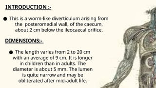
INTRODUCTION :-
● Thisis a worm-like diverticulum arising from
the posteromedial wall, of the caecum,
about 2 cm below the ileocaecal orifice.
DIMENSIONS:-
● The length varies from 2 to 20 cm
with an average of 9 cm. It is longer
in children than in adults. The
diameter is about 5 mm. The lumen
is quite narrow and may be
obliterated after mid-adult life.
3.
● POSITIONS:-
1.The appendixmay pass upwards and to the right. This is
paracolic or 11 o'clock position.
2 It may lie behind the caecum or colon, known as retrocaecal or
12 o'clock position. This is the commonest position of appendix,
about 65%.
3 The appendix may pass upwards and to the left. It points
towards the spleen. This is the splenic or 2 o'clock position. The
appendix may lie in front of the ileum (preileal) or behind the
ileum (postileal). The preileal type is most dangerous type.
4 It may pass horizontally to the left (as if pointing to the sacral
promontory called promontoric or 3 o'clock position.
4.
● POSITIONS:-
5 Itmay descend into the pelvis called pelvic
or 4 o'clock position. This is the second most
common position about 30%.
6 It may lie below the caecum (subcaecal) and
may point towards the inguinal ligament
called as midinguinal or 6 o'clock position
CLINICAL ANATOMY OFAPPENDIX
● Inflammation of the appendix is
known as appendicitis seen in
adolescent age. In this condition, it is
usuallynecessary to remove the
appendix. The operation for removal
of the appendix is called
appendicectomy.
● APPENDICITIS:-
7.
CLINICAL ANATOMY OFAPPENDIX
● With increasing inflammation,
pain is felt in the right iliac
fossa. This is caused by
involvement of the parietal
● Pain caused by appendicitis is first
felt in the reigon of umblicus.This is
called referred pain.
8.
CLINICAL ANATOMY OFAPPENDIX
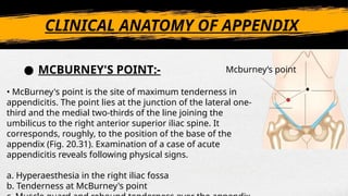
Mcburney’s point
• McBurney's point is the site of maximum tenderness in
appendicitis. The point lies at the junction of the lateral one-
third and the medial two-thirds of the line joining the
umbilicus to the right anterior superior iliac spine. It
corresponds, roughly, to the position of the base of the
appendix (Fig. 20.31). Examination of a case of acute
appendicitis reveals following physical signs.
a. Hyperaesthesia in the right iliac fossa
b. Tenderness at McBurney's point
● MCBURNEY'S POINT:-
9.
CLINICAL ANATOMY OFAPPENDIX
• Appendicular dyspepsia: Chronic appendicitis
produces dyspepsia resembling disease of
stomach, duodenum or gallbladder. It is due
passage of infected lymph to the subpyloric
nodes which cause irritation of pylorus. There is
history of earlier acute appendicitis.
● APPENDICULAR DYSPEPSIA:-