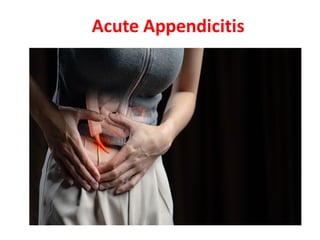
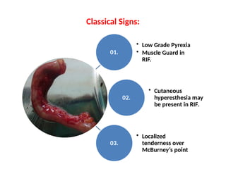
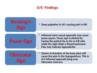
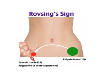
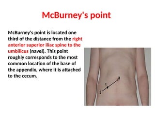
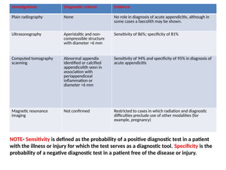
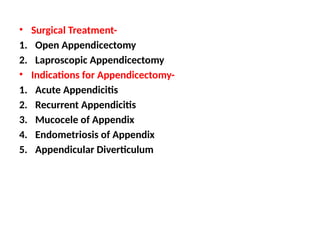
Anatomical Structure of Appendix-
The vermiform appendix is a tubular structure attached to the base of the
caecum. It is approximately 8-10 cm long in adults and represents the
underdeveloped distal end of the large caecum seen in other animals. In
humans it is regarded as a vestigial organ, and acute inflammation of this
structure is called acute appendicitis.
1. Retrocaecal / retrocolic (75%) —Right loin pain is often present, with
tenderness on examination. Muscular rigidity and tenderness to deep
palpation are often absent because of protection from the overlying
caecum. The psoas muscle may be irritated in this position, leading to
hip flexion and exacerbation of the pain on hip extension (psoas stretch
sign)
Subcaecal and pelvic (1.5%,21%
resp.)- Suprapubic pain and
urinary frequency may
predominate. Diarrhoea may be
present as a result of irritation of
the rectum. Abdominal
tenderness may be lacking, but
rectal or vaginal tenderness may
be present on the right.
Microscopic hematuria and
leucocytes may be present on
urine analysis.
Pre-ileal and post-ileal (1%,5%
resp.)—Signs and symptoms may
be lacking. Vomiting may be more
prominent, and diarrhea may
result from irritation of the distal
ileum.
Anatomical Positions of the Appendix
Etio-pathogenesis-
1. Obstruction of the appendiceal lumen. This can be from an
appendicolith or some other mechanical etiologies. - Appendiceal
tumors such as carcinoid tumors, appendiceal adenocarcinoma,
intestinal parasites, and hypertrophied lymphatic tissue are all known
causes of appendiceal obstruction and appendicitis.
2. When the appendiceal lumen gets obstructed, bacteria build up in the
appendix and cause acute inflammation with perforation and abscess
formation.
3. The appendix contains aerobic and anaerobic bacteria, including
Escherichia coli and Bacteroides. However, recent studies utilizing next-
generation sequencing revealed a significantly higher number of
bacterial phyla in patients with complicated perforated appendicitis.
4. Obstruction in appendix Increase intraluminal and intramural
pressure small vessel occlusion and lymphatic stasis. Once
obstructed, the appendix fills with mucus and becomes distended
lymphatic and vascular compromise advances Ischemia &
Necrosis of wall of Appendix Bacterial Overgrowth.
(Common organisms include Escherichia coli, Peptostreptococcus,
Bacteroides , and Pseudomonas).
5. Once significant inflammation and necrosis occur, the appendix is at
risk of perforation localized abscess and sometimes frank
peritonitis. The most common position of the appendix is retrocecal.
While the anatomical position of the root of the appendix is mostly
constant, tail positions may vary.
6. Possible positions include a. Retrocecal,
b. Subcecal,
c. Pre-and post-ileal
d. Pelvic.
Classical Symptoms:
01
• Peri-umbilical Colicky pain which later which later shifts to the
right iliac fossa. Coughing on sudden movement exacerbates
the pain.
02
• Fever – Usually there is mild rise in temp.
03
• Nausea and vomiting
Classical Signs:
01.
• Low Grade Pyrexia
• Muscle Guard in
RIF.
02.
• Cutaneous
hyperesthesia may
be present in RIF.
03.
• Localized
tenderness over
McBurney’s point
O/E- Findings:
• Deep palpation in LIF; causing pain in RIF.
Rovsing’s
Sign
• Inflamed retro-caecal appendix may cause
psoas spasm. Psoas sign is elicited by
having the patient lie on his or left side
while the right thigh is flexed backward.
Pain may indicate appendicitis.
Psoas Sign
• Flexion & Rotation of the knee joint will
cause the pain in the hypogastrium. This is
d/t inflamed appendix lying over
Obturator internus.
Obturator
Sign
Psoas Sign
Obturator Sign
McBurney's point
McBurney's point is located one
third of the distance from the right
anterior superior iliac spine to the
umbilicus (navel). This point
roughly corresponds to the most
common location of the base of
the appendix, where it is attached
to the cecum.
• Deep tenderness at McBurney's point, known as McBurney's sign, is a sign
of acute appendicitis.The clinical sign of referred pain in the epigastrium
when pressure is applied is also known as Aaron's sign. Specific localization
of tenderness to McBurney's point indicates that inflammation is no longer
limited to the lumen of the bowel (which localizes pain poorly), and is
irritating the lining of the peritoneum at the place where the peritoneum
comes into contact with the appendix.
• Tenderness at McBurney's point suggests the evolution of acute
appendicitis to a later stage, and thus, the increased likelihood of rupture.
Other abdominal processes can also sometimes cause tenderness at
McBurney's point. Thus, this sign is highly useful but neither necessary nor
sufficient to make a diagnosis of acute appendicitis.
• The anatomical position of the appendix is highly variable (for example in
retrocaecal appendix, an appendix behind the caecum), which also limits the
use of this sign, as many cases of appendicitis do not cause point tenderness
at McBurney's point. For most open appendectomies the incision is made at
McBurney's point.
Investigations
Investigations Diagnostic criteria Evidence
Plain radiography None No role in diagnosis of acute appendicitis, although in
some cases a faecolith may be shown.
Ultrasonography Aperistaltic and non-
compressible structure
with diameter >6 mm
Sensitivity of 86%; specificity of 81%
Computed tomography
scanning
Abnormal appendix
identified or calcified
appendicolith seen in
association with
periappendiceal
inflammation or
diameter >6 mm
Sensitivity of 94% and specificity of 95% in diagnosis of
acute appendicitis
Magnetic resonance
imaging
Not confirmed Restricted to cases in which radiation and diagnostic
difficulties preclude use of other modalities (for
example, pregnancy)
NOTE- Sensitivity is defined as the probability of a positive diagnostic test in a patient
with the illness or injury for which the test serves as a diagnostic tool. Specificity is the
probability of a negative diagnostic test in a patient free of the disease or injury.
• Surgical Treatment-
1. Open Appendicectomy
2. Laproscopic Appendicectomy
• Indications for Appendicectomy-
1. Acute Appendicitis
2. Recurrent Appendicitis
3. Mucocele of Appendix
4. Endometriosis of Appendix
5. Appendicular Diverticulum
Homoeopathic Management:
1. Belladonna
2. Mag. Phos
3. Colocynth
4. Nux Vomica
5. Calc. Carb

Acute Appendicitis and Homoeopathy......

  • 2.
    Anatomical Structure ofAppendix- The vermiform appendix is a tubular structure attached to the base of the caecum. It is approximately 8-10 cm long in adults and represents the underdeveloped distal end of the large caecum seen in other animals. In humans it is regarded as a vestigial organ, and acute inflammation of this structure is called acute appendicitis. 1. Retrocaecal / retrocolic (75%) —Right loin pain is often present, with tenderness on examination. Muscular rigidity and tenderness to deep palpation are often absent because of protection from the overlying caecum. The psoas muscle may be irritated in this position, leading to hip flexion and exacerbation of the pain on hip extension (psoas stretch sign)
  • 3.
    Subcaecal and pelvic(1.5%,21% resp.)- Suprapubic pain and urinary frequency may predominate. Diarrhoea may be present as a result of irritation of the rectum. Abdominal tenderness may be lacking, but rectal or vaginal tenderness may be present on the right. Microscopic hematuria and leucocytes may be present on urine analysis. Pre-ileal and post-ileal (1%,5% resp.)—Signs and symptoms may be lacking. Vomiting may be more prominent, and diarrhea may result from irritation of the distal ileum.
  • 4.
  • 5.
    Etio-pathogenesis- 1. Obstruction ofthe appendiceal lumen. This can be from an appendicolith or some other mechanical etiologies. - Appendiceal tumors such as carcinoid tumors, appendiceal adenocarcinoma, intestinal parasites, and hypertrophied lymphatic tissue are all known causes of appendiceal obstruction and appendicitis. 2. When the appendiceal lumen gets obstructed, bacteria build up in the appendix and cause acute inflammation with perforation and abscess formation. 3. The appendix contains aerobic and anaerobic bacteria, including Escherichia coli and Bacteroides. However, recent studies utilizing next- generation sequencing revealed a significantly higher number of bacterial phyla in patients with complicated perforated appendicitis. 4. Obstruction in appendix Increase intraluminal and intramural pressure small vessel occlusion and lymphatic stasis. Once obstructed, the appendix fills with mucus and becomes distended lymphatic and vascular compromise advances Ischemia & Necrosis of wall of Appendix Bacterial Overgrowth.
  • 6.
    (Common organisms includeEscherichia coli, Peptostreptococcus, Bacteroides , and Pseudomonas). 5. Once significant inflammation and necrosis occur, the appendix is at risk of perforation localized abscess and sometimes frank peritonitis. The most common position of the appendix is retrocecal. While the anatomical position of the root of the appendix is mostly constant, tail positions may vary. 6. Possible positions include a. Retrocecal, b. Subcecal, c. Pre-and post-ileal d. Pelvic.
  • 7.
    Classical Symptoms: 01 • Peri-umbilicalColicky pain which later which later shifts to the right iliac fossa. Coughing on sudden movement exacerbates the pain. 02 • Fever – Usually there is mild rise in temp. 03 • Nausea and vomiting
  • 8.
    Classical Signs: 01. • LowGrade Pyrexia • Muscle Guard in RIF. 02. • Cutaneous hyperesthesia may be present in RIF. 03. • Localized tenderness over McBurney’s point
  • 9.
    O/E- Findings: • Deeppalpation in LIF; causing pain in RIF. Rovsing’s Sign • Inflamed retro-caecal appendix may cause psoas spasm. Psoas sign is elicited by having the patient lie on his or left side while the right thigh is flexed backward. Pain may indicate appendicitis. Psoas Sign • Flexion & Rotation of the knee joint will cause the pain in the hypogastrium. This is d/t inflamed appendix lying over Obturator internus. Obturator Sign
  • 11.
  • 12.
  • 13.
    McBurney's point McBurney's pointis located one third of the distance from the right anterior superior iliac spine to the umbilicus (navel). This point roughly corresponds to the most common location of the base of the appendix, where it is attached to the cecum.
  • 14.
    • Deep tendernessat McBurney's point, known as McBurney's sign, is a sign of acute appendicitis.The clinical sign of referred pain in the epigastrium when pressure is applied is also known as Aaron's sign. Specific localization of tenderness to McBurney's point indicates that inflammation is no longer limited to the lumen of the bowel (which localizes pain poorly), and is irritating the lining of the peritoneum at the place where the peritoneum comes into contact with the appendix. • Tenderness at McBurney's point suggests the evolution of acute appendicitis to a later stage, and thus, the increased likelihood of rupture. Other abdominal processes can also sometimes cause tenderness at McBurney's point. Thus, this sign is highly useful but neither necessary nor sufficient to make a diagnosis of acute appendicitis. • The anatomical position of the appendix is highly variable (for example in retrocaecal appendix, an appendix behind the caecum), which also limits the use of this sign, as many cases of appendicitis do not cause point tenderness at McBurney's point. For most open appendectomies the incision is made at McBurney's point.
  • 15.
    Investigations Investigations Diagnostic criteriaEvidence Plain radiography None No role in diagnosis of acute appendicitis, although in some cases a faecolith may be shown. Ultrasonography Aperistaltic and non- compressible structure with diameter >6 mm Sensitivity of 86%; specificity of 81% Computed tomography scanning Abnormal appendix identified or calcified appendicolith seen in association with periappendiceal inflammation or diameter >6 mm Sensitivity of 94% and specificity of 95% in diagnosis of acute appendicitis Magnetic resonance imaging Not confirmed Restricted to cases in which radiation and diagnostic difficulties preclude use of other modalities (for example, pregnancy) NOTE- Sensitivity is defined as the probability of a positive diagnostic test in a patient with the illness or injury for which the test serves as a diagnostic tool. Specificity is the probability of a negative diagnostic test in a patient free of the disease or injury.
  • 16.
    • Surgical Treatment- 1.Open Appendicectomy 2. Laproscopic Appendicectomy • Indications for Appendicectomy- 1. Acute Appendicitis 2. Recurrent Appendicitis 3. Mucocele of Appendix 4. Endometriosis of Appendix 5. Appendicular Diverticulum
  • 17.
    Homoeopathic Management: 1. Belladonna 2.Mag. Phos 3. Colocynth 4. Nux Vomica 5. Calc. Carb