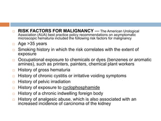
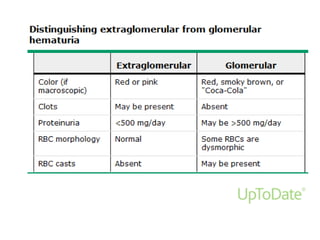
6. • Woman vs Man
• Premenopausal vs post menopausal
• Pregnancy vs non pregnancy
Uncomplicated UTIs in Adult
7. Definition
acute cystitis and acute
pyelonephritis in otherwise healthy
individuals
mostly in women without structural
and functional abnormalities within
the urinary tract, kidney diseases,
17. Recurrent (uncomplicated) UTIs
in women
Diagnosis
Recurrent UTIs are common among
young, healthy women, even though
they generally have anatomically and
physiologically normal urinary tracts .
Recurrent UTIs need to be diagnosed
by urine culture .
Excretory urography, cystography and
cystoscopy are not routinely
recommended for evaluation of
women with recurrent UTIs.
18. Prevention
Antimicrobial prophylaxis
Antimicrobial prophylaxis for prevention of recurrent UTI should be
considered only after counselling and behavioural modification has
been attempted (LE: 4, GR: A).
Before any prophylaxis regimen is initiated, eradication of a
previous UTI should be confirmed by a negative urine culture 1-2
weeks after treatment (LE: 4, GR: A).
Continuous or postcoital antimicrobial prophylaxis should be
considered to prevent recurrent uncomplicated cystitis in women in
whom non-antimicrobial measures have been unsuccessful (35)
(LE: 1a, GR: A).
The choice of antibiotics should be based upon the identification
and susceptibility pattern of the organism that causes the UTI and
the patient’s history of drug allergies. Drug regimens are shown in
Tables 3.3 and 3.4.
Recurrent (uncomplicated) UTIs
in women
21. Immunoactive prophylaxis
OM-89 (Uro-Vaxomâ) is sufficiently well-
documented and has been shown to be
more effective than placebo in several
randomised trials.
recommended for immunoprophylaxis in
female patients with recurrent
uncomplicated UTI (37,38) (LE: 1a, GR: B).
Recurrent (uncomplicated) UTIs
in women
22. Prophylaxis with probiotics
Accessibility of clinically proven probiotics
for UTI prophylaxis is currently not
universal.
Only the specifically in studies tested
Lactobacillus strains should be used for
prophylaxis.
Recurrent (uncomplicated) UTIs
in women
23. Prophylaxis with cranberry
Vaccinium macrocarpon is useful in reducing the
rate of lower UTIs in women (40,41) (LE: 1b, GR:
C).
For everyday practice, the daily consumption of
cranberry products, giving a minimum of 36 mg/day
proanthocyanindin A (the active compound), is
recommended (LE: 1b, GR: C).
The best approach is to use those compounds that
have demonstrated clear bioactivity in urine.
Recurrent (uncomplicated) UTIs
in women
24. UTIs in pregnancy
UTIs are common during pregnancy.
Most women acquire bacteriuria before
pregnancy
20-40% of women with asymptomatic
bacteriuria develop pyelonephritis during
pregnancy.
25. Definition of significant bacteriuria
Without symptom
2 consecutive voided urine specimens grow > 105
cfu/mL of the same bacterial species on quantitative
culture;
or a single catheterised specimen grows > 105
cfu/mL of a uropathogen (17) (LE: 2a, GR: A).
With symptom
Voided or catheterised urine specimen grows > 103
cfu/mL of a uropathogen (LE: 4, GR: B).
UTIs in pregnancy
26. Screening
Pregnant women should be screened for
bacteriuria during the first trimester (42) (LE: 1a,
GR: A).
UTIs in pregnancy
28. 3.6.5 Follow-up
Urine cultures should be obtained soon after
completion of therapy for asymptomatic bacteriuria
and symptomatic UTI in pregnancy (LE: 4, GR: A).
UTIs in pregnancy
29. Prophylaxis
Postcoital prophylaxis should be considered in
pregnant women with a history of frequent UTIs
before onset of pregnancy, to reduce their risk of UTI
(44) (LE: 2b, GR: B).
UTIs in pregnancy
31. Complicated UTI
Prolonged antibiotic therapy (7-10 days) should be
considered in this patient population (LE: 4, GR: B).
When indicated, ultrasonography or magnetic
resonance imaging (MRI) should be used
preferentially to avoid radiation risk to the foetus (LE:
4, GR: B).
UTIs in pregnancy
42. Urine cultures
Significant bacteriuria in a complicated UTI is defined by counts of
> 105 cfu/mL woman and > 104 cfu/mL MEN , in the MSU
straight catheter urine :> 104 cfu/mL. For an asymptomatic patient,
Asymptomatic PT: two consecutive urine cultures (at least 24 h
apart) yielding > 105 cfu/mL of the same microorganism
Pyuria :> 10 white blood cells (WBC) per high-power field (x400) in
the resuspended sediment of a centrifuged aliquot of urine or per
mm3 in unspun urine.
A dipstick method can also be used for routine assessment,
including a leukocyte esterase test, haemoglobin and probably a
nitrite reaction.
COMPLICATED UTIs
43. Microbiology
Spectrum and antibiotic resistance
diversity of microorganisms with a higher prevalence of resistance
against antimicrobials, and higher rates of treatment failure if the
underlying abnormality cannot be corrected.
E. coli, Proteus, Klebsiella, Pseudomonas and Serratia sp. and
enterococcienterococci are the usual strains found in cultures.
Enterobacteriaceae predominate (60- 75%) (6-8)
E. coli as the most common pathogen; particularly if the UTI is a
first infection.
Otherwise, the bacterial spectrum may vary over time and from one
hospital to another.
COMPLICATED UTIs
44. COMPLICATED UTIs
Complicated UTIs associated with urinary stones
Increased risk of urease-producing organisms,
Stag horn calculus disease
88% were found to have a UTI at the time of diagnosis
82% of patients infected with urease-producing organisms
The enzyme, urease, splits urea into carbon dioxide and ammonia.
increase in ammonia in the urine injures the glycosaminoglycan
layer, which in turn increases bacterial adherence and enhances
the formation of struvite crystals.
These aggregate to form renal stones and incrustations on urinary
catheters
The pathogenic potential of coagulase-negative staphylococci and
non-group D streptococci
46. Duration of antibiotic therapy
• Treatment for 7-14 days is generally
recommended
but the duration should be closely related to the
treatment of the underlying abnormality.
• prolongation for up to 21 days, according to the
clinical situation, is necessary
COMPLICATED UTIs
47. Follow-up after treatment
The greater likelihood of the
involvement of resistant
microorganisms in complicated
UTIs.
recurrent infection
urine cultures must be obtained for
the identification of the
microorganisms and the evaluation
of susceptibility testing.
COMPLICATED UTIs
57. Diabetes mellitus and UTI
Asymptomatic bacteriuria is common in
diabetic women
Organism :
Enterobacteriaceae that originate in the lower
urogenital tract.
Klebsiella infection is particularly common (25%
compared with 12% in non-diabetics).
Candida spp.
If untreated renal function impairment
58. Risk
diabetic nephropathy
autonomic neuropathy that causes voiding
dysfunction.
Impaired host resistance
Glycosuria inhibits phagocytosis and perhaps
cellular immunity, and encourages bacterial
adherence.
59. gas-forming
organisms,(emphysematouspyelonephritis)
acute pyogenic infiltration with micro-abscesses
and the development of acute renal failure.
The origin of the organisms may be
haematogenous.
intrarenal abscess that ruptures, which leads to a
perinephric collection and a psoas abscess.
Papillary necrosis permanent renal
parenchymal scarring
60. HIV and UTI
HIV infection can cause nephropathy( both acute
and chronic)
thrombotic microangiopathy, immune- mediated
glomerulonephritis and nephropathy due to virus-
induced cellular damage, primarily to the glomerular
epithelial cell
Prevention: corticosteroids, ACE inhibitors
HIV infection is therefore no longer a
contraindication to renal replacement therapy.
granulomatous infections :reduced cellular and
humoral immunity.
62. RISK OF INFECTION FOLLOWING
TRANSPLANTATION
Epidemiologic exposures
Community-acquired pathogens
Reactivation of infections
Nosocomial infections
Donor-derived infections
Net state of immunosuppression
63. TIMING OF INFECTION
POSTTRANSPLANTATION
First month after transplantation
Donor-derived infections
Recurrent infection
Infection may have been present in
the donor or in the recipient prior to
transplantation
Infectious complications related to
surgery
64. One to six months after transplantation
Major infections due to opportunistic pathogens
include:
Pneumocystis jirovecii
Latent infections, such as the protozoal diseases
including toxoplasmosis, leishmaniasis, and Chagas
disease
The geographic or endemic fungal infections caused
by Histoplasma capsulatum, Coccidioides spp,
Cryptococcus gattii, and, rarely, Blastomyces
dermatitidi
TIMING OF INFECTION
POSTTRANSPLANTATION
65. Viral pathogens, particularly the herpes group viruses
but also hepatitis B (HBV) and hepatitis C (HCV).
New viruses are recognized as opportunistic
pathogens with the use of more sensitive molecular
assays (eg, BK polyomavirus, human herpesvirus
[HHV]-6, -7, and -8
Tuberculosis and, increasingly, nontuberculous
mycobacteria
Gastrointestinal parasites (Cryptosporidium and
Microsporidium) and viruses (cytomegalovirus [CMV],
rotavirus) may be associated with diarrhea.
TIMING OF INFECTION
POSTTRANSPLANTATION
66. More than six months after
transplantation
stable and reduced levels of immunosuppression.
community-acquired pneumonias due to
respiratory viruses, the pneumococcus,
Legionella, or other common pathogens.
TIMING OF INFECTION
POSTTRANSPLANTATION
69. BK polyoma virus
Background
Polyoma family
Cause nephropathy
renal allografts with hemorrhagic cystitis, asymptomatic
viruria, interstitial nephritis, ureteric obstruction, and rising
creatinine values in renal transplant recipients
Screening:
Urine BK NAT (nucleic acid testing) ,identify the presence
of DNA and RNA
Diagnosis
Renal biopsy
70. When ?
monthly for the first 3–6 months after
transplantation (2D);
every 3 months until the end of the first post-
transplant year (2D);
unexplained rise in serum creatinine (2D);
And after treatment for acute rejection
71. treatment
• reducing immunosuppressive medications when BKV plasma
NAT is persistently greater than 10 000 copies/mL (107 copies/L).
(2D)
BK polyoma virus
72. treatment
antiviral therapy: cidofovir, leflunomide and/or
ciprofloxacin
there are no definitive data confirming their
effectiveness
BK polyoma virus
73. CMV
Clinical presentation
fever, leukopenia and atypical lymphocytosis
Or tissue-invasive infections (e.g. hepatitis,
pneumonitis and enteritis)
Risk for CMV after transplantation is strongly
depen- dent on donor (D) and recipient (R)
serology
D+/R−, D+/R+ or D−/R+ at risk for developing
CMV infection and disease
and D+/R− at highest risk for severe CMV
disease
The incidence of CMV disease in D−/R− is <5%.
74.
75. Diagnosis
NAT or culture.
screening
negative CMV is defined by the absence of CMV
IgG and IgM.
positive for CMV is defined as being CMV IgG-
positive.
76. CMV
treatment
recommend that all patients with serious (including
most patients with tissue invasive) CMV disease be
treated with intravenous ganciclovir. (1D)
recommend that CMV disease in adult KTRs that is
not serious (e.g. episodes that are associated with
mild clinical symptoms) be treated with either
intravenous ganciclovir or oral valganciclovir. (1D)
recommend that all CMV disease in pediatric KTRs be
treated with intravenous ganciclovir. (1D)
suggest continuing therapy until CMV is no longer
detectable by plasma NAT or pp65 antigenemia. (2D)
77. reducing immunosuppressive medication in
life-threatening CMV disease, and CMV
disease that persists in the face of treatment,
until CMV disease has resolved. (2D)
suggest monitoring graft function closely
during CMV disease. (2D)
CMV
78. CMV
Prevention
chemoprophylaxis for CMV infection with oral
ganciclovir or valganciclovir for at least 3 months
after transplantation, (1B) and for 6 weeks after
treatment with a T-cell–depleting antibody. (1C)
except when donor and recipient both have
negative CMV serologies
79. EPSTEIN-BARR VIRUS
Primary EBV (human herpes virus 4)
clinical syndromes associated with EBV and
lymphoproliferation, which range from self-
limited, polyclonal proliferation to malignancies
containing clonal chromosomal abnormalities
increased incidence of PTLD in KTRs
80. Definitive: biopsy affected tissue
EBV viral load
detectable and elevated in the vast majority of
KTRs with EBV disease
The EBV viral load becomes positive before the
development of EBV disease.
Reducing immunosuppressive medication may
prevent EBV disease and PTLD.
EPSTEIN-BARR VIRUS
81. Treatment
Reducing immunosuppressive medication may
prevent EBV disease and PTLD.
The potential role of antiviral therapy as a
preemptive response to a rising viral load is
controversial
EBV-seronegative patients with an increasing
EBV load have immunosuppressive medication
reduced. (2D)
recommend that patients with EBV disease,
including PTLD, have a reduction or cessation of
immunosuppressive medication. (1C)
EPSTEIN-BARR VIRUS
82. prevention
suggest monitoring high-risk (donor EBV
seropositive/recipient seronegative) KTRs for EBV
by NAT (2C)
once in the first week after transplantation (2D);
then at least monthly for the first 3–6 months after
transplantation (2D);
then every 3 months until the end of the first post-
transplant year (2D);
and additionally after treatment for acute rejection.
(2D)
EPSTEIN-BARR VIRUS
83. HERPES SIMPLEX VIRUS 1, 2
AND VARICELLA ZOSTER VIRUS
Superficial HSV 1, 2 infection be treated (1B) with
an appropriate oral antiviral agent (e.g. acyclovir,
valacyclovir, or famci- clovir) until all lesions have
resolved
Systemic HSV 1, 2 infection be treated (1B) with
intravenous acyclovir and a reduction in
immunosuppressive medication. (1D)
intravenous acyclovir continue until the patient has a
clinical response, (1B) then switch to an appropriate
oral antiviral agent (e.g. acyclovir, valacyclovir, or
famciclovir) to complete a total treatment duration of
14–21 days. (2D)
84. prophylactic antiviral agent for KTRs
experiencing frequent recurrences of HSV 1,2
infection. (2D)
prevention of primary varicella zoster with
active varicella zoster infection (1D):
varicella zoster immunoglobulin (or in- travenous
immunoglobulin) within 96 hours of exposure (1D)
immunoglobulin is not available or more than 96 h
have passed, a 7-day course of oral acyclovir
begun 7–10 days after varicella exposure. (2D)
HERPES SIMPLEX VIRUS 1, 2
AND VARICELLA ZOSTER VIRUS
85. Adenovirus :hemorrhagic nephritis/cystitis
picture diagnosed by culture or antigen
detection/immunofluorescence
Parvovirus B19 :anemia unresponsive to
erythropoietin or with myocarditis
86. PCP
Pneumocystis jirovecii (formally known as
Pneumocystis carinii)
opportunistic fungal pathogen known to cause
life-threatening pneumonia in
immunocompromised patients, including KTR
definitive diagnosis
demonstration of organisms in lung tissue or
lower respiratory tract secretions.
87. PCP
recommend that all KTRs receive PCP
prophylaxis with daily trimethoprim–
sulfamethoxazole for 3–6 months after
transplantation. (1B)
suggest that all KTRs receive PCP prophylaxis
with daily trimethoprim– sulfamethoxazole for
at least 6 weeks during and after treatment for
acute rejection
89. PCP
Treatment
recommend that KTRs with PCP diagnosed by
bronchial alveolar lavage and/ or lung biopsy be
treated with high-dose intravenous trimethoprim–
sulfamethoxa- zole, corticosteroids, and a
reduction in immunosuppressive medication. (1C)
recommend treatment with corticosteroids for
KTRs with moderate to severe PCP (as defined
by PaO2 <70 mm Hg in room air or an alveolar
gradient of >35 mm Hg). (1C)
duration of treatment is 2–3 weeks
90. TUBERCULOSIS
TB prophylaxis and treatment regimens be the
same in KTRs as would be used in the local,
general popu- lation who require therapy. (2D)
recommend monitoring CNI and mTORi blood
levels in patients receiving rifampin. (1C)
Consider substituting rifabutin for rifampin to
minimize interacions with CNIs and mTORi.
92. CANDIDA spp
increased risk for oral and esophageal
infections
prophylaxis with oral clotrimazole lozenges,
nystatin, or fluconazole for 1– 3 months after
transplantation, and for 1 month after
treatment with an antilym- phocyte antibody.
(2C)
93. Common after organ transplantation
Risk 25-80 %
Decrease by prophylaxis ATB , low dose
immunosuppressive
Kidney allograft pyelonephritis may be
associated with bacteremia, metastatic
spread, impaired graft function and even
death.
KTRs with clinical and laboratory
evidence suggestive of kidney allograft
pyelonephritis should be hospitalized and
treated with intravenous antibiotics.
UTI in renal transplantation
94. Donor organ infection
Screen for viral and bacterial infection
Bladder catheter and ureteric stent :
microorganism producing bioflim
cover FB
UTI in renal transplantation
97. Treatment of UTI in renal transplant recipient
Fluoroquinolones : ATB of choice , penetrating to
renal parenchyma
If recurrent infection : evaluation anatomical
cause,prolong course of ATB
Schistosomiasis : Praziquantil and oxaninoquine
1 mo
UTI in renal transplantation
102. Gross hematuria
red or brown urine.
little as 1 mL of blood per liter of
urine can induce a visible color
change
In addition, the intermittent
excretion of red to brown urine can
be seen in a variety of clinical
conditions other than bleeding into
the urinary tract
105. A recent upper respiratory infection raises the possibility of postinfectious
glomerulonephritis or IgA nephropathy or sometimes hereditary nephritis.
A positive family history of renal disease, as in hereditary nephritis, polycystic kidney
disease, or sickle cell disease.
Unilateral flank pain, which may radiate to the groin, suggests ureteral obstruction due
to a calculus or blood clot, but can occasionally be seen with malignancy. Flank pain
that is persistent or recurrent can also occur in the rare loin pain-hematuria syndrome.
Symptoms of prostatic obstruction in older men such as hesitancy and dribbling. The
cellular proliferation in benign prostatic hyperplasia (BPH)
Recent vigorous exercise or trauma in the absence of another possible cause.
History of a bleeding disorder or bleeding from multiple sites due to excessive
anticoagulant therapy.
Cyclic hematuria in women that is most prominent during and shortly after
menstruation, suggesting endometriosis of the urinary tract
Travel or residence in areas endemic for Schistosoma haematobium or tuberculosis.
Sterile pyuria with hematuria, which may occur with renal tuberculosis, analgesic
nephropathy and other interstitial diseases.
106. RISK FACTORS FOR MALIGNANCY — The American Urological
Association (AUA) best practice policy recommendations on asymptomatic
microscopic hematuria included the following risk factors for malignancy
Age >35 years
Smoking history in which the risk correlates with the extent of
exposure
Occupational exposure to chemicals or dyes (benzenes or aromatic
amines), such as printers, painters, chemical plant workers
History of gross hematuria
History of chronic cystitis or irritative voiding symptoms
History of pelvic irradiation
History of exposure to cyclophosphamide
History of a chronic indwelling foreign body
History of analgesic abuse, which is also associated with an
increased incidence of carcinoma of the kidney
110. Transient hematuria can also occur with
urinary tract infection (eg, cystitis or
prostatitis).
Dysuria: pyuria and bacteriuria
Painless : hematuria from bladder cancer.
transient hematuria occurs in patients over age
40 increased risk of malignancy