Download to read offline


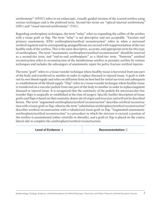
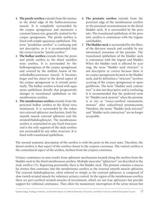
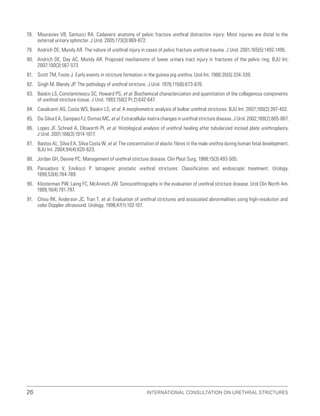
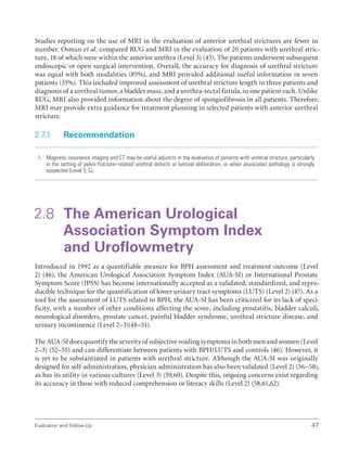
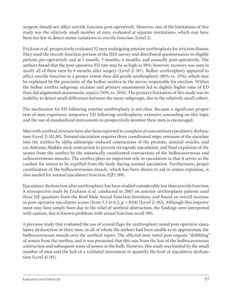
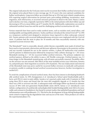
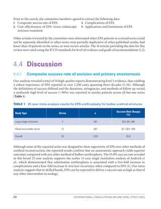
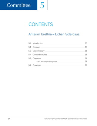
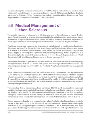
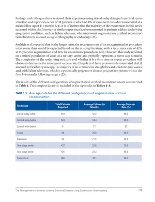
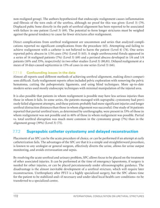
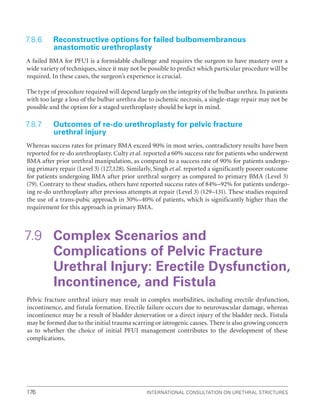
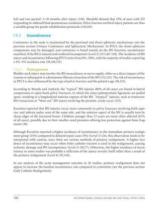
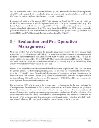
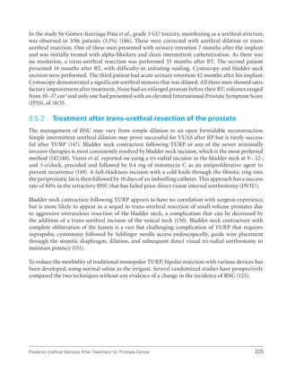
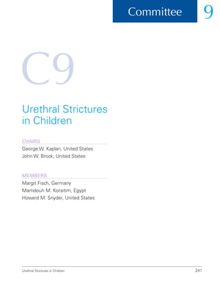
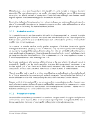
![XVIII
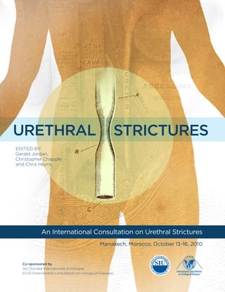
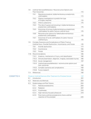
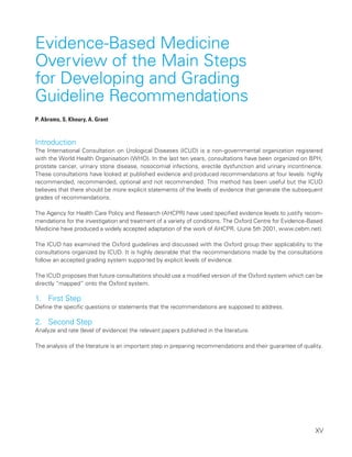
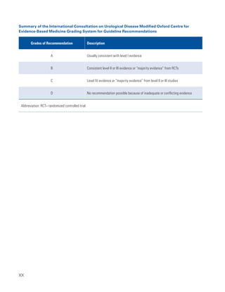
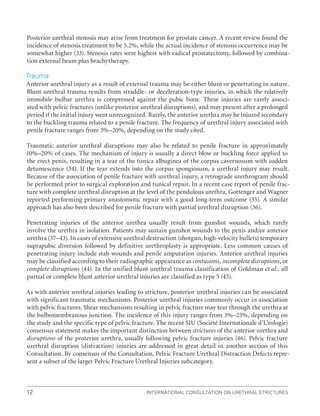
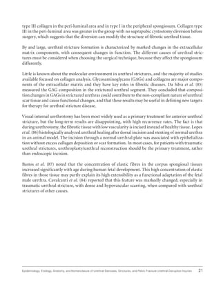
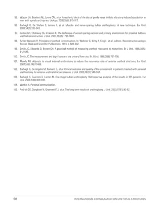
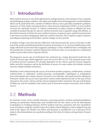
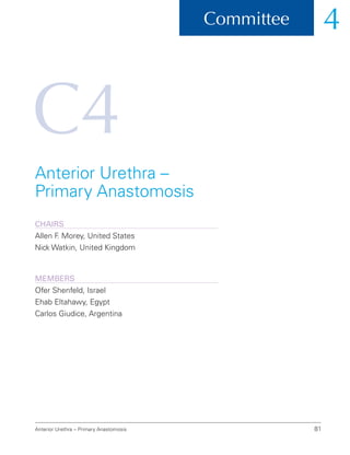
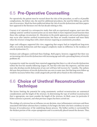
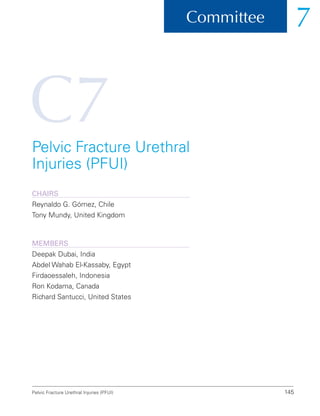
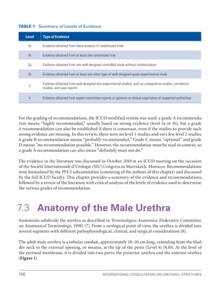
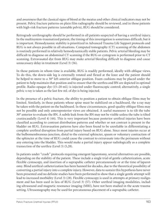
Level of
Evidence
Criteria
I
Incorporates Oxford 1a, 1b
Usually involves:
‚
‚ meta-analysis of trials (randomized controlled trials [RCTs]) or,
‚
‚ a good-quality RCT or,
‚
‚ “all or none” studies in which treatment is not an option (e.g. in vesicovaginal fistula)
II
Incorporates Oxford 2a, 2b and 2c
Includes:
‚
‚ low-quality RCT (e.g. 80% follow-up),
‚
‚ meta-analysis (with homogeneity) of good-quality prospective cohort studies
May include a single group when individuals who develop the condition are compared with others from
within the original cohort group.
There can be parallel cohorts, where those with the condition in the first group are compared with those
in the second group
III
Incorporates Oxford 3a, 3b and 4
Includes:
‚
‚ good-quality retrospective case-control studies, where a group of patients who have a condition
are matched appropriately (e.g. for age, sex, etc.) with control individuals who do not have the condition
‚
‚ good-quality case series, where a complete group of patients, all with the same condition, disease or
therapeutic intervention, are described without a comparison control group
IV
Incorporates Oxford 4
Includes expert opinion, where the opinion is based not on evidence but on “first principles”
(e.g. physiological or anatomical) or bench research.
The Delphi process can be used to give expert opinion greater authority:
‚
‚ involves a series of questions posed to a panel
‚
‚ answers are collected into a series of “options”
‚
‚ these “options” are serially ranked; if a 75% agreement is reached, then a Delphi consensus statement
can be made
6.2 Grades of recommendation
The ICUD will use the four grades from the Oxford system. As with levels of evidence, the grades of evidence
may apply either positively (procedure is recommended) or negatively (procedure is not recommended). Where
there is disparity of evidence, for example if there were three well-conducted RCTs indicating that Drug A was
superior to placebo, but one RCT whose results show no difference, then there has to be an individual judg-
ment as to the grade of recommendation given and the rationale explained.
Grade A recommendation usually depends on consistent level I evidence and often means that the recom-
mendation is effectively mandatory and placed within a clinical-care pathway. However, there will be occasions
where excellent evidence (level I) does not lead to a Grade A recommendation, for example, if the therapy is
prohibitively expensive, dangerous or unethical. Grade A recommendation can follow from Level II evidence.
However, a Grade A recommendation needs a greater body of evidence if based on anything except Level I
evidence.
Grade B recommendation usually depends on consistent level 2/3 studies, or “majority evidence” from RCTs.
Grade C
recommendation usually depends on level 4 studies or “majority eviden
ce” from level 2/3 studies or
Delphi processed expert opinion.
Grade D “
No recommendation possible” would be used where the evidence is inadequate or conflicting and
when expert opinion is delivered without a formal analytical process, such as by Delphi.](https://image.slidesharecdn.com/urethralstrictures2010-221127135159-2095864b/85/urethral_strictures_2010-pdf-19-320.jpg)


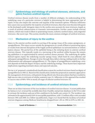
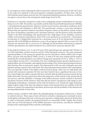
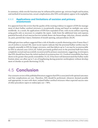
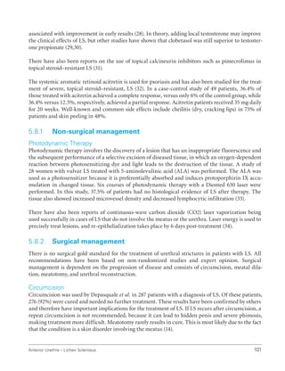
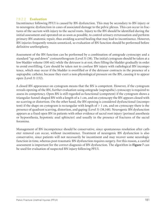
![International Consultation on Urethral Strictures
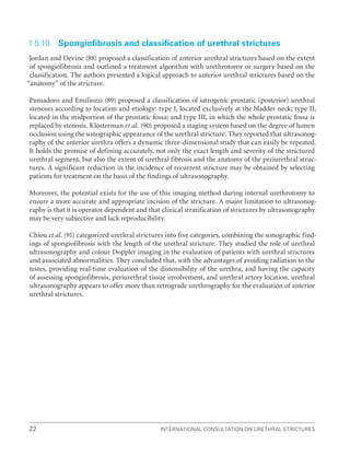
6
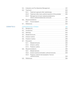
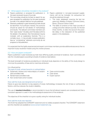
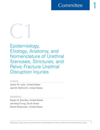
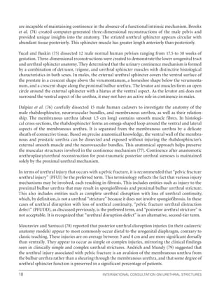
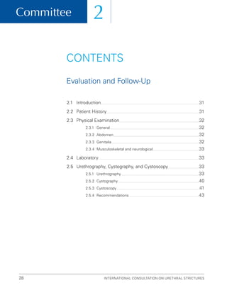
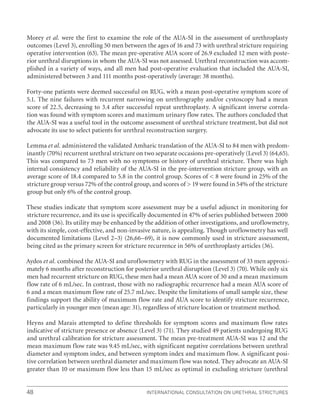
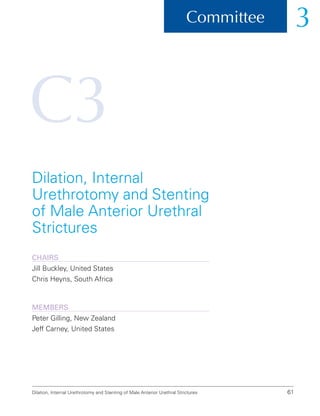
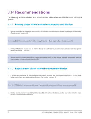
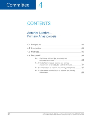
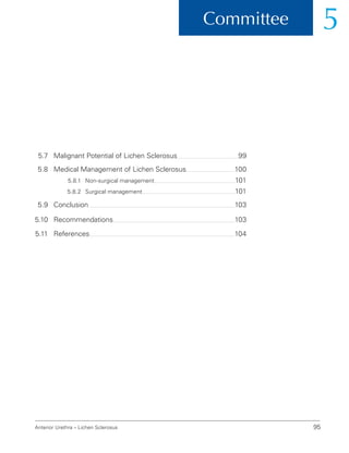
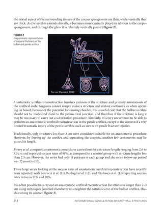
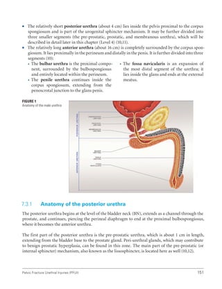
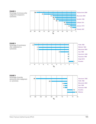
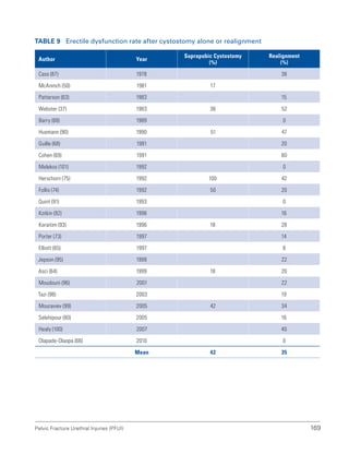
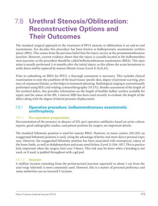
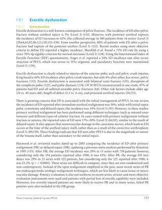
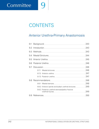
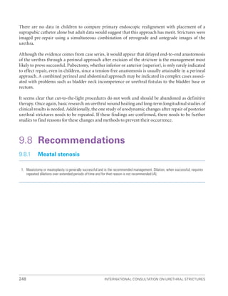
1.3.1 Quality of the references
Level 1: Meta-analyses of randomized controlled trials (RCTs) or good-quality prospective RCTs.
(Oxford 1a,b) [0 references]
Level 2: Clinical studies in which the data were collected prospectively and retrospective analyses
based on clearly reliable data. Types of studies so classified include: observational studies, cohort
studies, prevalence studies, low-quality RCTs, and case-control studies. (Oxford 2a,b,c) [0references]
Level 3: Studies based on retrospectively collected data. Evidence in this class includes case-control
studies, clinical case series, and database or registry reviews. (Oxford 3a,b,c) [~90 references]
Level 4: Expert opinion (Oxford 4)
1.4 Recommendations
There is insufficient Level 1 and 2 evidence/data to support any grade A or B recommendations, but
there is Level 3 evidence to support grade C recommendations regarding the epidemiology, anatomy,
and nomenclature of urethral stenoses, urethral strictures, and pelvic fracture urethral injuries.
1.5 Scientific Foundations
1.5.1
Nomenclature pertinent to urethral stenoses, strictures, and
pelvic fracture urethral injuries
Urethral “stricture” is the preferred term to describe an abnormal narrowing of any segment of the
urethra surrounded by corpus spongiosum, and specifically implies varying degrees of spongio-
fibrosis. The term “spongiofibrosis” refers to scarring of the corpus spongiosum of varying degrees.
Urethral “stricture disease” implies the underlying etiology. The Consultation Committee recom-
mends that urethral terminology should be anatomical; therefore, the preferred term to describe
urethral narrowing/obliteration is urethral “stricture.” The term “stricture disease” should be
reserved as a second-tier term. The term “stenosis” is reserved for narrowing of the membranous
urethra not secondary to pelvic fracture urethral injury, the prostatic urethra, and the bladder neck,
as they are not invested by corpus spongiosum. Importantly, the term “stenosis” does not imply
spongiofibrosis.
Urethral “calibration” refers to the measurement of the calibre (diameter) of the urethral lumen
by various techniques. Urethral “dilation” refers to the stretching or enlargement of the urethral
lumen by various techniques. The Consultation recognizes that the term “dilatation” is used inter-
changeably. “Urethrotomy” is the general term to describe the incision of urethral epithelium and
underlying spongiosum by either endoscopic or open techniques. “Internal urethrotomy” refers
to an endoscopic urethrotomy performed with or without visual guidance. “Direct vision internal](https://image.slidesharecdn.com/urethralstrictures2010-221127135159-2095864b/85/urethral_strictures_2010-pdf-35-320.jpg)


![23
Epidemiology, Etiology, Anatomy, and Nomenclature of Urethral Stenoses, Strictures, and Pelvic Fracture Urethral Disruption Injuries
1.6 References
1. Turner-Warwick R. Principles of urethral reconstruction. In: Webster GD, Kirby R, King L, et al., editors. Reconstructive urology.
Oxford: Blackwell Scientific Publications; 1993. p. 609-642.
2. Devine CJ, Devine PC, Felderman TP, et al. Classification and standardization of urethral strictures [abstract #325]. Presented at:
The 78th American Urologic Association Annual Meeting; 1983 Apr 17-21; Las Vegas.
3. Jordan GH, Devine CJ, Devine PC. An anatomical approach to urethral stricture disease [abstract]. J Urol. 1986;135:210A.
4. Jordan GH. Management of anterior urethral stricture disease. In: Webster GD, Kirby R, King L, et al., editors. Reconstructive
urology. Oxford: Blackwell Scientific Publications; 1993. p. 703-723.
5. Santucci RA, Joyce GF, Wise M. Male urethral stricture disease. J Urol. 2007;177(5):1667-1674.
6. Anger JT, Santucci RA, Grossberg AL, et al. The morbidity of urethral stricture disease among male Medicare beneficiaries. BMC
Urol. 2010;10:3.
7. Fenton AS, Morey AF, Aviles R, et al. Anterior urethral strictures: Etiology and characteristics. Urology. 2005;65(6):1055-1058.
8. Lumen N, Hoebeke P, Willemsen P, et al. Etiology of urethral stricture disease in the 21st century. J Urol. 2009;182(3):983-987.
9. McConnell JD, Barry MJ, Bruskewitz RC. Benign prostatic hyperplasia: Diagnosis and treatment (Quick reference guide for
clinicians). Rockville (MD): Agency for Health Care Policy and Research, Public Health Service, US Department of Health and
Human Services; 1994. Publication No.: AHCPR 94-0583. p. 1-17.
10. Jorgensen PE, Weis N, Bruun E. Etiology of urethral stricture following transurethral prostatectomy. Scand J Urol Nephrol.
1986;20(4):253-255.
11. Zheng W, Vilos G, McCulloch S, et al. Electrical burn of urethra as a cause of stricture after transurethral resection. J Endourol.
2000;14(2):225-228.
12. Sall M, Bruskewitz RC. Prostatic urethral strictures after transurethral microwave thermal therapy for benign prostatic
hyperplasia. Urology. 1997;50(6):983-985.
13. Biering-Sorensen F, Nielsen K, Vest Hansen H. Urethral epithelial cells on the surface of hydrophilic catheters after intermittent
catheterization: Cross-over study with two catheters. Spinal Cord. 1999; 37(4): 299-300.
14. Wyndaele JJ, Maes D. Clean intermittent self-catheterization: A 12-year followup. J Urol. 1990;143(5):906-908.
15. Albers P, Fichtner J, Bruhl P, et al. Long-term results of internal urethrotomy. J Urol. 1996;156(5):1611-1614.
16. Heyns CF, Steenkamp JW, DeKock MLS, et al. Treatment of male urethral strictures: Is repeated dilation or internal urethrotomy
useful? J Urol. 1998;160(2):356-358.
17. Barbagli G, Azzaro F, Menchi I, et al. Bacteriologic, histologic and ultrasonographic findings in strictures recurring after
urethrotomy. Scand J Urol Nephrol. 1995;29(2):193-195.
18. Kjaergaard B, Walter S, Bartholin J, et al. Prevention of urethral stricture recurrence using clean intermittent self-catheterization.
Br J Urol. 1994;73(6):692-695.
19. Badlani GH, Press SM, Defalco A, et al. UroLume endourethral prosthesis for the treatment of urethral stricture disease: Long-
term results of the North American multicenter UroLume trial. Urology. 1995;45(5):846-856.
20. Parikh AM, Milroy EJG. Precautions and complications in the use of the UroLume wallstent. Eur Urol. 1995;27(1):1-7.
21. Bailey DM, Foley SJ, McFarlane JP, et al. Histological changes associated with long-term urethral stents. Br J Urol.
1998;81(5):745-749.
22. Jordan GH. UroLume endoprosthesis for the treatment of recurrent bulbous urethral stricture. AUA Update Series. 2000;19(3):18-23.
23. Lal R, Bhatnagar V, Mitra DK. Urethral strictures after fulguration of posterior urethral valves. J Pediatr Surg. 1998;33(3):518-519.
24. Misra D, Chana J, Drake DP, et al. Operative trauma to the genitourinary tract in the treatment of anorectal malformations: 15
years’ experience. Urology. 1996;47(4):559-562.](https://image.slidesharecdn.com/urethralstrictures2010-221127135159-2095864b/85/urethral_strictures_2010-pdf-52-320.jpg)


![International Consultation on Urethral Strictures
58
48. Yalla SV, Sullivan MP, Lecamwasam HS, et al. Correlation of American Urological Association symptom index with obstructive
and nonobstructive prostatism. J Urol. 1995;153(3 Pt 1):674-679.
49. Chancellor MB, Rivas DA, Keeley FX, et al. Similarity of the American Urological Association Symptom Index among men with
benign prostate hyperplasia (BPH), urethral obstruction not due to BPH and detrusor hyperreflexia without outlet obstruction.
Br J Urol. 1994;74(2):200-203.
50. Ko DS, Fenster HN, Chambers K, et al. The correlation of multichannel urodynamic pressure-flow studies and American Urological
Association symptom index in the evaluation of benign prostatic hyperplasia. J Urol. 1995;154(2 Pt 1):396-398.
51. Roberts RO, Jacobsen SJ, Jacobsen DJ, et al. Natural history of prostatism: High American Urological Association Symptom
scores among community-dwelling men and women with urinary incontinence. Urology. 1998;51(2):213-219.
52. Chai TC, Belville WD, McGuire EJ, et al. Specificity of the American Urological Association voiding symptom index: Comparison
of unselected and selected samples of both sexes. J Urol. 1993;150(5 Pt 2):1710-1713.
53. Chancellor MB, Rivas DA. American Urological Association symptom index for women with voiding symptoms: Lack of index
specificity for benign prostate hyperplasia. J Urol. 1993;150(5 Pt 2):1706-1708.
54. Groutz A, Blaivas JG, Fait G, et al. The significance of the American Urological Association symptom index score in the evaluation
of women with bladder outlet obstruction. J Urol. 2000;163(1):207-211.
55. Scarpero HM, Fiske J, Xue X, et al. American Urological Association Symptom Index for lower urinary tract symptoms in women:
Correlation with degree of bother and impact on quality of life. Urology. 2003;61(6):1118-1122.
56. Plante M, Corcos J, Gregoire I, et al. The international prostate symptom score: Physician versus self-administration in the
quantification of symptomatology. Urology. 1996;47(3):326-328.
57. Barry MJ, Fowler FJ, Chang Y, et al. The American Urological Association symptom index: Does mode of administration affect its
psychometric properties? J Urol. 1995;154(3):1056-1059.
58. Netto Júnior NR, de Lima ML. The influence of patient education level on the International Prostatic Symptom Score. J Urol.
1995;154(1):97-99.
59. Sagnier PP, Richard F, Botto H, et al. Adaptation and validation in the French language of the International Score of Symptoms of
Benign Prostatic Hypertrophy [Article in French]. Prog Urol. 1994;4(4):532-538.
60. Arocho R, Kason NM, Colon B, et al. Translation and validation of the American Urological Association symptom index into
Spanish. Clin Ther. 1995;17(4):777-785.
61. MacDiarmid SA, Goodson TC, Holmes TM, et al. An assessment of the comprehension of the American Urological Association
Symptom Index. J Urol. 1998;159(3):873-874.
62. Johnson TV, Abbasi A, Ehrlich SS, et al. Patient misunderstanding of the individual questions of the American Urological
Association symptom score. J Urol. 2008;179(6):2291-2294.
63. Morey AF, McAninch JW, Duckett CP, et al. American Urological Association symptom index in the assessment of urethroplasty
outcomes. J Urol. 1998;159(4):1192-1194.
64. Lemma B, Taye M, Hawando T, et al. International prostate symptom score as a clinical outcome measure for Ethiopian patients
with urethral stricture. Ethiop Med J. 2004;42(2):277-281.
65. Lemma B, Bakke A, Taye M, et al. Validation of the Amharic translation of international prostate symptom score in Ethiopian
patients. Ethiop J Health Dev. 2001;15(3):203-208.
66. Drach GW, Layton TN, Binard WJ. Male peak urinary flow rate: Relationships to volume voided and age. J Urol. 1979;122(2):210-
214.
67. Bukurov NS, Stefanovic KB, Marinkovic JM. Uroflow via stenotic urethra. Int Urol Nephrol. 1992;24(2):55-63.
68. Rosier P, de la Rosette JJ, Koldewijn EL, et al. Variability of pressure-flow analysis parameters in repeated cystometry in patients
with benign prostatic hyperplasia. J Urol. 1995;153(5):1520-1525.
69. Reynard JM, Yang Q, Donovan JL, et al. The ICS, ‘BPH’ Study: Uroflowmetry, lower urinary tract symptoms and bladder outlet
obstruction. Br J Urol. 1998;82(5):619-623.](https://image.slidesharecdn.com/urethralstrictures2010-221127135159-2095864b/85/urethral_strictures_2010-pdf-87-320.jpg)
![Evaluation and Follow-Up 59
70. Aydos MM, Memis A, Yakupoglu YK, et al. The use and efficacy of the American Urological Association Symptom Index in
assessing the outcome of urethroplasty for post-traumatic complete posterior urethral strictures. BJU Int. 2001;88(4):382-384.
71. Heyns CF, Marais DC. Prospective evaluation of the American Urological Association symptom index and peak urinary flow rate
for the followup of men with known urethral stricture disease. J Urol. 2002;168(5):2051-2054.
72. Jorgensen JB, Jensen KM-E, Klarskov P, et al. Intra- and inter- observer variations in classification of urinary flow curve patterns.
Neurourol Urodyn. 1990;9(5):535-539.
73. Klarskov P. Urologic diagnosis by urinary flow measurements with the Glostrup flowmeter [Article in Danish]. Ugeskr Laeger.
1974;136(48):2684-2686.
74. Shoukry I, Susset JG, Elhilali MM, et al. Role of uroflowmetry in the assessment of lower urinary tract obstruction in adult males.
Br J Urol. 1975;47(5):599-566.
75. Gacci M, Del Popolo G, Artibani W, et al. Visual assessment of uroflowmetry curves: Description and interpretation by
urodynamists. World J Urol. 2007;25(3):333-337.
76. Erickson BA, Breyer BN, McAninch JW. The use of uroflowmetry to diagnose recurrent stricture after urethral reconstructive
surgery. J Urol. 2010;184(4):1386-1390.
77. Garibay JT, Reid C, Gonzalez R. Functional evaluation of the results of hypospadias surgery with uroflowmetry. J Urol. 1995;
154(2 Pt 2):835-836.
78. Kaya C, Kucuk E, Ilktac A, et al. Value of urinary flow patterns in the follow-up of children who underwent Snodgrass operation.
Urol Int. 2007;78(3):245-248.
79. Dunsmuir WD, Fenely M, Corry DA, et al. The day-to-day variation (test-retest reliability) of residual urine measurement. Br J
Urol. 1996;77(2):192-193.
80. Mochtar CA, Kiemeney LA, van Riemsdijk MM, et al. Post-void residual urine volume is not a good predictor of the need for
invasive therapy among patients with benign prostatic hyperplasia. J Urol. 2006;175(1):213-216.
81. Kessler TM, Fisch M, Heitz M, et al. Patient satisfaction with the outcome of surgery for urethral stricture. J Urol. 2002;167(6):2507-
2511.
82. Coursey JW, Morey AF, McAninch JW, et al. Erectile function after anterior urethroplasty. J Urol. 2001;166(6): 2273-2276.
83. Erickson BA, Wysock JS, McVary KT, et al. Erectile function, sexual drive, and ejaculatory function after reconstructive surgery
for anterior urethral stricture disease. BJU Int. 2007;99(3):607-611.
84. O’Leary MP, Fowler FJ, Lenderking WR, et al. A brief male sexual function inventory for urology. Urology. 1995;46(5):697-706.
85. Rosen RC, Riley A, Wagner G, et al. The international index of erectile function (IIEF): A multidimensional scale for assessment of
erectile dysfunction. Urology. 1997;49(6):822-830.
86. Rosen RC, Cappelleri JC, Smith MD, et al. Development and evaluation of an abridged, 5-item version of the International Index
of Erectile Function (IIEF-5) as a diagnostic tool for erectile dysfunction. Int J Impot Res. 1999;11(6):319-326.
87. Anger JT, Sherman ND, Webster GD. The effect of bulbar urethroplasty on erectile function. J Urol. 2007;178(1 Pt 1):1009-1011.
88. Erickson BA, Granieri MA, Meeks JJ, et al. Prospective analysis of erectile dysfunction after anterior urethroplasty: Incidence
and recovery of function. J Urol. 2010;183(2):657-661.
89. Erickson BA, Granieri MA, Meeks JJ, et al. Prospective analysis of ejaculatory function after anterior urethral reconstruction.
J Urol. 2010;184(1):238-242.
90. Yang CC, Bradley WE. Reflex innervation of the bulbocavernosus muscle. BJU Int. 2000;85(7):857-863.
91. Rosen RC, Catania J, Pollack L, et al. Male Sexual Health Questionnaire (MSHQ): Scale development and psychometric validation.
Urology. 2004;64(4):777-782.
92. Vijayan P, Sundin T. Island patch urethroplasty: Effects on urinary flow and ejaculation. Br J Urol. 1983;55(1):69-72.
93. Anger JT, Sherman ND, Webster GD. Ejaculatory profiles and fertility in men after posterior urethroplasty for pelvic fracture-
urethral distraction defect injuries. BJU Int. 2008;102(3):351-353.
94. Yang CC, Bradley WE. Somatic innervation of the human bulbocavernosus muscle. Clin Neurophysiol. 1999;110(3):412-418.](https://image.slidesharecdn.com/urethralstrictures2010-221127135159-2095864b/85/urethral_strictures_2010-pdf-88-320.jpg)

![International Consultation on Urethral Strictures
76
3.15 References
1. Sachse H. Treatment of urethral stricture: Transurethral slit in view using sharp section [Article in German]. Fortschr Med. 1974;
92(1):12-15.
2. Pansadoro V, Emiliozzi P. Internal urethrotomy in the management of anterior urethral strictures: Long-term followup. J Urol. 1996;
156(1):73-75.
3. Steenkamp JW, Heyns CF, de Kock ML. Internal urethrotomy versus dilation as treatment for male urethral strictures:
A prospective, randomized comparison. J Urol. 1997;157(1):98-101.
4. Albers P, Fichtner J, Brühl P, et al. Long-term results of internal urethrotomy. J Urol. 1996;156(5):1611-1614.
5. Naudé AM, Heyns CF. What is the place of internal urethrotomy in the treatment of urethral stricture disease? Nat Clin Pract Urol.
2005;2(11):538-545.
6. Milroy EJ, Chapple CR, Cooper JE, et al. A new treatment for urethral strictures. Lancet. 1988;1(8600):1424-1427.
7. Smith PJ, Roberts JB, Ball AJ, et al. Long-term results of optical urethrotomy. Br J Urol. 1983;55(6):698-700.
8. Heyns CF, Steenkamp JW, De Kock ML, et al. Treatment of male urethral strictures: Is repeated dilation or internal urethrotomy
useful? J Urol. 1998;160(2):356-358.
9. Santucci R, Eisenberg L. Urethrotomy has a much lower success rate than previously reported. J Urol. 2010;183(5):1859-1862.
10. Bullock TL, Brandes SB. Adult anterior urethral strictures: A national practice patterns survey of board certified urologists in the
United States. J Urol. 2007;177(2):685-690.
11. Anger JT, Buckley JC, Santucci RA, et al. Trends in stricture management among male Medicare beneficiaries: Underuse of
urethroplasty? Urology. 2011; 77(2): 481-5.
12. Steenkamp JW, Heyns CF, de Kock ML. Outpatient treatment for male urethral strictures–Dilatation versus internal urethrotomy.
S Afr J Surg. 1997;35(3):125-130.
13. Altinova S, Turkan S. Optical urethrotomy using topical anesthesia. Int Urol Nephrol. 2007;39(2):511-512.
14. Ather MH, Zehri AA, Soomro K, et al. The safety and efficacy of optical urethrotomy using a spongiosum block with sedation:
A comparative nonrandomized study. J Urol. 2009;181(5):2134-2138.
15. Tonkin JB, Jordan GH. Management of distal anterior urethral strictures. Nat Rev Urol. 2009;6(10):533-538.
16. Barbagli G, De Angelis M, Romano G, et al. Long-term followup of bulbar end-to-end anastomosis: A retrospective analysis of
153 patients in a single center experience. J Urol. 2007;178(6):2470-2473.
17. Santucci RA, Mario LA, McAninch JW. Anastomotic urethroplasty for bulbar urethral stricture: Analysis of 168 patients.
J Urol. 2002;167(4):1715-1719.
18. Andrich DE, Mundy AR. Urethral strictures and their surgical treatment. BJU Int. 2000;86(5):571-580.
19. Wong SS, Narahari R, O’Riordan A, et al. Simple urethral dilatation, endoscopic urethrotomy, and urethroplasty for urethral
stricture disease in adult men. Cochrane Database Syst Rev. 2010(4):CD006934.
20. Stormont TJ, Suman VJ, Oesterling JE. Newly diagnosed bulbar urethral strictures: Etiology and outcome of various treatments.
J Urol. 1993;150(5 Pt 2):1725-1728.
21. Hizli F, Berkmen F, Gunes MN, et al. Outcomes of internal urethrotomy after transurethral resection related urethral strictures and
literature review [Article in Turkish]. Turk Uroloji Dergisi. 2005;31(3):417-422.
22. Greenwell TJ, Castle C, Andrich DE, et al. Repeat urethrotomy and dilation for the treatment of urethral stricture are neither
clinically effective nor cost-effective. J Urol. 2004;172(1):275-277.
23. Mandhani A, Chaudhury H, Kapoor R, et al. Can outcome of internal urethrotomy for short segment bulbar urethral stricture be
predicted? J Urol. 2005;173(5):1595-1597.](https://image.slidesharecdn.com/urethralstrictures2010-221127135159-2095864b/85/urethral_strictures_2010-pdf-105-320.jpg)
![Dilation, Internal Urethrotomy and Stenting of Male Anterior Urethral Strictures 77
24. Martov AG, Ergakov DV, Saliukov RV, et al. Long-term results of endoscopic treatment of urethral strictures [Article in Russian].
Urologiia. 2007(5):27-32.
25. Zehri AA, Ather MH, Afshan Q. Predictors of recurrence of urethral stricture disease following optical urethrotomy. Int J Surg. 2009;
7(4):361-364.
26. Graversen PH, Rosenkilde P, Colstrup H. Erectile dysfunction following direct vision internal urethrotomy. Scand J Urol Nephrol. 1991;
25(3):175-178.
27. Schneider T, Sperling H, Lummen G, et al. Sachse internal urethrotomy. Is erectile dysfuction a possible complication?
[Article in German]. Urologe A. 2001;40(1):38-41.
28. Wright JL, Wessells H, Nathens AB, et al. What is the most cost-effective treatment for 1 to 2-cm bulbar urethral strictures?
Societal approach using decision analysis. Urology. 2006;67(5):889-893.
29. Rourke KF, Jordan GH. Primary urethral reconstruction: The cost minimized approach to the bulbous urethral stricture. J Urol. 2005;
173(4):1206-1210.
30. Ogbonna BC. Managing many patients with a urethral stricture: A cost-benefit analysis of treatment options. Br J Urol. 1998;
81(5):741-744.
31. Matsuoka K, Inoue M, Iida S, et al. Endoscopic antegrade laser incision in the treatment of urethral stricture. Urology. 2002;
60(6):968-972.
32. Kamp S, Knoll T, Osman MM, et al. Low-power holmium:YAG laser urethrotomy for treatment of urethral strictures: Functional
outcome and quality of life. J Endourol. 2006;20(1):38-41.
33. Xiao J, Wu B, Chen LW, et al. Holmium laser urethrotomy for male urethral stricture [Article in Chinese]. Zhonghua Nan Ke Xue. 2008;
14(8):734-736.
34. Hossain AZ, Khan SA, Hossain S, et al. Holmium laser urethrotomy for urethral stricture. Bangladesh Med Res Counc Bull. 2004;
30(2):78-80.
35. Guo FF, Lu H, Wang GJ, et al. Efficacy of transurethral 2 microm laser urethrotomy in the treatment of urethral stricture and
atresia [Article in Chinese]. Zhonghua Yi Xue Za Zhi. 2008;88(18):1270-1272.
36. Guo FF, Lu H, Wang GJ, et al. Transurethral 2-microm laser in the treatment of urethral stricture. World J Urol. 2010;28(2):173-175.
37. Dogra PN, Ansari MS, Gupta NP, et al. Holmium laser core-through urethrotomy for traumatic obliterative strictures of urethra:
Initial experience. Urology. 2004;64(2):232-235.
38. Dogra PN, Nabi G. Nd-YAG laser core-through urethrotomy in obliterative posttraumatic urethral strictures in children.
Pediatr Surg Int. 2003;19(9-10):652-655.
39. Gürdal M, Tekin A, Yücebaş E, et al. Contact neodymium: YAG laser ablation of recurrent urethral strictures using a side-firing
fiber. J Endourol. 2003;17(9):791-794.
40. Kamal BA. The use of the diode laser for treating urethral strictures. BJU Int. 2001;87(9):831-833.
41. Morey A. Urethral stricture is now an open surgical disease. J Urol. 2009;181(3):953-954.
42. Mangera A, Chapple C. Management of anterior urethral stricture: An evidence-based approach. Curr Opin Urol. 2010;20(6):453-458.
43. Culty T, Boccon-Gibod L. Anastomotic urethroplasty for posttraumatic urethral stricture: Previous urethral manipulation has a
negative impact on the final outcome. J Urol. 2007;177(4):1374-1377.
44. Harriss DR, Beckingham IJ, Lemberger RJ, et al. Long-term results of intermittent low-friction self-catheterization in patients
with recurrent urethral strictures. Br J Urol. 1994;74(6):790-792.
45. Kjaergaard B, Walter S, Bartholin J, et al. Prevention of urethral stricture recurrence using clean intermittent self-catheterization.
Br J Urol. 1994;73(6):692-695.
46. Tunc M, Tefekli A, Kadioglu A, et al. A prospective, randomized protocol to examine the efficacy of postinternal urethrotomy
dilations for recurrent bulbomembranous urethral strictures. Urology. 2002;60(2):239-244.](https://image.slidesharecdn.com/urethralstrictures2010-221127135159-2095864b/85/urethral_strictures_2010-pdf-106-320.jpg)
![International Consultation on Urethral Strictures
78
47. Lauritzen M, Greis G, Sandberg A, et al. Intermittent self-dilatation after internal urethrotomy for primary urethral strictures:
A case-control study. Scand J Urol Nephrol. 2009;43(3):220-225.
48. Pitkamaki KK, Tammela TL, Kontturi MJ. Recurrence of urethral stricture and late results after optical urethrotomy: Comparison
of strictures caused by toxic latex catheters and other causes. Scand J Urol Nephrol. 1992;26(4):327-331.
49. Hebert PW. The treatment of urethral stricture: Transurethral injection of triamcinolone. J Urol. 1972;108(5):745-747.
50. Mundy AR. Adjuncts to visual internal urethrotomy to reduce the recurrence rate of anterior urethral strictures. Eur Urol. 2007;
51(6):1467-1468.
51. Mazdak H, Meshki I, Ghassami F. Effect of mitomycin C on anterior urethral stricture recurrence after internal urethrotomy.
Eur Urol. 2007;51(4):1089-1092.
52. Mazdak H, Izadpanahi MH, Ghalamkari A, et al. Internal urethrotomy and intraurethral submucosal injection of triamcinolone in
short bulbar urethral strictures. Int Urol Nephrol. 2010;42(3):565-568.
53. Breyer BN, McAninch JW, Whitson JM, et al. Multivariate analysis of risk factors for long-term urethroplasty outcome. J Urol. 2010;
183(2):613-617.
54. Roehrborn CG, McConnell JD. Analysis of factors contributing to success or failure of 1-stage urethroplasty for urethral stricture
disease. J Urol. 1994;151(4):869-874.
55. Barbagli G, Palminteri E, Lazzeri M, et al. Long-term outcome of urethroplasty after failed urethrotomy versus primary repair.
J Urol. 2001;165(6 Pt 1):1918-1919.
56. Beier-Holgersen R, Brasso K, Nordling J, et al. The “Wallstent”: A new stent for the treatment of urethral strictures. Scand J Urol
Nephrol. 1993;27(2):247-250.
57. Milroy E, Allen A. Long-term results of Urolume® urethral stent for recurrent urethral strictures. J Urol. 1996;155(3):904-908.
58. Morgia G, Saita A, Morana F, et al. Endoprosthesis implantation in the treatment of recurrent urethral stricture: A multicenter
study. Sicilian-Calabrian Urology Society. J Endourol. 1999;13(8):587-590.
59. De Vocht TF, van Venrooij GE, Boon TA. Self-expanding stent insertion for urethral strictures: A 10-year follow-up. BJU Int.
2003;91(7):627-630.
60. Hussain M, Greenwell TJ, Shah J, et al. Long-term results of a self-expanding wallstent in the treatment of urethral stricture.
BJU Int. 2004;94(7):1037-1039.
61. Shah DK, Paul EM, Badlani GH. 11-year outcome analysis of endourethral prosthesis for the treatment of recurrent bulbar urethral
stricture. J Urol. 2003;170(4 Pt 1):1255-1258.
62. Chapple CR, Bhargava S. Management of the failure of a permanently implanted urethral stent - A therapeutic challenge.
Eur Urol. 2008;54(3):665-670.
63. Palminteri E, Gacci M, Berdondini E, et al. Management of urethral ste nt failure for recurrent anterior urethral strictures. Eur Urol. 2010;
57(4):615-621.
64. Palminteri E. Stents and urethral strictures: A lesson learned? Eur Urol. 2008;54(3):498-500.
65. Yachia D, Beyar M. Temporarily implanted urethral coil stent for the treatment of recurrent urethral strictures: A preliminary
report. J Urol. 1991;146(4):1001-1004.
66. Yachia D, Beyar M. New, self-expanding, self-retaining temporary coil stent for recurrent urethral strictures near the external
sphincter. Br J Urol. 1993;71(3):317-321.
67. Jordan G, van der Burght M. Memokath™044 - A removable stent for treatment of recurrent bulbar urethral strictures. Results
of the North American study group’s multi-centre trial [abstract #1103324]. Presented at: The 26th Congress of the European
Association of Urology; 2011 Mar 18-22; Vienna, Austria.
68. Isotalo T, Tammela TL, Talja M, et al. A bioabsorbable self-expandable, self-reinforced poly-l-lactic acid urethral stent for recurrent
urethral strictures: A preliminary report. J Urol. 1998;160(6 Pt 1):2033-2036.](https://image.slidesharecdn.com/urethralstrictures2010-221127135159-2095864b/85/urethral_strictures_2010-pdf-107-320.jpg)


![Anterior Urethra – Primary Anastomosis 91
4.7 References
1. Andrich DE, Dunglison N, Greenwell TJ, et al. The long-term results of urethroplasty. J Urol. 2003;170(1): 90-92.
2. Azoury BS, Freiha FS. Excision of urethral stricture and end to end anastomosis. Urology. 1976;8(2):138-140.
3. Barbagli G, Guazzoni G, Massimo L. One-stage bulbar urethroplasty: Retrospective analysis of the results in 375 patients.
Eur Urol. 2008;53(4):828-833.
4. Elgammal M. Straddle injuries to the bulbar urethra: Management and outcome in 53 patients. Int Braz J Urol. 2009;35(4):450-458.
5. Eltahawy EA, Virasoro R, Schlossberg, SM, et al. Long-term followup for excision and primary anastomosis for anterior urethral
strictures. J Urol. 2007;177(5):1803-1806.
6. Martinez-Piñeiro JA, Carcamo P, García Matres MJ, et al. Excision and anastomotic repair for urethral stricture disease:
Experience with 150 cases. Eur Urol. 1997;32(4):433-441.
7. Lumen N, Hoebeke P, Oosterlinck W. Urethroplasty for urethral strictures: Quality assessment of an in-home algorithm. Int J Urol.
2010;17(2):167-174.
8. Micheli E, Ranieri A, Peracchia G, et al. End-to-end urethroplasty: Long-term results. BJU Int. 2002;90(1):68-71.
9. Morey AF, Kizer WS. Proximal bulbar urethroplasty via extended anastomotic approach–What are the limits? J Urol.
2006;175(6):2145-2149.
10. Santucci RA, Mario LA, McAninch JW. Anastomotic urethroplasty for bulbar urethral stricture: Analysis of 168 patients. J Urol.
2002;167(4):1715-1719.
11. Terlecki RP, Steele MC, Valadez C, et al. Grafts are unnecessary for proximal bulbar reconstruction. J Urol. 2010;184(6):2395-2399.
12. Lindell O, Borkowski J, Noll F, et al. Urethral stricture repair: Results in 179 patients. Scand J Urol Nephrol. 1993;27(2):241-245.
13. Kessler TM, Schreiter F, Kralidis G, et al. Long-term results of surgery for urethral stricture: A statistical analysis. J Urol.
2003;170(3):840-844.
14. DeCastro B, Anderson SB, Morey AF. End-to-end reconstruction of synchronous urethral strictures. J Urol. 2002;167(3):1389.
15. Panagakis A, Smith JC, Williams JL. One-stage excision urethroplasty for stricture. Br J Urol. 1978;50(6):410-414.
16. Lewis JB, Wolgast KA, Ward JA, et al. Outpatient anterior urethroplasty: Outcome analysis and patient selection criteria. J Urol.
2002;168(3):1024-1026.
17. Sachse H. Treatment of urethral stricture: Transurethral slit in view using sharp section [Article in German]. Fortschr Med.
1974;92(1):12-15.
18. Pansadoro V, Emiliozzi P. Internal urethrotomy in the management of anterior urethral strictures: Long-term followup. J Urol.
1996;156(1):73-75.
19. Santucci R, Eisenberg L. Urethrotomy has a much lower success rate than previously reported. J Urol. 2010;183(5):1859-1862.
20. Bullock TL, Brandes SB. Adult anterior strictures: A national practice patterns survey of board certified urologists in the United
States. J Urol. 2007;177(2):685-690.
21. Rourke KF, Jordan GH. Primary urethral reconstruction: The cost minimized approach to the bulbous urethral stricture. J Urol.
2005;173(4):1206-1210.
22. Wright JL, Wessells H, Nathens AB, et al. What is the most cost-effective treatment for 1 to 2-cm bulbar urethral strictures?
Societal approach using decision analysis. Urology. 2006;67(5):889-893.
23. Anema JG, Morey AF, McAninch JW. Complications related to the high lithotomy position during urethral reconstruction. J Urol.
2000;164(2):360-363.
24. Barbagli G, De Angelis M, Romano G, et al. Long-term followup of bulbar end-to-end anastomosis: A retrospective analysis of
153 patients in a single center experience. J Urol. 2007;178(6):2470-2473.
25. Coursey JW, Morey AF, McAninch JW, et al. Erectile function after anterior urethroplasty. J Urol. 2001;166(6):2273-2276.](https://image.slidesharecdn.com/urethralstrictures2010-221127135159-2095864b/85/urethral_strictures_2010-pdf-120-320.jpg)


![International Consultation on Urethral Strictures
104
5.11 References
1. Weir RF. Ichthyosis of the tongue and vulva. NY State J Med. 1875;21:240-256.
2. Stühmer A. Balanitis xerotica obliterans (post operationem) und ihre beziehungen zur kraurosis glandis et preaeputii penis
[Article in German]. Arch Dermatol Syphilol. 1928; 156: 613-623.
3. Freeman C, Laymon CW. Balanitis xerotica obliterans. Arch Dermatol Syphilol. 1941;44(4):547-559.
4. Laymon CW, Freeman C. Relationship of balanitis xerotica obliterans to lichen sclerosus et atrophicus. Arch Dermatol Syphilol.
1944;49: 57-59.
5. Friedrich EG Jr. New nomenclature for vulvar disease. Obstet Gynecol. 1976;47:122-124.
6. OyamaN,ChanI,NeillSM,etal.Autoantibodiestoextracellularmatrixprotein1inlichensclerosus.Lancet.2003;362(9378):118-123.
7. Sander CS, Ali I, Dean D, et al. Oxidative stress is implicated in the pathogenesis of lichen sclerosus. Br J Dermatol.
2004;151(3):627-635.
8. Sherman V, McPhearson T, Baldo M, et al. The high rate of familial lichen sclerosus suggests a genetic contribution: An
observational cohort study. J Eur Acad Dermatol Venereol. 2010;24(9):1031-1034.
9. De Vito JR, Merogi AJ, Vo T, et al. Role of Borrelia burgdorferi in the pathogenesis of morphea/scleroderma and lichen sclerosus
et atrophicus: A PCR study of thirty-five cases. J Cutan Pathol. 1996;23(4):350-358.
10. Edmonds E, Mavin S, Francis N, et al. Borrelia burgdorferi is not associated with genital lichen sclerosus in men. Br J Dermatol.
2009;160(2):459-460.
11. Wallace HJ. Lichen sclerosus et atrophicus. Trans St Johns Hosp Dermatol Soc. 1971;57(1): 9-30.
12. Kizer WS, Prarie T, Morey AF. Balanitis xerotica obliterans: Epidemiologic distribution in an equal access health care system.
South Med J. 2003;96(1): 9-11.
13. Meffert JJ, Davis BM, Grimwood RE. Lichen sclerosus. J Am Acad Dermatol. 1995;32(3):393-416.
14. Depasquale I, Park AJ, Bracka A. The treatment of balanitis xerotica obliterans. BJU Int. 2000;86(4):459-465.
15. Riddell L, Edwards A, Sherrard J. Clinical features of lichen sclerosus in men attending a department of genitourinary medicine.
Sex Transm Infect. 2000;76(4):311-313.
16. Mollet I, Ongenae K, Naeyaert JM. Origin, clinical presentation, and diagnosis of hypomelanotic skin disorders. Dermatol Clin.
2007;25(3):363-371.
17. Carlson JA, Grabowski R, Mu XC, et al. Possible mechanisms of hypopigmentation in lichen sclerosus. Am J Dermatopathol.
2002;24(2):97-107.
18. Oyama N, Chan I, Neill SM, et al. Development of antigen-specific ELISA for circulating autoantibodies to extracellular matrix
protein 1 in lichen sclerosus. J Clin Invest. 2004;113(11):1550-1559.
19. Neill SM, Lessana-Leibowitch M, Pelisse M, et al. Lichen sclerosus, invasive squamous cell carcinoma, and human papillomavirus.
Am J Obstet Gynecol. 1990;162(6):1633-1634.
20. Powell J, Robson A, Cranston D, et al. High Incidence of lichen sclerosus in patients with squamous cell carcinoma of the penis.
Br J Dermatol. 2001;145(1):85-89.
21. Velazquez EF, Cubilla AL. Lichen sclerosus in 68 patients with squamous cell carcinoma of the penis: Frequent atypias and
correlation with special carcinoma variants suggests a precancerous role. Am J Surg Pathol. 2003;27(11):1448-1453.
22. Pietrzak P, Hadway P, Corbishley CM, et al. Is the association between balanitis xerotica obliterans and penile carcinoma
underestimated? BJU Int. 2006;98(1):74-76.
23. Barbagli G, Palminteri E, Mirri F, et al. Penile carcinoma in patients with genital lichen sclerosus: A multicenter survey. J Urol.
2006;175(4):1359-1363.](https://image.slidesharecdn.com/urethralstrictures2010-221127135159-2095864b/85/urethral_strictures_2010-pdf-133-320.jpg)
![105
Anterior Urethra – Lichen Sclerosus
24. Kiss A, Csontia A, Pirot L, et al. The response of balanitis xerotica obliterans to local steroid application compared with placebo
in children. J Urol. 2001;165(1):219-220.
25. Val I, Almeida G. An overview of lichen sclerosus. Clin Obstet Gynecol. 2005;48(4):808-817.
26. Pandher BS, Rustin MHA, Kaisary AV. Treatment of balanitis xerotica obliterans with topical tacrolimus. J Urol. 2003;170(3):923.
27. Pasieczny TA. The treatment of balanitis xerotica obliterans with testosterone propionate ointment. Acta Derm Venereol.
1977;57(3):275-277.
28. SecrestCL,RollinsR,HardinBM.Abreakthroughinthetreatmentofbalanitisxeroticaobliterans[abstract].JUrol.2008;179(4):258.
29. Ayhan A, Guven S, Guvendag Guven ES, et al. Topical testosterone versus clobetasol for vulvar lichen sclerosus. Int J Gynaecol
Obstet. 2007;96(2):117-121.
30. Powell JJ, Wojnarowska F. Lichen sclerosus. Lancet. 1999;353(9166):1777-1783.
31. Yesudian PD. The role of calcineurin inhibitors in the management of lichen sclerosus. Am J Clin Dermatol. 2009;10(5): 313-318.
32. Ioannides D, Lazaridou E, Apalla Z, et al. Acitretin for severe lichen sclerosus of male genitalia: A randomized, placebo controlled
study. J Urol. 2010;183(4):1395-1399.
33. Olejek A, Kozak-Darmas I, Kellas-Sleczka S, et al. Effectiveness of photodynamic therapy in the treatment of lichen sclerosus:
Cell changes in immunohistochemistry. Neuro Endocrinol Lett. 2009;30(4):547-551.
34. Rosemberg SK, Jacobs H. Continuous wave carbon dioxide treatment of balanitis xerotica obiliterans. Urology. 1982;19(5):
539-541.
35. Morey AF, Lin HC, DeRoda CA, et al. Fossa navicularis reconstruction: Impact of stricture length on outcomes and assessment of
extended meatotomy (first stage Johanson) maneuver. J Urol. 2007;177(1):184-187.
36. Malone P. A new technique for meatal stenosis in patients with lichen sclerosus. J Urol. 2004;172(3):949-952.
37. Venn SN, Mundy AR. Urethroplasty for balanitis xerotica obliterans. Br J Urol. 1998;81(5):735-737.
38. Virasora R, Eltahawy EA, Jordan GH. Long-term follow-up for reconstruction of strictures of the fossa navicularis with a single
technique. BJU Int. 2007;100(5):1143-1145.
39. DasSK,KumarA,SharmaGK,etal.Lingualmucosalgrafturethroplastyforanteriorurethralstrictures.Urology.2009;73(1):105-108.
40. Dubey D, Sehgal A, Srivastava A, et al. Buccal mucosal urethroplasty for balanitis xerotica obliterans related urethral strictures:
The outcome of 1 and 2-stage techniques. J Urol. 2005;173(2):463-466.
41. Kulkarni S, Barbagli G, Kirpekar D, et al. Lichen sclerosus of the male genitalia and urethra: Surgical options and results in a
multicenter international experience with 215 patients. Eur Urol. 2009;55(4):945-954.](https://image.slidesharecdn.com/urethralstrictures2010-221127135159-2095864b/85/urethral_strictures_2010-pdf-134-320.jpg)


![International Consultation on Urethral Strictures
124
6.14 References
1. Smith JC. Urethral resistance to micturition. Br J Urol. 1968;40(2):125-156.
2. Chapple C, Barbagli G, Jordan G, et al. Consensus statement on urethral trauma. BJU Int. 2004;93(9):1195-1202.
3. Beckert R, Gilbert P, Kreutzig T. Spongiosography: A valuable adjunct to the diagnosis of urethral strictures. J Urol.
1991;146(4):993-996.
4. Barbagli G, Azzaro F, Menchi I, et al. Bacteriologic, histologic and ultrasonographic findings in strictures recurring after
urethrotomy. A preliminary study. Scand J Urol Nephrol. 1995;29(2):193-195.
5. Davies TO, McCammon KA, Jordan GH. Bulbar urethral reconstruction: Does ultrasound add to preoperative planning? [abstract].
J Urol. 2009;181(4):16.
6. Coursey JW, Morey AF, McAninch JW, et al. Erectile function after anterior urethroplasty. J Urol. 2001;166(6):2273-2276.
7. Anger JT, Sherman ND, Webster GD. The effect of bulbar urethroplasty on erectile function. J Urol. 2007;178(3 Pt 1):1009-1011.
8. Erickson BA, Wysock JS, McVary KT, et al. Erectile function, sexual drive, and ejaculatory function after reconstructive surgery
for anterior urethral stricture disease. BJU Int. 2007;99(3):607-611.
9. Erickson BA, Granieri MA, Meeks JJ, et al. Prospective analysis of erectile dysfunction after anterior urethroplasty: Incidence
and recovery of function. J Urol. 2010;183(2):657-661.
10. Morey AF, Kizer WS. Proximal bulbar urethroplasty via extended anastomotic approach–What are the limits? J Urol.
2006;175(6):2145-2149.
11. Santucci RA, Mario LA, McAninch JW. Anastomotic urethroplasty for bulbar urethral stricture: Analysis of 168 patients. J Urol.
2002;167(4):1715-1719.
12. Barbagli G, Guazzoni G, Lazzeri M. One-stage bulbar urethroplasty: Retrospective analysis of the results in 375 patients. Eur Urol.
2008;53(4):828-833.
13. Eltahawy EA, Virasoro R, Schlossberg SM, et al. Long-term followup for excision and primary anastomosis for anterior urethral
strictures. J Urol. 2007;177(5):1803-1806.
14. Patterson JM, Chapple CR. Surgical techniques in substitution urethroplasty using buccal mucosa for the treatment of anterior
urethral strictures. Eur Urol. 2008;53(6):1162-1171.
15. Greenwell TJ, Venn SN, Mundy AR. Changing practice in anterior urethroplasty. BJU Int. 1999;83(6):631-635.
16. Wessells H, McAninch JW. Current controversies in anterior urethral stricture repair: Free-graft versus pedicled skin-flap
reconstruction. World J Urol. 1998;16(3):175-180.
17. Dubey D, Vijjan V, Kapoor R, et al. Dorsal onlay buccal mucosa versus penile skin flap urethroplasty for anterior urethral strictures:
Results from a randomized prospective trial. J Urol. 2007;178(6):2466-2469.
18. Jordan GH. Scrotal and perineal flaps for anterior urethral reconstruction. Urol Clin North Am. 2002;29(2):411-416, viii.
19. Vyas PR, Roth DR, Perlmutter AD. Experience with free grafts in urethral reconstruction. J Urol. 1987;137(3):471-474.
20. Kinkead TM, Borzi PA, Duffy PG, et al. Long-term followup of bladder mucosa graft for male urethral reconstruction. J Urol.
1994;151(4):1056-1058.
21. Xu YM, Qiao Y, Sa YL, et al. Urethral reconstruction using colonic mucosa graft for complex strictures. J Urol. 2009;182(3):1040-1043.
22. Bhargava S, Patterson JM, Inman RD, et al. Tissue-engineered buccal mucosa urethroplasty-Clinical outcomes. Eur Urol.
2008;53(6):1263-1269.
23. Jenkins BJ, Badenoch DF, Fowler CG, et al. Long-term results of treatment of urethral injuries in males caused by external trauma.
Br J Urol. 1992;70(1):73-75.](https://image.slidesharecdn.com/urethralstrictures2010-221127135159-2095864b/85/urethral_strictures_2010-pdf-153-320.jpg)
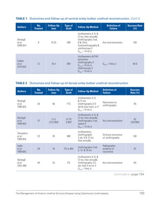
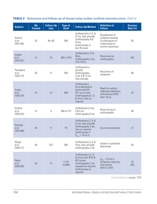
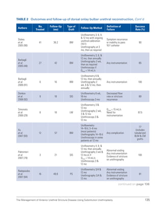
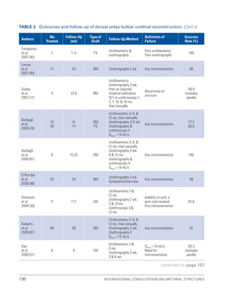
![125
The Management of Anterior Urethral Stricture Disease Using Substitution Urethroplasty
24. Barbagli G, Morgia G, Lazzeri M. Dorsal onlay skin graft bulbar urethroplasty: Long-term follow-up. Eur Urol. 2008;53(3):628-633.
25. Chapple CR, Goonesinghe SK, Nicholson T, et al. The importance of endoscopic surveillance in the follow up of patients with
urethral stricture disease. J Urol. 2002;167(Suppl):16.
26. Andrich DE, Dunglison N, Greenwell TJ, et al. The long-term results of urethroplasty. J Urol. 2003;170(1):90-92.
27. Humby G, Higgins T. A one-stage operation for hypospadias. Br J Surg. 1941;29(113):84-92.
28. Bürger RA, Müller SC, el-Damanhoury H, et al. The buccal mucosal graft for urethral reconstruction: A preliminary report. J Urol.
1992;147(3):662-664.
29. Simonato A, Gregori A, Lissiani A, et al. The tongue as an alternative donor site for graft urethroplasty: A pilot study. J Urol.
2006;175(2):589-592.
30. Simonato A, Gregori A, Ambruosi C, et al. Lingual mucosal graft urethroplasty for anterior urethral reconstruction. Eur Urol.
2008;54(1):79-85.
31. DasSK,KumarA,SharmaGK,etal.Lingualmucosalgrafturethroplastyforanteriorurethralstrictures.Urology.2009;73(1):105-108.
32. Barbagli G, De Angelis M, Romano G, et al. The use of lingual mucosal graft in adult anterior urethroplastyu: Surgical steps and
short-term outcome. Eur Urol. 2008;54(3):671-676.
33. Barbagli G, Romano G, De Angelis M, et al. Complications and patient satisfaction in 349 patients who underwent oral graft
harvesting from a single cheek. Eur Urol Supplement. 2010;9(2):141.
34. Wood DN, Allen SE, Andrich DE, et al. The morbidity of buccal mucosal graft harvest for urethroplasty and the effect of nonclosure
of the graft harvest site on postoperative pain. J Urol. 2004;172(2):580-583.
35. Muruganandam K, Dubey D, Gulia AK, et al. Closure versus nonclosure of buccal mucosal graft harvest site: A prospective
randomized study on post operative morbidity. Indian J Urol. 2009;25(1):72-75.
36. Kamp S, Knoll T, Osman M, et al. Donor-site morbidity in buccal mucosa urethroplasty: Lower lip or inner cheek? BJU Int.
2005;96(4):619-623.
37. El-Kassaby A, AbouShwareb T, Atala A. Randomized comparative study between buccal mucosal and acellular bladder matrix
grafts in complex anterior urethral strictures. J Urol. 2008;179(4):1432-1436.
38. Fiala R, Vidlar A, Vrtal R, et al. Porcine small intestinal submucosa graft for repair of anterior urethral strictures. Eur Urol.
2007;51(6):1702-1708.
39. Fiala R, Vidlar A, Vrtal R. Porcine small intestinal submucosa in the treatment of anterior urethral strictures. BJU Int.
2009;103(Suppl 4):12-46.
40. Hauser S, Bastian PJ, Fechner G, et al. Small intestine submucosa in urethral stricture repair in a consecutive series. Urology.
2006;68(2):263-266.
41. Bhargava S, Chapple CR, Bullock AJ, et al. Tissue-engineered buccal mucosa for substitution urethroplasty. BJU Int. 2004;
93(6):807-811.
42. Barbagli G, Selli C, Tosto A, et al. Dorsal free graft urethroplasty. J Urol. 1996;155(1):123-126.
43. Monseur J. Widening of the urethra using the supra-urethral layer [Article in French]. J Urol (Paris). 1980;86(6):439-449.
44. Mundy AR. Anastomotic urethroplasty. BJU Int. 2005;96(6):921-944.
45. El-Kassaby AW, El-Zayat TM, Azazy S, et al. One-stage repair of long bulbar urethral strictures using augmented Russell dorsal
strip anastomosis: Outcome of 234 cases. Eur Urol. 2008;53(2):420-424.
46. Abouassaly R, Angermeier KW. Augmented anastomotic urethroplasty. J Urol. 2007;177(6):2211-2215.
47. Rourke K. Outcomes and complications of urethral reconstruction using dorsal onlay augmented anastomosis with
buccal mucosa: Is this the evolving gold standard for treatment of the long segment bulbar urethral stricture? [abstract].
J Urol. 2009;181(4):13.](https://image.slidesharecdn.com/urethralstrictures2010-221127135159-2095864b/85/urethral_strictures_2010-pdf-154-320.jpg)
![International Consultation on Urethral Strictures
126
48. Barbagli G, Palminteri E, Guazzoni G, et al. Bulbar urethroplasty using buccal mucosa grafts placed on the ventral, dorsal or
lateral surface of the urethra: Are results affected by the surgical technique? J Urol. 2005;174(3):955-957.
49. Kulkarni S, Barbagli G, Sansalone S, et al. One-sided anterior urethroplasty: A new dorsal onlay graft technique. BJU Int.
2009;104(8):1150-1155.
50. Mangera A, Patterson JM, Chapple CR. A systematic review of graft augmentation urethroplasty techniques for the treatment
of anterior urethral strictures. Eur Urol. 2011;59(5):797-814.
51. Asopa HS, Garg M, Singhal GG, et al. Dorsal free graft urethroplasty for urethral stricture by ventral sagittal urethrotomy
approach. Urology. 2001;58(5):657-659.
52. Pisapati VL, Paturi S, Bethu S, et al. Dorsal buccal mucosal graft urethroplasty for anterior urethral stricture by Asopa technique.
Eur Urol. 2009;56(1):201-205.
53. Palminteri E, Manzoni G, Berdondini E, et al. Combined dorsal plus ventral double buccal mucosa graft in bulbar urethral
reconstruction. Eur Urol. 2008;53(1):81-89.
54. Andrich DE, Greenwell TJ, Mundy AR. The problems of penile urethroplasty with particular reference to 2-stage reconstructions.
J Urol. 2003;170(1):87-89.
55. Barbagli G, De Angelis M, Palminteri E, et al. Failed hypospadias repair presenting in adults. Eur Urol. 2006;49(5):887-894.
56. Andrich DE, Mundy A. Outcome of different management options for full-length anterior urethral strictures [abstract] J Urol.
2009;181(4):13.
57. Peterson AC, Palminteri E, Lazzeri M, et al. Heroic measures may not always be justified in extensive urethral stricture due to
lichen sclerosus (balanitis xerotica obliterans). Urology. 2004;64(3):565-568.
58. Smith JC. The measurement and significance of the urinary flow rate. Br J Urol. 1966;38(6):701-706.
59. Rollema HJ. Uroflowmetry. In: Krane RJ, Siroky MB, editors. Clinical neuro-urology. Boston: Brown and Co; 1991. p. 201-243.
60. Jackson MJ, Sciberras J, Mangera A, et al. Defining a patient-reported outcome measure for urethral stricture surgery. Eur Urol.
2011;60(1):60-68.
61. Barbagli G, Romano G, Sansalone S, et al. Italian validation of the English PROM-USS-Q questionnaire in patients undergoing
anterior urethroplasty [Article in Italian]. Urologia. 2011;78(2):98-107.
62. Morey AF, McAninch JW. When and how to use buccal mucosal grafts in adult bulbar urethroplasty. Urology. 1996;48(2):194-198.
63. Wessells H, McAninch JW. Use of free grafts in urethral stricture reconstruction. J Urol. 1996;155(6):1912-1915.
64. Pansadoro V, Emiliozzi P, Gaffi M, et al. Buccal mucosa urethroplasty for the treatment of bulbar urethral strictures. J Urol.
1999;161(5):1501-1503.
65. Andrich DE, Leach CJ, Mundy AR. The Barbagli procedure gives the best results for patch urethroplasty of the bulbar urethra.
BJU Int. 2001;88(4):385-389.
66. Andrich DE, Mundy AR. Substitution urethroplasty with buccal mucosal-free grafts. J Urol. 2001;165(4):1131-1133.
67. Meneghini A, Cacciola A, Cavarretta L, et al. Bulbar urethral stricture repair with buccal mucosa graft urethroplasty. Eur Urol.
2001;39(3):264-267.
68. Palminteri E, Lazzeri M, Guazzoni G, et al. New 2-stage buccal mucosal graft urethroplasty. J Urol. 2002;167(1):130-132.
69. Lewis JB, Wolgast KA, Ward JA, et al. Outpatient anterior urethroplasty: Outcome analysis and patient selection criteria. J Urol.
2002;168(3):1024-1026.
70. Kane CJ, Tarman GJ, Summerton DJ, et al. Multi-institutional experience with buccal mucosa onlay urethroplasty for bulbar
urethral reconstruction. J Urol. 2002;167(3):1314-1317.
71. Heinke T, Gerharz EW, Bonfig R, et al. Ventral onlay urethroplasty using buccal mucosa for complex stricture repair. Urology.
2003;61(5):1004-1007.
72. Pansadoro V, Emiliozzi P, Gaffi M, et al. Buccal mucosa urethroplasty in the treatment of bulbar urethral strictures. Urology.
2003;61(5):1008-1010.](https://image.slidesharecdn.com/urethralstrictures2010-221127135159-2095864b/85/urethral_strictures_2010-pdf-155-320.jpg)


![193
Pelvic Fracture Urethral Injuries (PFUI)
74. Follis HW, Koch MO, McDougal WS. Immediate management of prostatomembranous urethral disruptions. J Urol. 1992;
147(5):1259-1262.
75. Herschorn S, Thijssen A, Radomski SB. The value of immediate or early catheterization of the traumatized posterior urethra.
J Urol. 1992;148(5):1428-1431.
76. Londergan TA, Gundersen LH, van Every MJ. Early fluoroscopic realignment for traumatic urethral injuries. Urology.
1997;49(1):101-103.
77. Al-AliIH,HusainI.Disruptinginjuriesofthemembranousurethra–Thecaseforearlysurgeryandcathetersplinting.BrJUrol.1983;
55(6):716-720.
78. Podestá ML, Medel R, Castera R, et al. Immediate management of posterior urethral disruptions due to pelvic fracture:
Therapeutic alternatives. J Urol. 1997;157(4):1444-1448.
79. Singh BP, Andankar MG, Swain SK, et al. Impact of prior urethral manipulation on outcome of anastomotic urethroplasty for
post-traumatic urethral stricture. Urology. 2010;75(1):179-182.
80. Salehipour M, Khezri A, Askari R, et al. Primary realignment of posterior urethral rupture. Urol J. 2005;2(4):211-215.
81. Fowler JW, Watson G, Smith MF, et al. Diagnosis and treatment of posterior urethral injury. Br J Urol. 1986;58(2):167-173.
82. Deweerd JH. Management of injuries to the bladder, urethra and genitalia. Surg Clin North Am. 1959;39(4):973-987.
83. Gibson GR. Urological management and complications of fractured pelvis and ruptured urethra. J Urol. 1974;111(3):353-355.
84. Jackson DH, Williams JL. Urethral injury: A retrospective study. Br J Urol. 1974;46(6):665-676.
85. Crassweller PO, Farrow GA, Robson CJ, et al. Traumatic rupture of the supramembranous urethra. J Urol. 1977;118(5):770-771.
86. Coffield KS, Weems WL. Experience with management of posterior urethral injury associated with pelvic fracture. J Urol. 1977;
117(6):722-724.
87. Cass AS, Godec CJ. Urethral injury due to external trauma. Urology. 1978;11(6):607-611.
88. Barry, JM. Visual urethrotomy in the management of the obliterated membranous urethra. Urol Clin North Am. 1989;16(2):319-324.
89. Dhabuwala CB, Hamid S, Katsikas DM, et al. Impotence following delayed repair of prostatomembranous urethral disruption.
J Urol. 1990;144(3):677-678.
90. Husmann DA, Wilson WT, Boone TB, et al. Prostatomembranous urethral disruptions: Management by suprapubic cystostomy
and delayed urethroplasty. J Urol. 1990;144(1):76-78.
91. Quint HJ, Stanisic TH. Above and below delayed endoscopic treatment of traumatic posterior urethral disruptions. J Urol. 1993;
149(3):484-487.
92. Kotkin L, Koch MO. Impotence and incontinence after immediate realignment of posterior urethral trauma: Result of injury or
management? J Urol. 1996;155(5):1600-1603.
93. Koraitim MM. Pelvic fracture urethral injuries: Evaluation of various methods of management. J Urol. 1996;156(4):1288-1291.
94. Sahin H, Bircan MK, Akay AF, et al. Endoscopic treatment of complete posterior urethral obliteration. Acta Urol Belg. 1998;
66(4):21-24.
95. Jepson BR, Boullier JA, Moore RG, et al. Traumatic posterior urethral injury and early primary endoscopic realignment:
Evaluation of long-term follow-up. Urology. 1999;53(6):1205-1210.
96. Moudouni SM, Patard JJ, Manunta A, et al. Early endoscopic realignment of post-traumatic posterior urethral disruption.
Urology. 2001;57(4):628-632.
97. Ku JH, Kim ME, Jeon YS, et al. Management of bulbous urethral disruption by blunt external trauma: The sooner, the better?
Urology. 2002;60(4):579-583.
98. Tazi H, Ouali M, Lrhorfi MH, et al. Endoscopic realignment for post-traumatic rupture of posterior urethra [Article in French].
Prog Urol. 2003;13(6):1345-1350.
99. Mouraviev VB, Coburn M, Santucci RA. The treatment of posterior urethral disruption associated with pelvic fractures:
Comparative experience of early realignment versus delayed urethroplasty. J Urol. 2005;173(3):873-876.](https://image.slidesharecdn.com/urethralstrictures2010-221127135159-2095864b/85/urethral_strictures_2010-pdf-222-320.jpg)
![International Consultation on Urethral Strictures
194
100. Healy CE, Leonard DS, Cahill R, et al. Primary endourologic realignment of complete posterior urethral disruption. Ir Med J. 2007;
100(6):488-489.
101. Melekos MD, Pantazakos A, Daouaher H, et al. Primary endourologic re-establishment of urethral continuity after disruption of
prostatomembranous urethra. Urology. 1992;39(2):135-138.
102. Morey AF, McAninch JW. Reconstruction of posterior urethral disruption injuries: Outcome analysis in 82 patients. J Urol. 1997;
157(2):506-510.
103. Cooperberg MR, McAninch JW, Alsifaki NF, et al. Urethral reconstruction for traumatic posterior urethral disruption: Outcomes
of a 25-year experience. J Urol. 2007;178(5):2006-2010.
104. BiswasS,GnanasekaranI,IvaturyRR,etal.Exaggeratedlithotomyposition-relatedrhabdomyolysis.AmSurg.1997;63(4):361-364.
105. Bocca G, van Moorselaar JA, Feitz WF, et al. Compartment syndrome, rhabdomyolysis and risk of acute renal failure as
complications of the lithotomy position. J Nephrol. 2002;15(2):183-185.
106. Gabrielli A, Caruso L. Postoperative acute renal failure secondary to rhabdomyolysis from exaggerated lithotomy position.
J Clin Anesth. 1999;11(3):257-263.
107. Bildsten SA, Dmochowski RR, Spindel MR, et al. The risk of rhabdomyolysis and acute renal failure with the patient in the
exaggerated lithotomy position. J Urol. 1994;152(6 Pt 1):1970-1972.
108. Marion G. De la reconstitution de l`urètre par urétrorrhaphie circulaire avec dérivation de l’urine [Article in French]. J Urol Med Chir.
1912;1:523-528.
109. Paine D, Coombes W. Transpubic reconstruction of the urethra. Br J Urol. 1968;40(1):78-84.
110. Waterhouse K, Laungani G, Patil U. The surgical repair of membranous urethral strictures: Experience with 105 consecutive
cases. J Urol. 1980;123(4):500-505.
111. Turner-Warwick RT. A technique for posterior urethroplasty. J Urol. 1960;83:416-419.
112. Webster GD, Ramon J. Repair of pelvic fracture posterior urethral defects using an elaborated perineal approach: Experience
with 74 cases. J Urol. 1991;145(4):744-748.
113. Koraitim MM. Post-traumatic posterior urethral strictures: Preoperative decision making. Urology. 2004;64(2):228-231.
114. Andrich DE, O’Malley J, Summerton DJ, et al. The type of urethroplasty for a pelvic fracture urethral distraction defect cannot
be predicted preoperatively. J Urol. 2003;170(2 Pt 1):464-467.
115. Koraitim MM. On the art of anastomotic posterior urethroplasty: A 27-year experience. J Urol. 2005;173(1):135-139.
116. Koraitim MM. Predictors of surgical approach to repair pelvic fracture urethral distraction defects. J Urol. 2009;182(4):1435-1439.
117. Koraitim MM. Gapometry and anterior urethrometry in the repair of posterior urethral defects. J Urol. 2008;179(5):1879-1881.
118. Javanmard B, Hosseinee J, Rezaei A, et al. MP-10.11: Evaluation of supracrural rerouting success rate as a technique for resolution
of posterior urethral disruption defects. Urology. 2009;74(4):S90-S91.
119. Kizer WS, Armenakas NA, Brandes SB, et al. Simplified reconstruction of posterior urethral disruption defects: Limited role of
supracrural rerouting. J Urol. 2007;177(4):1378-1381.
120. Flynn BJ, Delvecchio FC, Webster GD. Perineal repair of pelvic fracture urethral distraction defects: Experience in 120 patients
during the last 10 years. J Urol. 2003;170(5):1877-1880.
121. Corriere JN. 1-stage delayed bulboprostatic anastomotic repair of posterior urethral rupture: 60 patients with 1-year followup.
J Urol. 2001;165:404-407.
122. Fu Q, Zhang J, Sa YL, et al. Transperineal bulboprostatic anastomosis in patients with simple traumatic posterior urethral
strictures: A retrospective study from a referral urethral center. Urology. 2009:74(5):1132-1136.
123. Tunc HM, Tefekli AH, Kaplancan T, et al. Delayed repair of post-traumatic posterior urethral distraction injuries: Long-term results.
Urology. 2000;55(6):837-841.
124. Kulkarni SB, Barbagli G, Kulkarni JS, et al. Posterior urethral stricture after pelvic fracture urethral distraction defects in
developing and developed countries, and choice of surgical technique. J Urol. 2010;183(3):1049-1054.](https://image.slidesharecdn.com/urethralstrictures2010-221127135159-2095864b/85/urethral_strictures_2010-pdf-223-320.jpg)


![234 International Consultation on Urethral Strictures
23. Levy JB, Ramchandani P, Berlin JW, et al. Vesicourethral healing following radical prostatectomy: Is it related to surgical
approach? Urology. 1994;44(6):888-892.
24. Gillitzer R, Melchior SW, Hampel C, et al. Specific complications of radical perineal prostatectomy: A single institution study of
more than 600 cases. J Urol. 2004;172(1):124-128.
25. Besarani D, Amoroso P, Kirby R. Bladder neck contracture after radical retropubic prostatectomy. BJU Int. 2004;94(9):1245-1247.
26. Gonzalgo ML, Pavlovich CP, Trock BJ, et al. Classification and trends of perioperative morbidities following laparoscopic radical
prostatectomy. J Urol. 2005;174(1):135-139.
27. Surya BV, Provet J, Johanson KE, et al. Anastomotic strictures following radical prostatectomy: Risk factors and management.
J Urol. 1990;143(4):755-758.
28. Dalkin BL. Endoscopic evaluation and treatment of anastomotic strictures after radical retropubic prostatectomy. J Urol.
1996;155(1):206-208.
29. Sano T, Iguchi R, Asaki S, et al. Relationship between type of suture and anastomotic stricture after radical prostatectomy
[Article in Japanese]. Hinyokika Kiyo. 2010;56(2):95-98.
30. Huang G, Lepor H. Factors predisposing to the development of anastomotic strictures in a single-surgeon series of radical
retropubic prostatectomies. BJU Int. 2006;97(2):255-258.
31. Kostakopoulos A, Argiropoulos V, Protogerou V, et al. Vesicourethral anastomotic strictures after radical retropubic
prostatectomy: The experience of a single institution. Urol Int. 2004;72(1):17-20.
32. Gallo L, Perdonà S, Autorino R, et al. Vesicourethral anastomosis during radical retropubic prostatectomy: Does the number of
sutures matter? Urology. 2007;69(3):547-551.
33. Igel TC, Wehle MJ. Vesicourethral reconstruction in radical retropubic prostatectomy: An alternative technique. J Urol.
1999;161(3):844-846.
34. Srougi M, Paranhos M, Leite KM, et al. The influence of bladder neck mucosal eversion and early urinary extravasation on patient
outcome after radical retropubic prostatectomy: A prospective controlled trial. BJU Int. 2005;95(6):757-760.
35. Ozu C, Hagiuda J, Nakagami Y, et al. Radical retropubic prostatectomy with running vesicourethral anastomosis and early
catheter removal: Our experience. Int J Urol. 2009;16(5):487-492.
36. Thiel DD, Igel TC, Brisson TE, et al. Outcomes with an alternative anastomotic technique after radical retropubic prostatectomy:
10-year experience. Urology. 2006;68(1):132-136.
37. Crew JP, Jephcott CR, Reynard JM. Radiation-induced haemorrhagic cystitis. Eur Urol. 2001;40(2):111-123.
38. deVries CR, Freiha FS. Hemorrhagic cystitis: A review. J Urol. 1990;143(1):1-9.
39. Zietman AL, DeSilvio ML, Slater JD, et al. Comparison of conventional-dose vs high-dose conformal radiation therapy in clinically
localized adenocarcinoma of the prostate: A randomized controlled trial. JAMA. 2005;294(10):1233-1239.
40. Zelefsky MJ, Fuks Z, Hunt M, et al. High dose radiation delivered by intensity modulated conformal radiotherapy improves the
outcome of localized prostate cancer. J Urol. 2001;166(3):876-881.
41. Pollack A, Zagars GK, Starkschall G, et al. Prostate cancer radiation dose response: Results of the M. D. Anderson phase III
randomized trial. Int J Radiat Oncol Biol Phys. 2002;53(5):1097-1105.
42. Lukka H, Warde P, Pickles T, et al. Controversies in prostate cancer radiotherapy: Consensus development. Can J Urol.
2001;8(4):1314-1322.
43. McVey GP, McPhail S, Fowler S, et al. Initial management of low-risk localized prostate cancer in the UK: Analysis of the British
Association of Urological Surgeons Cancer Registry. BJU Int. 2010;106(8):1161-1164.
44. Mate TP, Gottesman JE, Hatton J, et al. High dose-rate afterloading 192Iridium prostate brachytherapy: Feasibility report.
Int J Radiat Oncol Biol Phys. 1998;41(3):525-533.
45. Aström L, Pedersen D, Mercke C, et al. Long-term outcome of high dose rate brachytherapy in radiotherapy of localised prostate
cancer. Radiother Oncol. 2005;74(2):157-161.
46. Morton GC. The emerging role of high-dose-rate brachytherapy for prostate cancer. Clin Oncol (R Coll Radiol). 2005;17(4):219-227.](https://image.slidesharecdn.com/urethralstrictures2010-221127135159-2095864b/85/urethral_strictures_2010-pdf-263-320.jpg)
![239
Posterior Urethral Stenosis After Treatment for Prostate Cancer
137. Al-Singary W, Arya M, Patel HR. Bladder neck stenosis after transurethral resection of prostate: Does size matter? Urol Int.
2004;73(3):262-265.
138. Greene LF, Leary FJ. Contractures of the vesical neck following transurethral prostatic resection. Surg Gynecol Obstet.
1967;124(6):1277-1282.
139. Orandi A. Transurethral incision of the prostate. J Urol. 1973;110(2):229-231.
140. Kletscher BA, Oesterling JE. Transurethral incision of the prostate: A viable alternative to transurethral resection. Semin Urol.
1992;10(4):265-272.
141. Abrams P, Chapple C, Khoury S, et al. Evaluation and treatment of lower urinary tract symptoms in older men. J Urol.
2009;181(4):1779-1787.
142. McVary KT, Roehrborn CG, Avins AL, et al. Update on AUA guideline on the management of benign prostatic hyperplasia. J Urol.
2011;185(5):1793-1803.
143. Benoit RM, Naslund MJ, Cohen JK. Complications after prostate brachytherapy in the Medicare population. Urology.
2000;55(1):91-96.
144. Bullock TL, Brandes SB. Adult anterior urethral strictures: A national practice patterns survey of board certified urologists in the
United States. J Urol. 2007;177(2):685-690.
145. Elliott SP, McAninch JW, Chi T, et al. Management of severe urethral complications of prostate cancer therapy. J Urol. 2006;
176(6 Pt 1):2508-2513.
146. Gómez-Iturriaga Piña A, Crook J, Borg J, et al. Median 5 year follow-up of 125iodine brachytherapy as monotherapy in men
agedor=55 years with favorable prostate cancer. Urology. 2010;75(6):1412-1416.
147. Sikafi Z, Butler MR, Lane V, et al. Bladder neck contracture following prostatectomy. Br J Urol. 1985;57(3):308-310.
148. Wettlaufer JN, Kronmiller P. The management of post-prostatectomy vesical neck contracture. J Urol. 1976;116(4):482-483.
149. Vanni A, Zinman L, Buckley J. Management of recurrent bladder neck contractures with urethrotomy and mitomycin C [abstract].
J Urol. 2010;183(4):e426.
150. Kulb TB, Kamer M, Lingeman JE, et al. Prevention of post-prostatectomy vesical neck contracture by prophylactic vesical neck
incision. J Urol. 1987;137(2):230-231.
151. Aygün C, Peskircioglu L, Tekin MI, et al. Endoscopic treatment of complete bladder neck obstruction by transurethral Seldinger
technique. Int J Urol. 2001;8(8):455-456.
152. Rusnak B, Castaneda-Zuniga W, Kotula F, et al. An improved dilator system for percutaneous nephrostomies. Radiology.
1982;144(1):174.
153. Herschorn S, Carrington E. S-shaped coaxial dilators for male urethral strictures. Urology. 2007;69(6):1199-1201.
154. GiesyJD,FinnJC,HermannGD,etal.Coaxialballoondilationandcalibrationofurethralstrictures.AmJSurg.1984;147(5):611-614.
155. Bapat SS. A new endoscopic urethral dilator. J Urol. 1979;122(1):30-33.
156. Soloway MS. Optical dilator to obviate blind urethral dilatation prior to endoscopic resections. Urology. 1988;31(5):427-428.
157. Ramchandani P, Banner MP, Berlin JW, et al. Vesicourethral anastomotic strictures after radical prostatectomy: Efficacy of
transurethral balloon dilation. Radiology. 1994;193(2):345-349.
158. Geary ES, Dendinger TE, Freiha FS, et al. Incontinence and vesical neck strictures following radical retropubic prostatectomy.
Urology. 1995;45(6):1000-1006.
159. Popken G, Sommerkamp H, Schultze-Seemann W, et al. Anastomotic stricture after radical prostatectomy. Incidence, findings
and treatment. Eur Urol. 1998;33(4):382-386.
160. Hayashi T, Yoshinaga A, Ohno R, et al. Successful treatment of recurrent vesicourethral stricture after radical prostatectomy
with holmium laser: Report of three cases. Int J Urol. 2005;12(4):414-416.
161. Lagerveld BW, Laguna MP, Debruyne FM, et al. Holmium:YAG laser for treatment of strictures of vesicourethral anastomosis
after radical prostatectomy. J Endourol. 2005;19(4):497-501.](https://image.slidesharecdn.com/urethralstrictures2010-221127135159-2095864b/85/urethral_strictures_2010-pdf-268-320.jpg)
![240 International Consultation on Urethral Strictures
162. Eltahawy E, Gur U, Virasoro R, et al. Management of recurrent anastomotic stenosis following radical prostatectomy using
holmium laser and steroid injection. BJU Int. 2008;102(7):796-798.
163. Meulen T, Zambon JV, Janknegt RA. Treatment of anastomotic strictures and urinary incontinence after radical prostatectomy
with urolume wallstent and AMS 800 artificial sphincter. J Endourol. 1999;13(7):517-520.
164. Zivan I, Stein A. New modality for treatment of resistant anastomotic strictures after radical prostatectomy: UroLume urethral
stent. J Endourol. 2001;15(8):869-871.
165. Elliott DS, Boone TB. Combined stent and artificial urinary sphincter for management of severe recurrent bladder neck contracture
and stress incontinence after prostatectomy: A long-term evaluation. J Urol. 2001;165(2):413-415.
166. Magera JS Jr, Inman BA, Elliott DS. Outcome analysis of urethral wall stent insertion with artificial urinary sphincter placement
for severe recurrent bladder neck contracture following radical prostatectomy. J Urol. 2009;181(3):1236-1241.
167. Chiou RK, Howe S, Morton JJ, et al. Treatment of recurrent vesicourethral anastomotic stricture after radical prostatectomy with
endourethroplasty. Urology. 1996;47(3):422-425.
168. Kuyumcuoglu U, Eryildirim B, Tarhan F, et al. Antegrade endourethroplasty with free skin graft for recurrent vesicourethral
anastomotic strictures after radical prostatectomy. J Endourol. 2010;24(1):63-67.
169. Yurkanin JP, Dalkin BL, Cui H. Evaluation of cold knife urethrotomy for the treatment of anastomotic stricture after radical
retropubic prostatectomy. J Urol. 2001;165(5):1545-1548.
170. Anger JT, Raj GV, Delvecchio FC, et al. Anastomotic contracture and incontinence after radical prostatectomy: A graded approach
to management. J Urol. 2005;173(4):1143-1146.
171. Wessells H, Morey AF, McAninch JW. Obliterative vesicourethral strictures following radical prostatectomy for prostate cancer:
Reconstructive armamentarium. J Urol. 1998;160(4):1373-1375.
172. Schlossberg S, Jordan G, Schellhammer P. Repair of obliterative vesicourethral stricture after radical prostatectomy: A technique
for preservation of continence. Urology. 1995;45(3):510-513.
173. Theodoros C, Katsifotis C, Stournaras P, et al. Abdomino-perineal repair of recurrent and complex bladder neck-prostatic urethra
contractures. Eur Urol. 2000;38(6):734-740.
174. Simonato A, Gregori A, Lissiani A, et al. Two-stage transperineal management of posterior urethral strictures or bladder
neck contractures associated with urinary incontinence after prostate surgery and endoscopic treatment failures. Eur Urol.
2007;52(5):1499-1504.
175. Herschorn S. Management of post-radical prostatectomy complete bladder neck occlusion [abstract]. J Urol. 2007;177(4):14.
176. Ullrich NF, Wessells H. A technique of bladder neck closure combining prostatectomy and intestinal interposition for unsalvageable
urethral disease. J Urol. 2002;167(2 Pt 1):634-636.
177. Herschorn S, Thijssen AJ, Radomski SB. Experience with the hemi-Kock ileocystoplasty with a continent abdominal stoma.
J Urol. 1993;149(5):998-1001.
178. Rowland RG, Mitchell ME, Bihrle R, et al. Indiana continent urinary reservoir. J Urol. 1987;137(6):1136-1139.](https://image.slidesharecdn.com/urethralstrictures2010-221127135159-2095864b/85/urethral_strictures_2010-pdf-269-320.jpg)


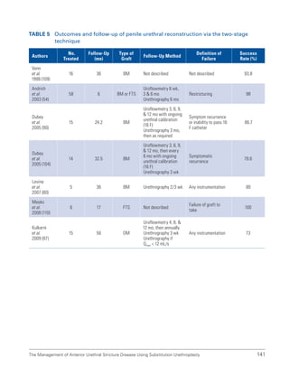
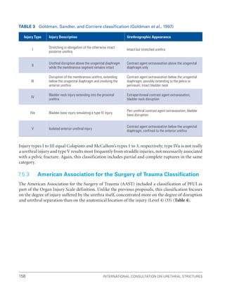
This document summarizes the proceedings of an international consultation on urethral strictures held in Marrakech, Morocco in 2010. It was co-sponsored by the Société Internationale d'Urologie and the International Consultation on Urological Diseases. The document contains recommendations from various committees that reviewed the epidemiology, evaluation, treatment, and management of urethral strictures. It addresses strictures in both the anterior and posterior urethra, as well as specific conditions like lichen sclerosus and strictures following treatment for prostate cancer.


![Hypothalamus short notes on location, function and disorders by Dr. Neha [PT]...](https://cdn.slidesharecdn.com/ss_thumbnails/hypothalamusbydr-260124142231-2b48143d-thumbnail.jpg?width=640&height=640&fit=bounds)


