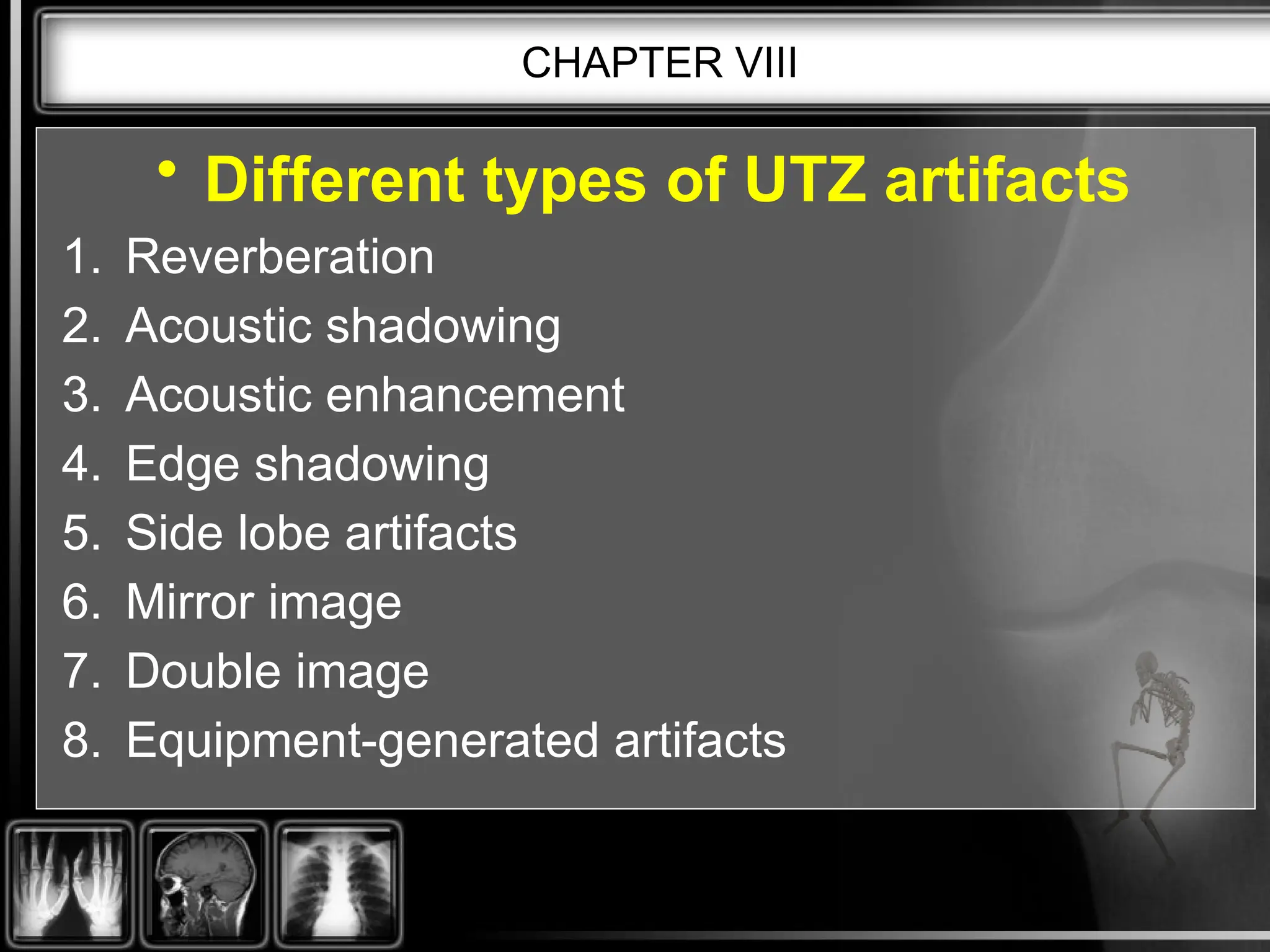
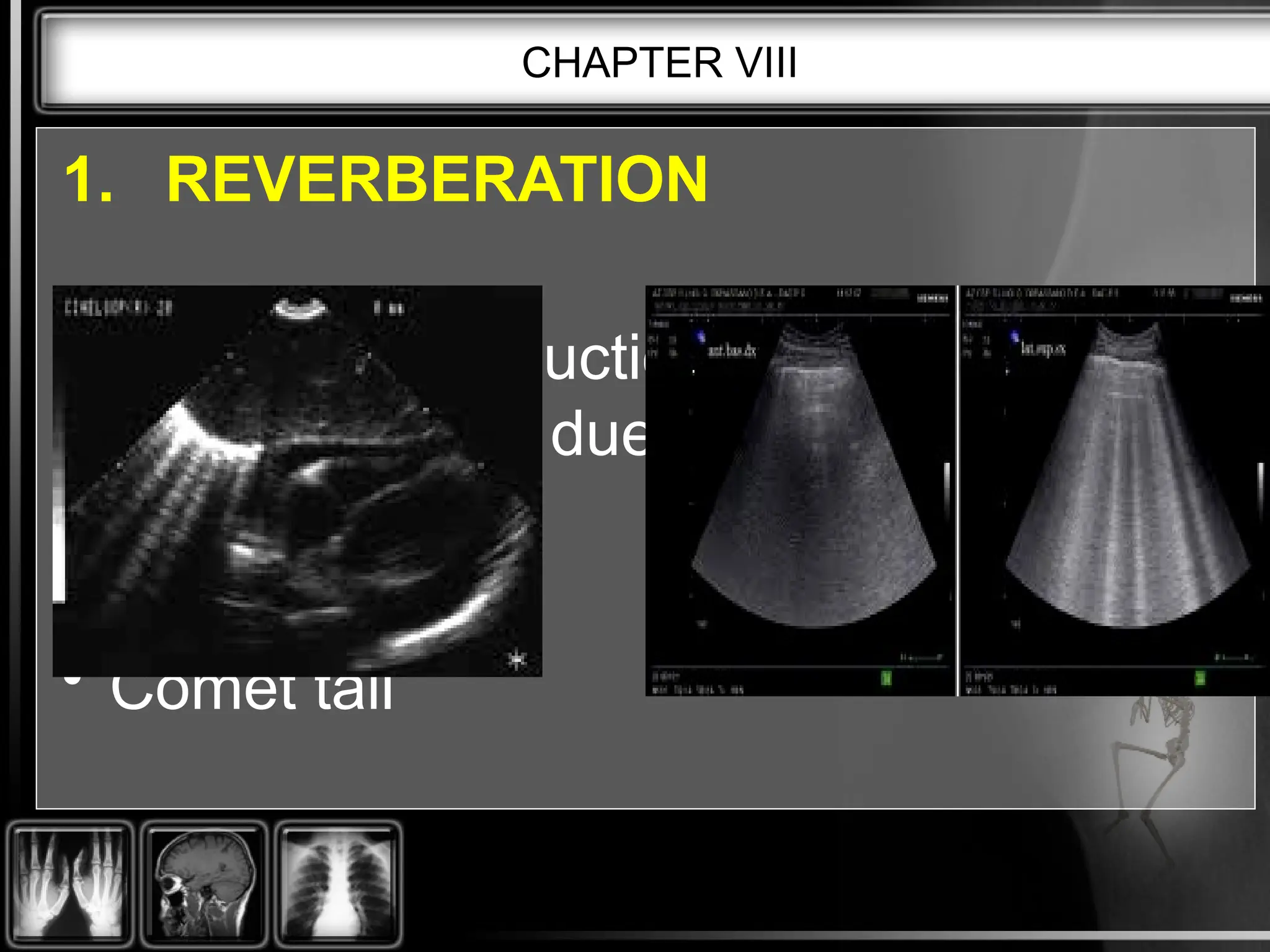
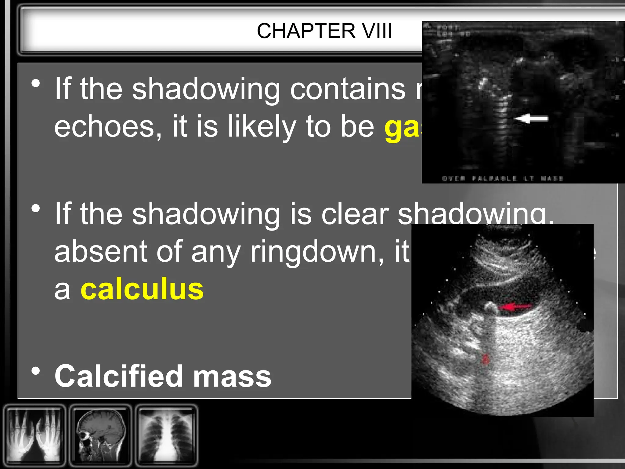
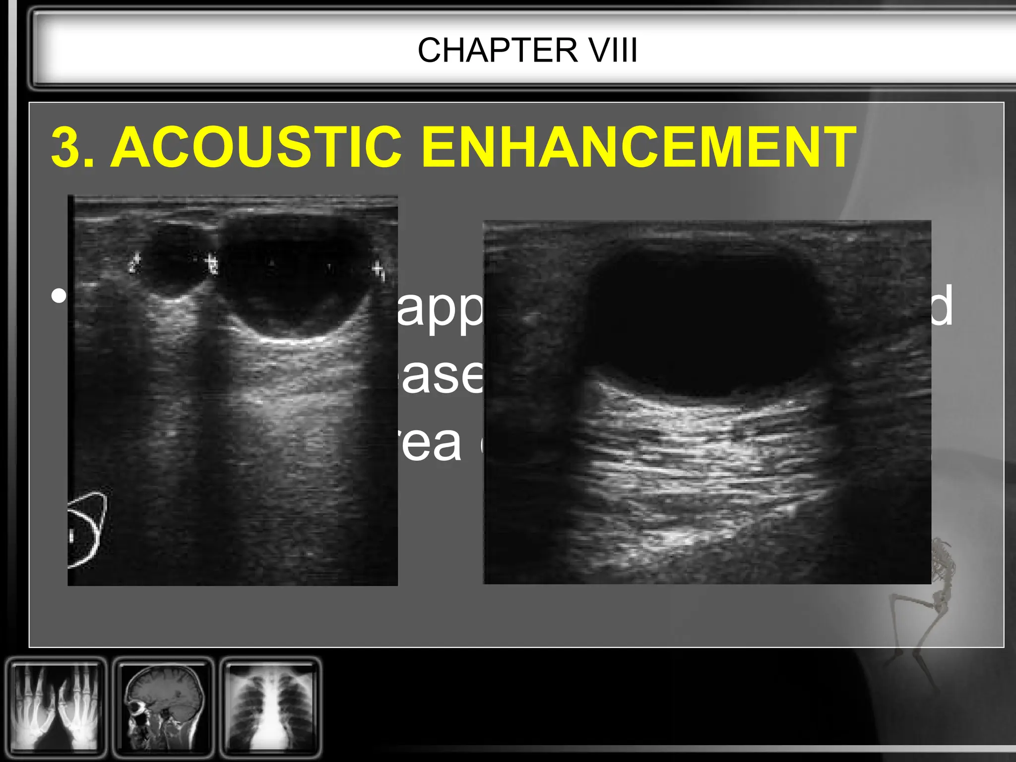
CHAPTER VIII:
ARTIFACTS
CHAPTER VIII
• An ultrasound artifact is an
additional, missing or distorted
image which does not conform to
the real image of the part being
examined.
CHAPTER VIII
• Different types of UTZ artifacts
1. Reverberation
2. Acoustic shadowing
3. Acoustic enhancement
4. Edge shadowing
5. Side lobe artifacts
6. Mirror image
7. Double image
8. Equipment-generated artifacts
CHAPTER VIII
1. REVERBERATION
• This is the production of spurious
(false) echoes due to repeated
reflections
• Comet tail
CHAPTER VIII
• Artifacts appearing as parallel, equally
spaced lines is called reverberation
• Reverberation artifacts are a result of
the presence of two or more strong
reflecting surfaces
• Shotgun wound
CHAPTER VIII
2. ACOUSTIC SHADOWING
• This appears as an area of low
amplitude echoes (hypoechoic or
anechoic) behind an area of strongly
attenuating tissue.
CHAPTER VIII
• If the shadowing contains ringdown
echoes, it is likely to be gas.
• If the shadowing is clear shadowing,
absent of any ringdown, it is likely to be
a calculus
• Calcified mass
CHAPTER VIII
3. ACOUSTIC ENHANCEMENT
• This artifact appears as a localized
area of increased echo amplitude
behind an area of low attenuation.
CHAPTER VIII
• Can commonly be seen distal to fluid filled
structures such as the urinary bladder, the
gallbladder or a cyst.
CHAPTER VIII
• Acoustic enhancement is caused
by weakly attenuating structures.
• Fluid filled mass
CHAPTER VIII
4. EDGE SHADOWING
• Occurring at the edges of rounded
structures will result in an edge
shadowing artifact.
CHAPTER VIII
5. SIDE LOBE ARTIFACT
• The energy within the ultrasound beam
exists as several side lobes radiating at a
number of angles from a central lobe.
CHAPTER VIII
6. MIRROR IMAGE ARTIFACT
• These artifacts result in a mirror image of a
structure.
• They arise due to specular reflection of
the beam at a large smooth interface.
CHAPTER VIII
• fluid-air interface.
• Diaphragm, Heart, Liver
CHAPTER VIII
7. DOUBLE IMAGE ARTIFACT
• This artifact is caused by refraction of the
beam and may occur in areas such as the
rectus abdominis muscle on the anterior
abdominal wall.
CHAPTER VIII
• In order to establish whether these echoes
are genuine, it is necessary to move the
transducer slightly to one side to avoid the
junction of the rectus abdominis muscles.
CHAPTER VIII
8. EQUIPMENT-GENERATED ARTIFACTS
• Incorrect use of the equipment controls can
lead to artifacts appearing.
CHAPTER VIII
• If too much gain is applied then the
electronic noise, inherent in all systems, will
also be amplified.
• If too little gain is applied, this can lead to
loss of relevant information, and incorrect
diagnosis may occur.
CHAPTER VIII
****END****

Ultrasound Artifacts_powerpoint presentation

  • 1.
  • 2.
    CHAPTER VIII • Anultrasound artifact is an additional, missing or distorted image which does not conform to the real image of the part being examined.
  • 3.
    CHAPTER VIII • Differenttypes of UTZ artifacts 1. Reverberation 2. Acoustic shadowing 3. Acoustic enhancement 4. Edge shadowing 5. Side lobe artifacts 6. Mirror image 7. Double image 8. Equipment-generated artifacts
  • 4.
    CHAPTER VIII 1. REVERBERATION •This is the production of spurious (false) echoes due to repeated reflections • Comet tail
  • 5.
    CHAPTER VIII • Artifactsappearing as parallel, equally spaced lines is called reverberation • Reverberation artifacts are a result of the presence of two or more strong reflecting surfaces • Shotgun wound
  • 6.
    CHAPTER VIII 2. ACOUSTICSHADOWING • This appears as an area of low amplitude echoes (hypoechoic or anechoic) behind an area of strongly attenuating tissue.
  • 7.
    CHAPTER VIII • Ifthe shadowing contains ringdown echoes, it is likely to be gas. • If the shadowing is clear shadowing, absent of any ringdown, it is likely to be a calculus • Calcified mass
  • 8.
    CHAPTER VIII 3. ACOUSTICENHANCEMENT • This artifact appears as a localized area of increased echo amplitude behind an area of low attenuation.
  • 9.
    CHAPTER VIII • Cancommonly be seen distal to fluid filled structures such as the urinary bladder, the gallbladder or a cyst.
  • 10.
    CHAPTER VIII • Acousticenhancement is caused by weakly attenuating structures. • Fluid filled mass
  • 11.
    CHAPTER VIII 4. EDGESHADOWING • Occurring at the edges of rounded structures will result in an edge shadowing artifact.
  • 12.
    CHAPTER VIII 5. SIDELOBE ARTIFACT • The energy within the ultrasound beam exists as several side lobes radiating at a number of angles from a central lobe.
  • 13.
    CHAPTER VIII 6. MIRRORIMAGE ARTIFACT • These artifacts result in a mirror image of a structure. • They arise due to specular reflection of the beam at a large smooth interface.
  • 14.
    CHAPTER VIII • fluid-airinterface. • Diaphragm, Heart, Liver
  • 15.
    CHAPTER VIII 7. DOUBLEIMAGE ARTIFACT • This artifact is caused by refraction of the beam and may occur in areas such as the rectus abdominis muscle on the anterior abdominal wall.
  • 16.
    CHAPTER VIII • Inorder to establish whether these echoes are genuine, it is necessary to move the transducer slightly to one side to avoid the junction of the rectus abdominis muscles.
  • 17.
    CHAPTER VIII 8. EQUIPMENT-GENERATEDARTIFACTS • Incorrect use of the equipment controls can lead to artifacts appearing.
  • 18.
    CHAPTER VIII • Iftoo much gain is applied then the electronic noise, inherent in all systems, will also be amplified. • If too little gain is applied, this can lead to loss of relevant information, and incorrect diagnosis may occur.
  • 19.