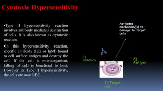
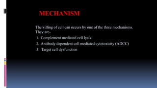
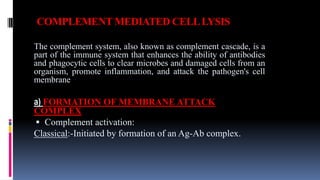
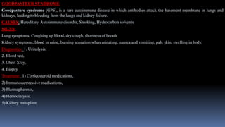
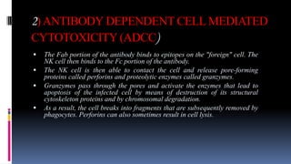
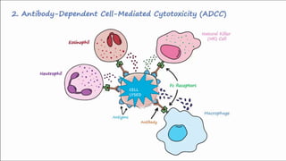
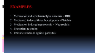
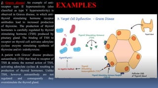
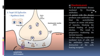
TYPE II HYPERSENSITIVITY
REACTION
AADHIRA R
MSC Biotechnology
1) Target
cell
2)
Antigen
3)
Antibody
Activates
mechanism(s) to
damage to target
cells
•Type II hypersensitivity reaction
involves antibody mediated destruction
of cells. It is also known as cytotoxic
reaction.
•In this hypersensitivity reaction,
specific antibody (IgG or IgM) bound
to cell surface antigen and destroy the
cell. If the cell is microorganism,
killing of cell is beneficial to host.
However in Type II hypersensitivity,
the cells are own RBC.
Cytotoxic Hypersensitivity
MECHANISM
The killing of cell can occurs by one of the three mechanisms.
They are-
1. Complement mediated cell lysis
2. Antibody dependent cell mediated cytotoxicity (ADCC)
3. Target cell dysfunction
COMPLEMENT MEDIATED CELLLYSIS
The complement system, also known as complement cascade, is a
part of the immune system that enhances the ability of antibodies
and phagocytic cells to clear microbes and damaged cells from an
organism, promote inflammation, and attack the pathogen's cell
membrane
a) FORMATION OF MEMBRANE ATTACK
COMPLEX
 Complement activation:
Classical:-Initiated by formation of an Ag-Ab complex.
C1 becomes activated when it binds to the ends of the antibodies. Only certain Ab can activate this
pathway(IgM and certain classes of IgG).
Once C1 is activated, it activates 2 complement proteins, C2 & C4 by cutting them in half. C2 is
cleaved into C2a & C2b. C4 is cleaved into C4a & C4b.Both C2a & C4a diffuses away. C2b & C4b
binds together on the surface of the bacteria forming C3 activation complex whose function is to
activate C3 proteins by cleaving into C3a & C3b. C3b is a opsonin. C3a increases the inflammatory
response by binding to mast cells and causing them to release histamine.
When C3b binds to C2b & C4b it forms a new complex referred to as C5 activation complex. C5
activation complex activates C5 proteins by cleaving them into C5a & C5b. C5a disperses away from
bacteria. C5b on the surface of bacteria binds to C6 which gets activated and binds to C7. C7 binds to
C8 which in turn binds to many C9’s. Together these proteins form a circular complex called the
Membrane Attack Complex (MAC).
MAC can put holes in the cytoplasmic membrane causing lysis.
C9
C5-C8
Membrane
Attack
Complex (MAC)
B) OPSONIZATION
• When antigen enters into host body, antibodies are produced.
• Antibody binds to antigen’s epitope through Fab region. Fc region of antibody remains free.
• Phagocytic cells such as Neutrophils, macrophages and monocytes have receptors that can bind to Fc region of
antibody. The receptor is known as FcR.
• Opsonin molecules usually bind, on one end, to the receptors present in the antigen and, on the other end, to
the receptors on the phagocytes.
• In this case antibody molecule directly cross links antigen (Microorganism or RBC or target cell) with
phagocytic cells. This cross-linkage activates phagocytic cells and increases the rate of phagocytosis.
• This increased rate of phagocytosis by binding of antibody to antigen is called Opsonization.
EXAMPLES
1) AUTOIMMUNE HEMOLYTIC ANAEMIA
Autoimmune hemolytic anemia (AIHA) occurs when antibodies directed
against the person's own red blood cells (RBCs) cause them to burst
(lyse), leading to an insufficient number of oxygen-carrying red blood
cells in the circulation. The lifetime of the RBCs is reduced from the
normal 100–120 days(later are recycled or consumed by macrophages)
to just a few days in serious cases. The intracellular components of the
RBCs are released into the circulating blood and into tissues, leading to
some of the characteristic symptoms of this condition. The antibodies are
usually directed against high-incidence antigens, therefore they also
commonly act on allogenic RBCs (RBCs originating from outside the
person themselves, e.g. in the case of a blood transfusion). AIHA is a
relatively rare condition, affecting one to three people per 100,000 per
year.
 Classification of the antibodies is based on their activity at different
temperatures and their etiology. Antibodies with high activity at physiological
temperature (approximately 37 °C) are termed warm autoantibodies. Cold
autoantibodies act best at temperatures of 0–4 °C. Patients with cold-type AIHA,
therefore, have higher disease activity but are rare.
 Patients may present with one or both types of autoantibodies; if both are present,
the disease is termed "mixed-type" AIHA.
 Warm-type AIHA are most common and shows a positive reaction with antisera
to IgG antibodies with or without complement activation (like phagocytosis).
Cases may also arise with complement alone or with IgA, IgM or a combination
of these three antibody classes and complement (MAC cytolysis).
 Cold-type AIHA usually reacts with antisera to complement and occasionally to
the above antibodies.
 Mixed-type can, like the others, present unusually with positive reactions to other
antisera.
SIGNS:
Spherocytes are found in immunologically-mediated hemolytic anemias.
CAUSES:
The primary illness is idiopathic (no cause).
Secondary causes of autoimmune hemolytic anaemia include:
 Leukaemia
 Blood cancers
 Mycoplasma pneumonia
 Hepatitis
 HIV
TREATMENT:
Warm-type AIHA is usually a more insidious disease, not treatable by simply
removing the underlying cause. Corticosteroids are first-line therapy.
Cold agglutinin disease is treated with avoidance of cold exposure. Or
plasmapheresis.
DIAGNOSIS: Coomb’s test, Direct antigen test.
MISMATCHED TRANSFUSED BLOOD CELLS
• ABO blood transfusion reaction is an example of type II hypersensitivity reaction. Human
RBCs contains A and/or B antigen as major antigen on the surface of RBC. Other minor
antigens such as Rh, Kell, Duffy etc. are also present. Antibodies to ABO antigen are
called isohemagglutinin (antibody that agglutinate RBC from other of same species) and
are usually of IgM class whereas antibodies to other minor antigen are of IgG class.
• An individual with blood group A recognizes B antigen like epitope (blood group B) as
foreign and produces isohemagglutinin (antibodies). The same individual does not produce
antibodies to A antigen as it is similar to self antigen, so that state of tolerance exists.
• If individual with blood group A is transfused with blood containing B antigen then
transfusion reaction occurs in which anti-B isohemagglutinin ( antibodies) binds with B-
blood cells and mediate destruction of transfused RBC by complement activation.
• Since lysis of RBC occurs in intravascular space, free
hemoglobin appears in urine. Hemoglobin may be converted
into bilirubin which is highly toxic to tissues.
• For treatment of this hypersensitivity reaction, transfusion
should be stopped immediately. Furthermore patients should be
given diuretic (increased passing of urine) to eliminate
hemoglobin in urine.
• Blood transfusion reaction occurs immediately in case of
mismatched ABO antigen. In this case transfused RBCs are
lysed by complement.
• In case of minor antigen (Rh) mismatched, delayed reaction
occurs and in this case transfused RBCs are lysed by
opsonization (IgG antibody) and no free hemoglobin appears
in urine.
Hemolytic disease of new born (Erythroblastosis fetalis)
• Hemolytic disease of newborn develops when maternal IgG antibodies specific for fetal blood
group crosses placenta and destroy fetal RBCs.
• The consequences of such transfer of antibody can be minor, serious or lethal to fetus.
• Serious hemolytic diseases of new born develops when Rh –ve mother conceive Rh +ve fetus,
which causes erythroblastosis fetalis.
• During pregnancy fetal RBCs are separated from mother’s circulation by a layer of cell in placenta
called trophoblast. During her 1st pregnancy with Rh +ve fetus, mother circulation is not exposed
to enough fetal RBC to activate Rh specific B cells for antibody production.
• At the time of delivery, large amount of fetal umbilical cord blood enter to mother’s circulation.
These fetal blood activates mother Rh specific B cells resulting in production of plasma cell and
memory cell. The plasma cell produce IgM antibodies which binds and destroy fetal RBCs from
mother’s circulation but the memory cell remains which threat any subsequent pregnancy with Rh
+ve fetus.
• Activation of memory cells in subsequent pregnancy with Rh +ve fetus causes production of IgG antibodies which
can cross placenta and destroy fetal RBCs.
• Mild to severe anemia develops in fetus and sometime fetal. The conversion of hemoglobin to bilirubin produces
additional threat to new born because bilirubin may accumulate in brain and damage it.
• First born infants are often not affected unless the mother has had previous mis-carriages or abortions, which
could have sensitized her system for developing antibodies.
• This hemolytic disease of new born can be prevented by injecting preformed antibodies against Rh antigen to
mother at around 28 weeks of pregnancy and within 24-48 hours of 1st The antibodies marketed as RhoGAM.
These antibodies bind to RBCs of fetus in mother circulation and clear before B cell activation.
SIGNS:
MOTHER : Polyhydramnios( Presence of excessive amniotic fluid surrounding the foetus) in mother.
BABY : Pale skin, active haemolysis, Jaundice develops in next few hours of delivery, mental retardation.
PREVENTION:
Rh immunoglobulin; Giving these Rh antibodies to an Rh -ve pregnant woman prevent her immune system from
producing its own anti Rh antibodies, which would attack the Rh +ve RBCs of the foetus.
TREATMENT: Affected baby should be treated with:
1. Aggressive hydration
2. Early phototherapy
3. Early exchange transfusion if required
– Removes bilirubin
– Removes antibodies
– Removes sensitized cells which are liable to be haemolysed
– Replace the RBC numbers- haemolysed
EXAMS & TESTS:
 A +ve Coomb’s test result: Direct(baby) and Indirect(mother) Coomb’s test.
 Foetal blood sampling (FBS) for Rh sensitization during pregnancy.
 High level of bilirubin in the baby’s cord blood.
GOODPASTEUR SYNDROME
Goodpasture syndrome (GPS), is a rare autoimmune disease in which antibodies attack the basement membrane in lungs and
kidneys, leading to bleeding from the lungs and kidney failure.
CAUSES: Hereditary, Autoimmune disorder, Smoking, Hydrocarbon solvents
SIGNS:
Lung symptoms; Coughing up blood, dry cough, shortness of breath
Kidney symptoms; blood in urine, burning sensation when urinating, nausea and vomiting, pale skin, swelling in body.
Diagnostics: 1. Urinalysis,
2. Blood test,
3. Chest Xray,
4. Biopsy
Treatment: 1) Corticosteroid medications,
2) Immunosuppressive medications,
3) Plasmapheresis,
4) Hemodialysis,
5) Kidney transplant
2)ANTIBODY DEPENDENT CELL MEDIATED
CYTOTOXICITY (ADCC)
 The Fab portion of the antibody binds to epitopes on the "foreign" cell. The
NK cell then binds to the Fc portion of the antibody.
 The NK cell is then able to contact the cell and release pore-forming
proteins called perforins and proteolytic enzymes called granzymes.
 Granzymes pass through the pores and activate the enzymes that lead to
apoptosis of the infected cell by means of destruction of its structural
cytoskeleton proteins and by chromosomal degradation.
 As a result, the cell breaks into fragments that are subsequently removed by
phagocytes. Perforins can also sometimes result in cell lysis.
CELL
LYSED
EXAMPLES
1. Medication induced haemolytic anaemia – RBC
2. Medicated induced thrombocytopenia - Platelets
3. Medication induced neutropenia - Neutrophils
4. Transplant rejection
5. Immune reactions against parasites
THROMBOCYTOPENIA
Thrombocytopenia is a condition characterized by abnormally low levels of platelets,
also known as thrombocytes, in the blood.
SIGNS: Thrombocytopenia usually has no symptoms and is picked up on a routine
complete blood count. Some individuals with thrombocytopenia may experience
external bleeding such as nosebleeds, or bleeding gums. A person with this disease
may also complain of malaise, fatigue, and general weakness.
CAUSE: Thrombocytopenia can be inherited or acquired.
DIAGNOSIS: Laboratory tests for thrombocytopenia might include full blood
count, kidney function, vitamin B12 levels, etc.
TREATMENT: Corticosteroids may be used to increase platelet production.
3) TARGET CELLDYSFUNCTION
 Not all type II reactions cause cell death. Instead antibody
may cause physiological changes underlying diseases.
 So, there are some non- cytotoxic type 2 hypersensitivity
reaction called antibody mediated cellular dysfunction or
target cell dysfunction.
1) Graves disease: An example of anti-
receptor type II hypersensitivity (also
classified as type V hypersensitivity) is
observed in Graves disease, in which anti-
thyroid stimulating hormone receptor
antibodies lead to increased production
of thyroxine. The production of thyroid
hormones is carefully regulated by thyroid
stimulating hormone (TSH) produced by
pituitary gland. The binding of TSH to
receptor on thyroid cell activates adenylate
cyclase enzyme stimulating synthesis of
thyroxine and tri- iodothyroxine.
A patient with Graves’ disease produces
autoantibody (TSI) that bind to receptor of
TSH & mimic the normal action of TSH,
activating adenylate cyclase & resulting in
production of thyroid hormones. Unlike
TSH, however autoantibody are not
regulated and consequently they
overstimulate the thyroid gland.
EXAMPLES
Acetylcholine
(ACH)receptors
2) Myasthenia gravis
•It is an autoimmune disease
mediated by blocking
antibodies.
•A patient with this disease
produces auto antibodies that
bind the acetylcholine
receptor on motor end plates
of muscles, blocking the
normal binding of acetyl
choline. The result is
progressive weakening the
skeletal muscles.
•It also inducing complement
mediated lysis of cells and
the antibodies cause the
destruction of the cells
bearing receptors.
3) Pernicious anemia
It is caused by auto- antibodies to intrinsic factor, a membrane bound intestinal protein on gastric parietal
cells which facilitate uptake of vitamin B12 from small intestine. Binding of auto-antibody to intrinsic
factor block absorption of vitamin B12. In absence of sufficient vitamin B12, which is necessary for proper
hematopoiesis, the number of functional mature RBC decrease below normal.
Type 2.pptx

Type 2.pptx

  • 1.
  • 2.
    1) Target cell 2) Antigen 3) Antibody Activates mechanism(s) to damageto target cells •Type II hypersensitivity reaction involves antibody mediated destruction of cells. It is also known as cytotoxic reaction. •In this hypersensitivity reaction, specific antibody (IgG or IgM) bound to cell surface antigen and destroy the cell. If the cell is microorganism, killing of cell is beneficial to host. However in Type II hypersensitivity, the cells are own RBC. Cytotoxic Hypersensitivity
  • 3.
    MECHANISM The killing ofcell can occurs by one of the three mechanisms. They are- 1. Complement mediated cell lysis 2. Antibody dependent cell mediated cytotoxicity (ADCC) 3. Target cell dysfunction
  • 4.
    COMPLEMENT MEDIATED CELLLYSIS Thecomplement system, also known as complement cascade, is a part of the immune system that enhances the ability of antibodies and phagocytic cells to clear microbes and damaged cells from an organism, promote inflammation, and attack the pathogen's cell membrane a) FORMATION OF MEMBRANE ATTACK COMPLEX  Complement activation: Classical:-Initiated by formation of an Ag-Ab complex.
  • 5.
    C1 becomes activatedwhen it binds to the ends of the antibodies. Only certain Ab can activate this pathway(IgM and certain classes of IgG). Once C1 is activated, it activates 2 complement proteins, C2 & C4 by cutting them in half. C2 is cleaved into C2a & C2b. C4 is cleaved into C4a & C4b.Both C2a & C4a diffuses away. C2b & C4b binds together on the surface of the bacteria forming C3 activation complex whose function is to activate C3 proteins by cleaving into C3a & C3b. C3b is a opsonin. C3a increases the inflammatory response by binding to mast cells and causing them to release histamine. When C3b binds to C2b & C4b it forms a new complex referred to as C5 activation complex. C5 activation complex activates C5 proteins by cleaving them into C5a & C5b. C5a disperses away from bacteria. C5b on the surface of bacteria binds to C6 which gets activated and binds to C7. C7 binds to C8 which in turn binds to many C9’s. Together these proteins form a circular complex called the Membrane Attack Complex (MAC). MAC can put holes in the cytoplasmic membrane causing lysis.
  • 6.
  • 7.
    B) OPSONIZATION • Whenantigen enters into host body, antibodies are produced. • Antibody binds to antigen’s epitope through Fab region. Fc region of antibody remains free. • Phagocytic cells such as Neutrophils, macrophages and monocytes have receptors that can bind to Fc region of antibody. The receptor is known as FcR. • Opsonin molecules usually bind, on one end, to the receptors present in the antigen and, on the other end, to the receptors on the phagocytes. • In this case antibody molecule directly cross links antigen (Microorganism or RBC or target cell) with phagocytic cells. This cross-linkage activates phagocytic cells and increases the rate of phagocytosis. • This increased rate of phagocytosis by binding of antibody to antigen is called Opsonization.
  • 9.
    EXAMPLES 1) AUTOIMMUNE HEMOLYTICANAEMIA Autoimmune hemolytic anemia (AIHA) occurs when antibodies directed against the person's own red blood cells (RBCs) cause them to burst (lyse), leading to an insufficient number of oxygen-carrying red blood cells in the circulation. The lifetime of the RBCs is reduced from the normal 100–120 days(later are recycled or consumed by macrophages) to just a few days in serious cases. The intracellular components of the RBCs are released into the circulating blood and into tissues, leading to some of the characteristic symptoms of this condition. The antibodies are usually directed against high-incidence antigens, therefore they also commonly act on allogenic RBCs (RBCs originating from outside the person themselves, e.g. in the case of a blood transfusion). AIHA is a relatively rare condition, affecting one to three people per 100,000 per year.
  • 11.
     Classification ofthe antibodies is based on their activity at different temperatures and their etiology. Antibodies with high activity at physiological temperature (approximately 37 °C) are termed warm autoantibodies. Cold autoantibodies act best at temperatures of 0–4 °C. Patients with cold-type AIHA, therefore, have higher disease activity but are rare.  Patients may present with one or both types of autoantibodies; if both are present, the disease is termed "mixed-type" AIHA.  Warm-type AIHA are most common and shows a positive reaction with antisera to IgG antibodies with or without complement activation (like phagocytosis). Cases may also arise with complement alone or with IgA, IgM or a combination of these three antibody classes and complement (MAC cytolysis).  Cold-type AIHA usually reacts with antisera to complement and occasionally to the above antibodies.  Mixed-type can, like the others, present unusually with positive reactions to other antisera.
  • 12.
    SIGNS: Spherocytes are foundin immunologically-mediated hemolytic anemias. CAUSES: The primary illness is idiopathic (no cause). Secondary causes of autoimmune hemolytic anaemia include:  Leukaemia  Blood cancers  Mycoplasma pneumonia  Hepatitis  HIV TREATMENT: Warm-type AIHA is usually a more insidious disease, not treatable by simply removing the underlying cause. Corticosteroids are first-line therapy. Cold agglutinin disease is treated with avoidance of cold exposure. Or plasmapheresis. DIAGNOSIS: Coomb’s test, Direct antigen test.
  • 13.
    MISMATCHED TRANSFUSED BLOODCELLS • ABO blood transfusion reaction is an example of type II hypersensitivity reaction. Human RBCs contains A and/or B antigen as major antigen on the surface of RBC. Other minor antigens such as Rh, Kell, Duffy etc. are also present. Antibodies to ABO antigen are called isohemagglutinin (antibody that agglutinate RBC from other of same species) and are usually of IgM class whereas antibodies to other minor antigen are of IgG class. • An individual with blood group A recognizes B antigen like epitope (blood group B) as foreign and produces isohemagglutinin (antibodies). The same individual does not produce antibodies to A antigen as it is similar to self antigen, so that state of tolerance exists. • If individual with blood group A is transfused with blood containing B antigen then transfusion reaction occurs in which anti-B isohemagglutinin ( antibodies) binds with B- blood cells and mediate destruction of transfused RBC by complement activation.
  • 16.
    • Since lysisof RBC occurs in intravascular space, free hemoglobin appears in urine. Hemoglobin may be converted into bilirubin which is highly toxic to tissues. • For treatment of this hypersensitivity reaction, transfusion should be stopped immediately. Furthermore patients should be given diuretic (increased passing of urine) to eliminate hemoglobin in urine. • Blood transfusion reaction occurs immediately in case of mismatched ABO antigen. In this case transfused RBCs are lysed by complement. • In case of minor antigen (Rh) mismatched, delayed reaction occurs and in this case transfused RBCs are lysed by opsonization (IgG antibody) and no free hemoglobin appears in urine.
  • 18.
    Hemolytic disease ofnew born (Erythroblastosis fetalis) • Hemolytic disease of newborn develops when maternal IgG antibodies specific for fetal blood group crosses placenta and destroy fetal RBCs. • The consequences of such transfer of antibody can be minor, serious or lethal to fetus. • Serious hemolytic diseases of new born develops when Rh –ve mother conceive Rh +ve fetus, which causes erythroblastosis fetalis. • During pregnancy fetal RBCs are separated from mother’s circulation by a layer of cell in placenta called trophoblast. During her 1st pregnancy with Rh +ve fetus, mother circulation is not exposed to enough fetal RBC to activate Rh specific B cells for antibody production. • At the time of delivery, large amount of fetal umbilical cord blood enter to mother’s circulation. These fetal blood activates mother Rh specific B cells resulting in production of plasma cell and memory cell. The plasma cell produce IgM antibodies which binds and destroy fetal RBCs from mother’s circulation but the memory cell remains which threat any subsequent pregnancy with Rh +ve fetus.
  • 19.
    • Activation ofmemory cells in subsequent pregnancy with Rh +ve fetus causes production of IgG antibodies which can cross placenta and destroy fetal RBCs. • Mild to severe anemia develops in fetus and sometime fetal. The conversion of hemoglobin to bilirubin produces additional threat to new born because bilirubin may accumulate in brain and damage it. • First born infants are often not affected unless the mother has had previous mis-carriages or abortions, which could have sensitized her system for developing antibodies. • This hemolytic disease of new born can be prevented by injecting preformed antibodies against Rh antigen to mother at around 28 weeks of pregnancy and within 24-48 hours of 1st The antibodies marketed as RhoGAM. These antibodies bind to RBCs of fetus in mother circulation and clear before B cell activation. SIGNS: MOTHER : Polyhydramnios( Presence of excessive amniotic fluid surrounding the foetus) in mother. BABY : Pale skin, active haemolysis, Jaundice develops in next few hours of delivery, mental retardation.
  • 20.
    PREVENTION: Rh immunoglobulin; Givingthese Rh antibodies to an Rh -ve pregnant woman prevent her immune system from producing its own anti Rh antibodies, which would attack the Rh +ve RBCs of the foetus. TREATMENT: Affected baby should be treated with: 1. Aggressive hydration 2. Early phototherapy 3. Early exchange transfusion if required – Removes bilirubin – Removes antibodies – Removes sensitized cells which are liable to be haemolysed – Replace the RBC numbers- haemolysed EXAMS & TESTS:  A +ve Coomb’s test result: Direct(baby) and Indirect(mother) Coomb’s test.  Foetal blood sampling (FBS) for Rh sensitization during pregnancy.  High level of bilirubin in the baby’s cord blood.
  • 22.
    GOODPASTEUR SYNDROME Goodpasture syndrome(GPS), is a rare autoimmune disease in which antibodies attack the basement membrane in lungs and kidneys, leading to bleeding from the lungs and kidney failure. CAUSES: Hereditary, Autoimmune disorder, Smoking, Hydrocarbon solvents SIGNS: Lung symptoms; Coughing up blood, dry cough, shortness of breath Kidney symptoms; blood in urine, burning sensation when urinating, nausea and vomiting, pale skin, swelling in body. Diagnostics: 1. Urinalysis, 2. Blood test, 3. Chest Xray, 4. Biopsy Treatment: 1) Corticosteroid medications, 2) Immunosuppressive medications, 3) Plasmapheresis, 4) Hemodialysis, 5) Kidney transplant
  • 23.
    2)ANTIBODY DEPENDENT CELLMEDIATED CYTOTOXICITY (ADCC)  The Fab portion of the antibody binds to epitopes on the "foreign" cell. The NK cell then binds to the Fc portion of the antibody.  The NK cell is then able to contact the cell and release pore-forming proteins called perforins and proteolytic enzymes called granzymes.  Granzymes pass through the pores and activate the enzymes that lead to apoptosis of the infected cell by means of destruction of its structural cytoskeleton proteins and by chromosomal degradation.  As a result, the cell breaks into fragments that are subsequently removed by phagocytes. Perforins can also sometimes result in cell lysis.
  • 24.
  • 25.
    EXAMPLES 1. Medication inducedhaemolytic anaemia – RBC 2. Medicated induced thrombocytopenia - Platelets 3. Medication induced neutropenia - Neutrophils 4. Transplant rejection 5. Immune reactions against parasites
  • 26.
    THROMBOCYTOPENIA Thrombocytopenia is acondition characterized by abnormally low levels of platelets, also known as thrombocytes, in the blood. SIGNS: Thrombocytopenia usually has no symptoms and is picked up on a routine complete blood count. Some individuals with thrombocytopenia may experience external bleeding such as nosebleeds, or bleeding gums. A person with this disease may also complain of malaise, fatigue, and general weakness. CAUSE: Thrombocytopenia can be inherited or acquired. DIAGNOSIS: Laboratory tests for thrombocytopenia might include full blood count, kidney function, vitamin B12 levels, etc. TREATMENT: Corticosteroids may be used to increase platelet production.
  • 28.
    3) TARGET CELLDYSFUNCTION Not all type II reactions cause cell death. Instead antibody may cause physiological changes underlying diseases.  So, there are some non- cytotoxic type 2 hypersensitivity reaction called antibody mediated cellular dysfunction or target cell dysfunction.
  • 29.
    1) Graves disease:An example of anti- receptor type II hypersensitivity (also classified as type V hypersensitivity) is observed in Graves disease, in which anti- thyroid stimulating hormone receptor antibodies lead to increased production of thyroxine. The production of thyroid hormones is carefully regulated by thyroid stimulating hormone (TSH) produced by pituitary gland. The binding of TSH to receptor on thyroid cell activates adenylate cyclase enzyme stimulating synthesis of thyroxine and tri- iodothyroxine. A patient with Graves’ disease produces autoantibody (TSI) that bind to receptor of TSH & mimic the normal action of TSH, activating adenylate cyclase & resulting in production of thyroid hormones. Unlike TSH, however autoantibody are not regulated and consequently they overstimulate the thyroid gland. EXAMPLES
  • 30.
    Acetylcholine (ACH)receptors 2) Myasthenia gravis •Itis an autoimmune disease mediated by blocking antibodies. •A patient with this disease produces auto antibodies that bind the acetylcholine receptor on motor end plates of muscles, blocking the normal binding of acetyl choline. The result is progressive weakening the skeletal muscles. •It also inducing complement mediated lysis of cells and the antibodies cause the destruction of the cells bearing receptors.
  • 31.
    3) Pernicious anemia Itis caused by auto- antibodies to intrinsic factor, a membrane bound intestinal protein on gastric parietal cells which facilitate uptake of vitamin B12 from small intestine. Binding of auto-antibody to intrinsic factor block absorption of vitamin B12. In absence of sufficient vitamin B12, which is necessary for proper hematopoiesis, the number of functional mature RBC decrease below normal.