SlideShare a Scribd company logo
Ultrasound Obstet Gynecol 2003; 22: 264–267
Published online 9 June 2003 in Wiley InterScience (www.interscience.wiley.com). DOI: 10.1002/uog.151
Turner’s syndrome in fetal life
E. SURERUS, I. C. HUGGON and L. D. ALLAN
Fetal Cardiology Unit, Harris Birthright Research Centre for Fetal Medicine, King’s College, London, UK
KEYWORDS: cardiac defects; echocardiography; fetus; first trimester; karyotype; Turner’s syndrome
ABSTRACT
Objective To compare the incidence and type of heart
disease found in association with 45X karyotype in fetal
life with postnatal life and to examine the outcome after
fetal diagnosis.
Methods Fifty-three fetuses with a 45X karyotype were
examined echocardiographically over a 4-year period
between 1999 and 2002. Of these, 47 were referred
because of increased nuchal translucency (NT).
Results A cardiac abnormality was detected in 33/53
(62.2%) fetuses. The most common diagnosis was
coarctation of the aorta in 24/53 (45.3%) fetuses,
followed by the hypoplastic left heart syndrome (HLHS)
in 7/53 (13.2%) fetuses. The mean NT was significantly
higher in fetuses with a heart defect than in those with
normal echocardiography. Termination of pregnancy was
carried out in 45/53 (84.9%) fetuses and intrauterine
death occurred in six cases. Two of four fetuses with a
mosaic karyotype are currently alive.
Conclusion Turner’s syndrome is associated with a
higher incidence of heart defects detected prenatally
when compared to postnatal reports. The commonest
associated heart defects detected prenatally are HLHS and
coarctation of the aorta, in contrast to postnatal life where
a bicuspid aortic valve is the most common diagnosis. The
typical intrauterine presentation of Turner’s syndrome
with a markedly increased NT or with hydrops and
with a typical 45X karyotype has an extremely poor
prognosis for intrauterine survival. Copyright  2003
ISUOG. Published by John Wiley & Sons, Ltd.
INTRODUCTION
Turner’s syndrome is a well-recognized clinical entity
in children and adults, which includes cardiovascular
abnormalities, webbing of the neck, short stature and
ovarian dysfunction. It is characteristically associated
with monosomy of the X chromosome on karyotyping,
but nearly half of the cases recognized postnatally have
other X chromosome anomalies, such as mosaicism, ring
formation or deletions1,2
. Structural heart disease is a
feature of Turner’s syndrome, occurring in 15–30% of
those presenting in postnatal life2–5. The most frequent
cardiac abnormalities are a bicuspid aortic valve, which
occurs in 14–19% of cases, or coarctation of the aorta in
around 4–10% of cases. More severe left heart disease,
including the hypoplastic left heart syndrome (HLHS),
is also reported6. The prognosis for girls presenting
with Turner’s syndrome beyond the neonatal period is
generally good with appropriate treatment. In particular,
intellectual development is usually normal. The endocrine
deficiencies can usually be overcome by supplementation7
.
In fetal life there is a typical group of obstetric
ultrasound findings associated with Turner’s syndrome,
including cystic hygroma and fetal hydrops, which are
most commonly found up to 21 weeks’ gestation8–10.
In addition, a 45X karyotype is commonly identified
at chorionic villus sampling (CVS) after the detection
of increased nuchal translucency (NT) at the 11–14-
week scan. Usually, the NT in Turner’s syndrome is
markedly increased above the normal range11, and may
be associated with additional features of fluid retention.
The prognosis following presentation in this fashion
appears to be very much worse than that associated
with postnatal presentation of Turner’s syndrome. There
is a known high rate of spontaneous fetal loss in utero,
with a suggested incidence of at least 80% of fetuses with
Turner’s syndrome dying between 10 weeks’ gestation
and term12, with some authors reporting a mortality of
99% from the first trimester8
. Presentation with hydrops
may itself predict fetal loss10,13
.
The objective of this study was to compare the incidence
and type of heart disease found in association with 45X
karyotype in fetal life with that reported in postnatal life
and to document the outcome after fetal diagnosis.
Correspondence to: Prof. L. D. Allan, Harris Birthright Research Centre, 3rd Floor Jubilee Wing, King’s College Hospital, Denmark Hill,
London SE5 9RS, UK (e-mail: la48@columbia.edu)
Accepted: 27 March 2003
Copyright  2003 ISUOG. Published by John Wiley & Sons, Ltd. ORIGINAL PAPER
Fetal Turner’s syndrome 265
METHODS
A retrospective search of our database between February
1999 and July 2002 identified 78 cases of Turner’s
syndrome after karyotyping at the Harris Birthright
Research Centre for Fetal Medicine. Of these, 61 cases
had been examined echocardiographically by abdominal
ultrasound. In eight cases the echocardiographic images
were not diagnostic, leaving 53 cases to be analyzed in
this study. Fifty-two patients had fetal echocardiography
prior to invasive testing for fetal karyotype and one after
a 45X karyotype had been detected. The ultrasound
examination was performed using an Acuson Aspen
(Acuson, Mountain View, CA, USA) with a C7 7-MHz
probe. Of these 53 cases, 47 were referred because of
increased NT > 4 mm, which is our arbitrary cut-off for
early specialized fetal cardiac evaluation. The gestational
age of this group ranged between 11 and 14 (mean, 12)
weeks. A further five cases were referred because of fetal
hydrops between 15 and 20 (mean, 18) weeks, and in
the remaining case echocardiography was performed in
a fetus in which the NT was 1.5 mm but a mosaic 45X
karyotype was detected at CVS for a previous history of
trisomy 18.
RESULTS
The diagnosis of cardiac normality or abnormality was
made at the first visit in 51 fetuses. In two fetuses, diag-
nostic images were not achieved at 11 and 12 weeks but
were obtained at 13 and 15 weeks, respectively. Of the 53
cases, 49 were 45X karyotype and four had mosaicism for
45X. The gestational age at diagnosis was < 14 weeks in
47/53 cases. A cardiac abnormality was detected in 33/53
(62.3%) cases (Figure 1). The most common diagnosis
was disproportion between the two ventricles and between
the aorta and pulmonary trunk suggestive of coarctation
of the aorta 24/53 (45.3%) cases, followed by the HLHS
in 7/53 (13.2%) cases (Figures 2 and 3). In addition, there
was one case of an atrioventricular septal defect (AVSD)
and one of isolated tricuspid regurgitation. Of the four
mosaic karyotypes, all had a normal heart scan.
Of the group of 53 pregnancies, termination of preg-
nancy (TOP) took place in 45 (84.9%) pregnancies, and
spontaneous intrauterine death occurred in six cases at
gestations ranging between 16 and 33 weeks (Table 1).
In five of those six patients, later echocardiography con-
firmed the earlier findings. Of those four cases with a
mosaic karyotype, two patients chose pregnancy inter-
ruption without further testing, and two continued the
pregnancy without amniocentesis, which was offered.
Both newborns are alive and well, with normal hearts
as predicted. Of these, one had a normal postnatal
karyotype and the remaining case showed no obvious
signs of Turner’s syndrome at birth but postnatal kary-
otyping revealed 45X in all cells analyzed. Postmortem
Turner’s syndrome
n = 53
Normal echocardiography
n = 20 (37.7%)
Abnormal echocardiography
n = 33 (62.3%)
Coarctation
n = 24 (45.3%)
HLHS
n = 7 (13.2%)
Other
n = 2 (3.7%)
Figure 1 Echocardiographic findings in the fetuses with Turner
syndrome. HLHS, hypoplastic left heart syndrome; Other,
atrioventricular septal defect and tricuspid regurgitation.
RV
LV
LV
RV
S
S
Figure 2 (a) Normal four-chamber view of the heart at 12 weeks and (b) hypoplastic left heart at 12 weeks in a four-chamber view. There
was no flow into the left ventricle on color flow mapping and reverse flow in the arch. LV, left ventricle; RV, right ventricle; S, spine.
Copyright  2003 ISUOG. Published by John Wiley & Sons, Ltd. Ultrasound Obstet Gynecol 2003; 22: 264–267.
266 Surerus et al.
LV
RV
S
*
Figure 3 Ventricular disproportion at 16 weeks suggestive of
coarctation of the aorta. *, pleural effusion; LV, left ventricle;
RV, right ventricle; S, spine.
Table 1 Outcome of pregnancy according to fetal karyotype
45X karyotype Mosaic karyotype Total
TOP 43 2 45
IUD 6 0 6
LB 0 2 2
Total 49 4 53
IUD, intrauterine death; LB, live birth; TOP, termination of
pregnancy.
examination confirmed the echocardiographic findings in
five cases, two of which were predicted to have coarcta-
tion, two with HLHS and one with an AVSD.
The mean NT in the group of 47 fetuses with increased
NT (over the 95th centile for the crown–rump length) was
9.8 (range, 4.1–17.1) mm. The mean NT was significantly
higher in those cases with heart disease detected prenatally
(10.9 mm), than in those without cardiac abnormalities
(8.0 mm) (F statistic = 24.04, P = 0.0001). In addition,
the NT was higher in those cases with monosomy
X than in those with mosaicism (10.0 vs. 4.8 mm)
(F statistic = 13.4, P = 0.000671). Student’s t-test was
used in the statistical analysis.
DISCUSSION
The relatively benign course for a case of Turner’s
syndrome detected postnatally appears to be in sharp
contrast to the prognosis of those cases observed in fetal
life7,14
. The group of Turner’s syndrome detected by
increased NT between 11 and 14 weeks or with hydrops
at later gestation are known to have a high rate of
spontaneous fetal loss10,13
and this was confirmed in
our series. Only one of the cases in our series was found
at invasive testing by chance because of a previous history
of trisomy 18. In contrast, Saenger states in his review14
that most prenatal diagnosis of Turner’s syndrome occurs
by chance after routine amniocentesis for maternal age.
Such cases may have quite a different prognosis from the
group seen by us at a referral center with increased NT
or hydrops. Thus, our findings may not be applicable to
cases found in different clinical settings.
In our study, a higher incidence of congenital heart
defects was found compared to those cases identified
postnatally (62% vs. 20%). Coarctation of the aorta
was found in 45% of cases and the HLHS in 13%
prenatally, in contrast to 4–7% and 1–2%, respectively,
reported postnatally15,16
. A limitation of our study is
the small number of postmortem examinations after
TOP or miscarriage to confirm the prenatal diagnosis
of heart defects, but in 10 cases with either postmortem
or later echocardiography, the findings were confirmed.
The low rate of postmortem examination was because
most patients are referred to us from other hospitals for
CVS with the TOP taking place locally after the result
has been obtained. In addition, as most terminations
took place prior to 14 weeks the mode of termination
was surgical, which did not provide an intact fetus for
examination. In a series of four fetuses seen prior to
14 weeks’ gestation with increased NT and Turner’s
syndrome, reported by Haak et al., the HLHS was
found in 3/4 cases17
. Although their numbers are small,
fetal echocardiography in the first trimester successfully
identified this abnormality, which they confirmed at
postmortem examination. Gembruch et al. described a
hypoplastic aortic arch and hypoplastic left ventricle as the
commonest cardiac anomalies associated with Turner’s
syndrome in their prenatal series of five cases18
.
Isolated bicuspid aortic valve constitutes the majority
(12–34%) of heart defects identified in postnatal
life3,5,15,19 but this lesion cannot generally be detected
during prenatal ultrasound and was not seen in our series.
The X ring karyotype was related to a prevalence of
33.3% of cardiac defects in postnatal life by Prandstraller
et al. but the most common abnormality found in
association with this karyotype pattern was a bicuspid
aortic valve3,15,20
.
There is also a difference in the karyotype found
prenatally from that found postnatally. In our series,
almost all cases were of monosomy X fetuses (92.5%),
with only a small proportion of mosaics (7.5%), and
no cases of more minor X chromosome defects were
seen. It is, of course, possible that some of the cases of
mosaicism were not truly fetal mosaicism but confined
to the placenta. In the two continuing pregnancies with
mosaicism the parents decided against amniocentesis. In
one a postnatal karyotype proved normal and the other
was a true case of 45X. Similar to our findings, Monney
et al. report rates of 84.5% and 13.5% for 45X and
mosaic karyotype, respectively, in their prenatal series8
.
In contrast, in postnatal series, Gotzsche et al. reported
58% 45X karyotypes, 35% mosaics and 7% of structural
abnormalities and Douchin et al. reported 50% 45X
and 50% mosaics or structural abnormalities2,21
. This
Copyright  2003 ISUOG. Published by John Wiley & Sons, Ltd. Ultrasound Obstet Gynecol 2003; 22: 264–267.
Fetal Turner’s syndrome 267
suggests that most of the children with mosaicism or
more minor X anomalies, who comprise about half the
postnatal series, may appear normal at the ultrasound
examination in intrauterine life and also that intrauterine
lethality for 45X karyotype is higher than in mosaics.
Gravholt et al. observed in their series of 100 fetuses that
the probability of a mosaic karyotype fetus to reach term
was significantly higher than a 45X fetus surviving12
. In
our series, none of the six fetuses with a 45X karyotype,
where there was expectant management, survived to
term, although there was a wide range in the timing of
spontaneous loss (16–33 weeks). Conversely, two mosaic
pregnancies continued and delivered live births. It is of
interest that of our four mosaics, although three were
detected for increased NT, the increase in measurement
was more modest in these cases than in the group with
monosomy X at 4.3, 6.7 and 6.8 mm, respectively.
In postnatal series, the nature of the karyotype is
also strongly associated with the severity of the cardiac
abnormality identified. Fetuses with 45X karyotype have
been reported to be more likely to have a heart defect,
and to have a more severe form of cardiac disease,
such as coarctation of the aorta, than the mosaics
(29–30% vs. 16.6–24.3%)3,15
. Structural abnormalities
in the X chromosome are generally associated with
milder abnormalities such as bicuspid aortic valve and
cardiac dysfunction3
. Gotzsche et al. report a series of 179
patients where there was a cardiovascular malformation
in 38% with 45X karyotypes as against 11% with a
mosaic karyotype. None of their patients with structural
abnormalities of the X chromosome had cardiovascular
malformations2
.
In conclusion, there is a higher incidence of congenital
heart disease diagnosed in cases of Turner’s syndrome
where prenatal identification occurs as a result of
abnormal ultrasound findings. These fetuses usually
present with more severe forms of heart defects and this is
associated with a specific fetal karyotype of monosomy X.
In addition, the typical intrauterine presentation of
Turner’s syndrome with a markedly increased NT or with
hydrops has an extremely poor prognosis for survival.
ACKNOWLEDGMENT
This study was supported by a grant from The Fetal
Medicine Foundation (Registered Charity 1037116).
REFERENCES
1. Hook EB, Warburton D. The distribution of chromosomal
genotypes associated with Turner’s syndrome: livebirth preva-
lence rates and evidence for diminished fetal mortality and
severity in genotypes associated with structural abnormalities
or mosaicism. Hum Genet 1983; 64: 24–27.
2. Gotzsche CO, Krag-Olsen B, Nielsen J, Sorensen KE, Kris-
tensen BO. Prevalence of cardiovascular malformations and
associations with karyotypes in Turner’s syndrome. Arch Dis
Child 1994; 71: 433–436.
3. Mazzanti L, Cacciari E. Congenital heart disease in patients
with Turner’s syndrome. J Pediatr 1998; 133: 688–692.
4. Landin-Wilhelmsen K, Bryman I, Wilhelmsen L. Cardiac mal-
formations and hypertension, but not metabolic risk factor, are
common in Turner syndrome. J Clin Endocrinol Metab 2001;
86: 4166–4170.
5. Hou JW, Hwu WL, Tsai WY, Lee JS, Wang TR, Lue HC.
Cardiovascular disorders in Turner’s syndrome and its
correlation to karyotype. J Formos Med Assoc 1993; 92:
188–189.
6. Sybert VP. Cardiovascular malformations and complications in
Turner syndrome. Pediatrics 1998; 101: E11.
7. Sas TC, de Muinck Keizer-Schrama SM. Turner’s syndrome: a
pediatric perspective. Horm Res 2001; 56: 38–43.
8. Monney C, Pescia G, Addor M-C. Le syndrome de Turner.
Schweiz Med Wochenschr 2000; 130: 1339–1343.
9. Twining P, Zuccollo J. The ultrasound markers of chromosomal
disease: a retrospective study. Br J Radiol 1993; 66:
408–414.
10. Brown BS. The ultrasonographic features of nonimmune
hydrops fetalis: a study of 30 successive patients. Can Assoc
Radiol J 1986; 37: 164–168.
11. Nicolaides KH, Azar G, Snijders RJ, Gosden CM. Fetal nuchal
oedema: associated malformations and chromosomal defects.
Fetal Diagn Ther 1992; 7: 123–131.
12. Gravholt CH, Juul S, Naeraa RW, Hansen J. Prenatal and
postnatal prevalence of Turner’s syndrome: a registry study.
BMJ 1996; 312: 16–21.
13. MacLeod AM, McHugo JM. Prenatal diagnosis of nuchal cystic
hygroma. Br J Radiol 1991; 64: 802–807.
14. Saenger P. Turner’s syndrome. N Engl J Med 1996; 335:
1749–1754.
15. Prandstraller D, Mazzanti L, Picchio FM, Magnani C, Berga-
maschi R, Perri A, Tsingos E, Cacciari E. Turner’s syndrome:
cardiologic profile according to the different chromosomal pat-
terns and long-term clinical follow up of 136 nonpreselected
patients. Pediatr Cardiol 1999; 20: 108–112.
16. Van Egmond H, Orye E, Praet M, Coppens M, Devloo-
Blancquaert A. Hypoplastic left heart syndrome and 45X
karyotype. Br Heart J 1988; 60: 69–71.
17. Haak MC, Bartelings MM, Gittrnberger-de Groot AC, Van
Vugt JM. Cardiac malformations in first-trimester fetuses
with increased nuchal translucency: ultrasound diagnosis and
postmortem morphology. Ultrasound Obstet Gynecol 2002;
20: 14–21.
18. Gembruch U, Baschat AA, Knopfle G, Hansmann M. Results of
chromosomal analysis in fetuses with cardiac anomalies as diag-
nosed by first- and early second-trimester echocardiography.
Ultrasound Obstet Gynecol 1997; 10: 391–396.
19. Miller MJ, Geffner ME, Lippe BM, Itami RM, Kaplan AS,
DiSessa TG, Isabel-Jones JB, Friedman WF. Echocardiography
reveals a high incidence of bicuspid aortic valve in Turner
syndrome. J Pediatr 1983; 102: 47–50.
20. Mazzanti L, Prandstraller D, Tassinari D, Rubino I, Santucci S,
Picchio FM, Forabosco A, Cacciari E. Heart disease in Turner’s
syndrome. Helv Paediatr Acta 1988; 43: 25–31.
21. Douchin S, Rossignol AM, Klein SK, Siche JP, Baguet JP,
Bost M. Heart malformations and vascular complications with
Turner’s syndrome. Prospective study of 26 patients. Arch Mal
Coeur Vaiss 2000; 93: 565–570.
Copyright  2003 ISUOG. Published by John Wiley & Sons, Ltd. Ultrasound Obstet Gynecol 2003; 22: 264–267.

More Related Content

What's hot

Hydatidiform mole
Hydatidiform moleHydatidiform mole
Hydatidiform moledanz17
 
Lindsay impacto
Lindsay impactoLindsay impacto
Lindsay impactogisa_legal
 
Diagnosis, characterization and outcome of congenitally
Diagnosis, characterization and outcome of congenitallyDiagnosis, characterization and outcome of congenitally
Diagnosis, characterization and outcome of congenitallygisa_legal
 
Use of antithrombotic agents during pregnancy
Use of antithrombotic agents during pregnancyUse of antithrombotic agents during pregnancy
Use of antithrombotic agents during pregnancygisa_legal
 
Peripartum cardiomyopathy presentation
Peripartum cardiomyopathy presentationPeripartum cardiomyopathy presentation
Peripartum cardiomyopathy presentation
namkha dorji
 
Cmwh 8-2015-039
Cmwh 8-2015-039Cmwh 8-2015-039
Cmwh 8-2015-039
gisa_legal
 
Novo metodo prediz rashkind em tgvb
Novo metodo prediz rashkind em tgvbNovo metodo prediz rashkind em tgvb
Novo metodo prediz rashkind em tgvb
gisa_legal
 
Prediction for rashkind procedure in fetuses with d tgvb
Prediction for rashkind procedure in fetuses with d tgvbPrediction for rashkind procedure in fetuses with d tgvb
Prediction for rashkind procedure in fetuses with d tgvb
gisa_legal
 
Revised fetal hydrops (immune and nonimmune)
Revised fetal hydrops (immune and nonimmune)Revised fetal hydrops (immune and nonimmune)
Revised fetal hydrops (immune and nonimmune)
Hale Teka
 
Endometriosis in the peri menopause/ post menopause
Endometriosis in the peri menopause/ post menopauseEndometriosis in the peri menopause/ post menopause
Endometriosis in the peri menopause/ post menopause
ArunSharma10
 
Outcome of prenatally diagnosed fetal heterotaxy: systematic review and meta-...
Outcome of prenatally diagnosed fetal heterotaxy: systematic review and meta-...Outcome of prenatally diagnosed fetal heterotaxy: systematic review and meta-...
Outcome of prenatally diagnosed fetal heterotaxy: systematic review and meta-...
Võ Tá Sơn
 
New frontiers in endometriosis.Prof Salah.Roshdy
New frontiers in endometriosis.Prof Salah.RoshdyNew frontiers in endometriosis.Prof Salah.Roshdy
New frontiers in endometriosis.Prof Salah.Roshdy
Salah Roshdy AHMED
 
Diagnosis of tetralogy of fallot and its variants in the
Diagnosis of tetralogy of fallot and its variants in theDiagnosis of tetralogy of fallot and its variants in the
Diagnosis of tetralogy of fallot and its variants in thegisa_legal
 
Fetal megacystis - more than luto - Bàng quang lớn ở thai nhi LUTO
Fetal megacystis - more than luto - Bàng quang lớn ở thai nhi LUTOFetal megacystis - more than luto - Bàng quang lớn ở thai nhi LUTO
Fetal megacystis - more than luto - Bàng quang lớn ở thai nhi LUTO
Võ Tá Sơn
 
Drs. Milam and Thomas's CMC X-Ray Mastery Project: March Cases
Drs. Milam and Thomas's CMC X-Ray Mastery Project: March CasesDrs. Milam and Thomas's CMC X-Ray Mastery Project: March Cases
Drs. Milam and Thomas's CMC X-Ray Mastery Project: March Cases
Sean M. Fox
 
Vai trò của chỉ số não rốn trong đánh giá sức khỏe thai nhi ở thai AGA và SGA
Vai trò của chỉ số não rốn trong đánh giá sức khỏe thai nhi ở thai AGA và SGA Vai trò của chỉ số não rốn trong đánh giá sức khỏe thai nhi ở thai AGA và SGA
Vai trò của chỉ số não rốn trong đánh giá sức khỏe thai nhi ở thai AGA và SGA
Võ Tá Sơn
 
Guía ISUOG sobre ecografía en embarazo gemelar
Guía ISUOG sobre ecografía en embarazo gemelarGuía ISUOG sobre ecografía en embarazo gemelar
Guía ISUOG sobre ecografía en embarazo gemelar
Tony Terrones
 
04 b marino malattie cardiache congenite e sindromi genetiche
04  b marino malattie cardiache  congenite e sindromi  genetiche 04  b marino malattie cardiache  congenite e sindromi  genetiche
04 b marino malattie cardiache congenite e sindromi genetiche PiccoloGrandeCuore
 
First trimester screening Down's
First trimester screening Down's First trimester screening Down's
First trimester screening Down's
ajay dhawle
 

What's hot (19)

Hydatidiform mole
Hydatidiform moleHydatidiform mole
Hydatidiform mole
 
Lindsay impacto
Lindsay impactoLindsay impacto
Lindsay impacto
 
Diagnosis, characterization and outcome of congenitally
Diagnosis, characterization and outcome of congenitallyDiagnosis, characterization and outcome of congenitally
Diagnosis, characterization and outcome of congenitally
 
Use of antithrombotic agents during pregnancy
Use of antithrombotic agents during pregnancyUse of antithrombotic agents during pregnancy
Use of antithrombotic agents during pregnancy
 
Peripartum cardiomyopathy presentation
Peripartum cardiomyopathy presentationPeripartum cardiomyopathy presentation
Peripartum cardiomyopathy presentation
 
Cmwh 8-2015-039
Cmwh 8-2015-039Cmwh 8-2015-039
Cmwh 8-2015-039
 
Novo metodo prediz rashkind em tgvb
Novo metodo prediz rashkind em tgvbNovo metodo prediz rashkind em tgvb
Novo metodo prediz rashkind em tgvb
 
Prediction for rashkind procedure in fetuses with d tgvb
Prediction for rashkind procedure in fetuses with d tgvbPrediction for rashkind procedure in fetuses with d tgvb
Prediction for rashkind procedure in fetuses with d tgvb
 
Revised fetal hydrops (immune and nonimmune)
Revised fetal hydrops (immune and nonimmune)Revised fetal hydrops (immune and nonimmune)
Revised fetal hydrops (immune and nonimmune)
 
Endometriosis in the peri menopause/ post menopause
Endometriosis in the peri menopause/ post menopauseEndometriosis in the peri menopause/ post menopause
Endometriosis in the peri menopause/ post menopause
 
Outcome of prenatally diagnosed fetal heterotaxy: systematic review and meta-...
Outcome of prenatally diagnosed fetal heterotaxy: systematic review and meta-...Outcome of prenatally diagnosed fetal heterotaxy: systematic review and meta-...
Outcome of prenatally diagnosed fetal heterotaxy: systematic review and meta-...
 
New frontiers in endometriosis.Prof Salah.Roshdy
New frontiers in endometriosis.Prof Salah.RoshdyNew frontiers in endometriosis.Prof Salah.Roshdy
New frontiers in endometriosis.Prof Salah.Roshdy
 
Diagnosis of tetralogy of fallot and its variants in the
Diagnosis of tetralogy of fallot and its variants in theDiagnosis of tetralogy of fallot and its variants in the
Diagnosis of tetralogy of fallot and its variants in the
 
Fetal megacystis - more than luto - Bàng quang lớn ở thai nhi LUTO
Fetal megacystis - more than luto - Bàng quang lớn ở thai nhi LUTOFetal megacystis - more than luto - Bàng quang lớn ở thai nhi LUTO
Fetal megacystis - more than luto - Bàng quang lớn ở thai nhi LUTO
 
Drs. Milam and Thomas's CMC X-Ray Mastery Project: March Cases
Drs. Milam and Thomas's CMC X-Ray Mastery Project: March CasesDrs. Milam and Thomas's CMC X-Ray Mastery Project: March Cases
Drs. Milam and Thomas's CMC X-Ray Mastery Project: March Cases
 
Vai trò của chỉ số não rốn trong đánh giá sức khỏe thai nhi ở thai AGA và SGA
Vai trò của chỉ số não rốn trong đánh giá sức khỏe thai nhi ở thai AGA và SGA Vai trò của chỉ số não rốn trong đánh giá sức khỏe thai nhi ở thai AGA và SGA
Vai trò của chỉ số não rốn trong đánh giá sức khỏe thai nhi ở thai AGA và SGA
 
Guía ISUOG sobre ecografía en embarazo gemelar
Guía ISUOG sobre ecografía en embarazo gemelarGuía ISUOG sobre ecografía en embarazo gemelar
Guía ISUOG sobre ecografía en embarazo gemelar
 
04 b marino malattie cardiache congenite e sindromi genetiche
04  b marino malattie cardiache  congenite e sindromi  genetiche 04  b marino malattie cardiache  congenite e sindromi  genetiche
04 b marino malattie cardiache congenite e sindromi genetiche
 
First trimester screening Down's
First trimester screening Down's First trimester screening Down's
First trimester screening Down's
 

Viewers also liked

História ex e diag diferencial das cardiopatias na infância
História ex e diag diferencial das cardiopatias na infânciaHistória ex e diag diferencial das cardiopatias na infância
História ex e diag diferencial das cardiopatias na infânciagisa_legal
 
Detecção pré natal de cc resultado de programa preliminar
Detecção pré natal de cc   resultado de programa preliminarDetecção pré natal de cc   resultado de programa preliminar
Detecção pré natal de cc resultado de programa preliminar
gisa_legal
 
Artigo expert review 2010
Artigo expert review 2010Artigo expert review 2010
Artigo expert review 2010gisa_legal
 
Abordagem crianca sopro
Abordagem crianca soproAbordagem crianca sopro
Abordagem crianca soprogisa_legal
 
Analgesia e sedação em crianças
Analgesia e sedação em criançasAnalgesia e sedação em crianças
Analgesia e sedação em criançasgisa_legal
 
Sopro inocente
Sopro inocenteSopro inocente
Sopro inocentegisa_legal
 
Importância clínica rx no diag de cc
Importância clínica rx no diag de ccImportância clínica rx no diag de cc
Importância clínica rx no diag de cc
gisa_legal
 
Cc em um serviço de referência 2010
Cc em um serviço de referência   2010Cc em um serviço de referência   2010
Cc em um serviço de referência 2010gisa_legal
 
Tecnica eco fetal lindsay
Tecnica eco fetal lindsayTecnica eco fetal lindsay
Tecnica eco fetal lindsaygisa_legal
 
Clin pediatr 2010-bronzetti-713
Clin pediatr 2010-bronzetti-713Clin pediatr 2010-bronzetti-713
Clin pediatr 2010-bronzetti-713
gisa_legal
 
Avaliação cardiológica de crianças com suspeita cardíaca
Avaliação cardiológica de crianças com suspeita cardíacaAvaliação cardiológica de crianças com suspeita cardíaca
Avaliação cardiológica de crianças com suspeita cardíacagisa_legal
 
Has na infância jped 2003
Has na infância jped 2003Has na infância jped 2003
Has na infância jped 2003gisa_legal
 
Ecg tabelas ped1
Ecg tabelas ped1Ecg tabelas ped1
Ecg tabelas ped1
gisa_legal
 
Síndrome da morte súbita do lactente
Síndrome da morte súbita do lactenteSíndrome da morte súbita do lactente
Síndrome da morte súbita do lactentegisa_legal
 
Cc no adulto III
Cc no adulto IIICc no adulto III
Cc no adulto IIIgisa_legal
 
Erros diag na avaliação inicial de cardiopatia em crianças
Erros diag na avaliação inicial de cardiopatia em criançasErros diag na avaliação inicial de cardiopatia em crianças
Erros diag na avaliação inicial de cardiopatia em criançasgisa_legal
 
Cuidados ao viajar de avião com o recém nascido
Cuidados ao viajar de avião com o recém nascidoCuidados ao viajar de avião com o recém nascido
Cuidados ao viajar de avião com o recém nascidogisa_legal
 
Desnutrição e mcp dilatada idiopática
Desnutrição e mcp dilatada idiopáticaDesnutrição e mcp dilatada idiopática
Desnutrição e mcp dilatada idiopáticagisa_legal
 
Prevalência de CC em down
Prevalência de CC em downPrevalência de CC em down
Prevalência de CC em downgisa_legal
 

Viewers also liked (20)

História ex e diag diferencial das cardiopatias na infância
História ex e diag diferencial das cardiopatias na infânciaHistória ex e diag diferencial das cardiopatias na infância
História ex e diag diferencial das cardiopatias na infância
 
Detecção pré natal de cc resultado de programa preliminar
Detecção pré natal de cc   resultado de programa preliminarDetecção pré natal de cc   resultado de programa preliminar
Detecção pré natal de cc resultado de programa preliminar
 
Artigo expert review 2010
Artigo expert review 2010Artigo expert review 2010
Artigo expert review 2010
 
Abordagem crianca sopro
Abordagem crianca soproAbordagem crianca sopro
Abordagem crianca sopro
 
Analgesia e sedação em crianças
Analgesia e sedação em criançasAnalgesia e sedação em crianças
Analgesia e sedação em crianças
 
Sopro inocente
Sopro inocenteSopro inocente
Sopro inocente
 
CoAo
CoAoCoAo
CoAo
 
Importância clínica rx no diag de cc
Importância clínica rx no diag de ccImportância clínica rx no diag de cc
Importância clínica rx no diag de cc
 
Cc em um serviço de referência 2010
Cc em um serviço de referência   2010Cc em um serviço de referência   2010
Cc em um serviço de referência 2010
 
Tecnica eco fetal lindsay
Tecnica eco fetal lindsayTecnica eco fetal lindsay
Tecnica eco fetal lindsay
 
Clin pediatr 2010-bronzetti-713
Clin pediatr 2010-bronzetti-713Clin pediatr 2010-bronzetti-713
Clin pediatr 2010-bronzetti-713
 
Avaliação cardiológica de crianças com suspeita cardíaca
Avaliação cardiológica de crianças com suspeita cardíacaAvaliação cardiológica de crianças com suspeita cardíaca
Avaliação cardiológica de crianças com suspeita cardíaca
 
Has na infância jped 2003
Has na infância jped 2003Has na infância jped 2003
Has na infância jped 2003
 
Ecg tabelas ped1
Ecg tabelas ped1Ecg tabelas ped1
Ecg tabelas ped1
 
Síndrome da morte súbita do lactente
Síndrome da morte súbita do lactenteSíndrome da morte súbita do lactente
Síndrome da morte súbita do lactente
 
Cc no adulto III
Cc no adulto IIICc no adulto III
Cc no adulto III
 
Erros diag na avaliação inicial de cardiopatia em crianças
Erros diag na avaliação inicial de cardiopatia em criançasErros diag na avaliação inicial de cardiopatia em crianças
Erros diag na avaliação inicial de cardiopatia em crianças
 
Cuidados ao viajar de avião com o recém nascido
Cuidados ao viajar de avião com o recém nascidoCuidados ao viajar de avião com o recém nascido
Cuidados ao viajar de avião com o recém nascido
 
Desnutrição e mcp dilatada idiopática
Desnutrição e mcp dilatada idiopáticaDesnutrição e mcp dilatada idiopática
Desnutrição e mcp dilatada idiopática
 
Prevalência de CC em down
Prevalência de CC em downPrevalência de CC em down
Prevalência de CC em down
 

Similar to Turner in fetal life lindsay

Ebstein e displasia de vt em fetos
Ebstein e displasia de vt em fetosEbstein e displasia de vt em fetos
Ebstein e displasia de vt em fetosgisa_legal
 
Turner's Syndrome
Turner's SyndromeTurner's Syndrome
Turner's Syndrome
Apollo Hospitals
 
A new ultrasound marker of tetralogy of fallot
A new ultrasound marker of tetralogy of fallotA new ultrasound marker of tetralogy of fallot
A new ultrasound marker of tetralogy of fallotTony Terrones
 
Diagnosisoftetralogyoffallotanditsvariantsinthe 130818165058-phpapp01
Diagnosisoftetralogyoffallotanditsvariantsinthe 130818165058-phpapp01Diagnosisoftetralogyoffallotanditsvariantsinthe 130818165058-phpapp01
Diagnosisoftetralogyoffallotanditsvariantsinthe 130818165058-phpapp01accoll
 
A Case Analysis of Turner’s syndrome for Neonate_Crimson Publishers
A Case Analysis of Turner’s syndrome for Neonate_Crimson PublishersA Case Analysis of Turner’s syndrome for Neonate_Crimson Publishers
A Case Analysis of Turner’s syndrome for Neonate_Crimson Publishers
Crimsonpublishers-IGRWH
 
exome sequencing improves genetic diagnosis of fetal increased nuchal translu...
exome sequencing improves genetic diagnosis of fetal increased nuchal translu...exome sequencing improves genetic diagnosis of fetal increased nuchal translu...
exome sequencing improves genetic diagnosis of fetal increased nuchal translu...
Võ Tá Sơn
 
The thymic thoracic ratio in fetal heart defects 22 q 11
The thymic thoracic ratio in fetal heart defects 22 q 11The thymic thoracic ratio in fetal heart defects 22 q 11
The thymic thoracic ratio in fetal heart defects 22 q 11
gisa_legal
 
Clinical profile of paediatric patients with rheumatic heart disease at moi t...
Clinical profile of paediatric patients with rheumatic heart disease at moi t...Clinical profile of paediatric patients with rheumatic heart disease at moi t...
Clinical profile of paediatric patients with rheumatic heart disease at moi t...Alexander Decker
 
Single umbilical artery and chd
Single umbilical artery and chdSingle umbilical artery and chd
Single umbilical artery and chdTony Terrones
 
Pericardite recorrente
Pericardite recorrentePericardite recorrente
Pericardite recorrentegisa_legal
 
Prenatal evaluation and postnatal early outcomes of fetal
Prenatal evaluation and postnatal early outcomes of fetalPrenatal evaluation and postnatal early outcomes of fetal
Prenatal evaluation and postnatal early outcomes of fetalJegon Varakala
 
Ekman joelsson et-al-2015-acta_paediatrica_2
Ekman joelsson et-al-2015-acta_paediatrica_2Ekman joelsson et-al-2015-acta_paediatrica_2
Ekman joelsson et-al-2015-acta_paediatrica_2
gisa_legal
 
Pediatric radiology
Pediatric radiologyPediatric radiology
Pediatric radiology
Brian Wells, MD, MS, MPH
 
TURNER’S SYNDROME.pptx
TURNER’S SYNDROME.pptxTURNER’S SYNDROME.pptx
TURNER’S SYNDROME.pptx
sanskritisubedi2007
 
E0342023026
E0342023026E0342023026
E0342023026
inventionjournals
 
18 impatto dell’ecocardiografia fetale
18 impatto dell’ecocardiografia fetale18 impatto dell’ecocardiografia fetale
18 impatto dell’ecocardiografia fetalePiccoloGrandeCuore
 
92 Moorehead Bovine Arch
92 Moorehead Bovine Arch92 Moorehead Bovine Arch
92 Moorehead Bovine ArchTejas Kashyap
 
Gardiner et al-2016-ultrasound_in_obstetrics_&amp;amp;_gynecology (1)
Gardiner et al-2016-ultrasound_in_obstetrics_&amp;amp;_gynecology (1)Gardiner et al-2016-ultrasound_in_obstetrics_&amp;amp;_gynecology (1)
Gardiner et al-2016-ultrasound_in_obstetrics_&amp;amp;_gynecology (1)
gisa_legal
 

Similar to Turner in fetal life lindsay (20)

Ebstein e displasia de vt em fetos
Ebstein e displasia de vt em fetosEbstein e displasia de vt em fetos
Ebstein e displasia de vt em fetos
 
Turner's Syndrome
Turner's SyndromeTurner's Syndrome
Turner's Syndrome
 
A new ultrasound marker of tetralogy of fallot
A new ultrasound marker of tetralogy of fallotA new ultrasound marker of tetralogy of fallot
A new ultrasound marker of tetralogy of fallot
 
Diagnosisoftetralogyoffallotanditsvariantsinthe 130818165058-phpapp01
Diagnosisoftetralogyoffallotanditsvariantsinthe 130818165058-phpapp01Diagnosisoftetralogyoffallotanditsvariantsinthe 130818165058-phpapp01
Diagnosisoftetralogyoffallotanditsvariantsinthe 130818165058-phpapp01
 
A Case Analysis of Turner’s syndrome for Neonate_Crimson Publishers
A Case Analysis of Turner’s syndrome for Neonate_Crimson PublishersA Case Analysis of Turner’s syndrome for Neonate_Crimson Publishers
A Case Analysis of Turner’s syndrome for Neonate_Crimson Publishers
 
exome sequencing improves genetic diagnosis of fetal increased nuchal translu...
exome sequencing improves genetic diagnosis of fetal increased nuchal translu...exome sequencing improves genetic diagnosis of fetal increased nuchal translu...
exome sequencing improves genetic diagnosis of fetal increased nuchal translu...
 
The thymic thoracic ratio in fetal heart defects 22 q 11
The thymic thoracic ratio in fetal heart defects 22 q 11The thymic thoracic ratio in fetal heart defects 22 q 11
The thymic thoracic ratio in fetal heart defects 22 q 11
 
Clinical profile of paediatric patients with rheumatic heart disease at moi t...
Clinical profile of paediatric patients with rheumatic heart disease at moi t...Clinical profile of paediatric patients with rheumatic heart disease at moi t...
Clinical profile of paediatric patients with rheumatic heart disease at moi t...
 
Clinical profile of paediatric patients with rheumatic heart disease at moi t...
Clinical profile of paediatric patients with rheumatic heart disease at moi t...Clinical profile of paediatric patients with rheumatic heart disease at moi t...
Clinical profile of paediatric patients with rheumatic heart disease at moi t...
 
Single umbilical artery and chd
Single umbilical artery and chdSingle umbilical artery and chd
Single umbilical artery and chd
 
Pericardite recorrente
Pericardite recorrentePericardite recorrente
Pericardite recorrente
 
Prenatal evaluation and postnatal early outcomes of fetal
Prenatal evaluation and postnatal early outcomes of fetalPrenatal evaluation and postnatal early outcomes of fetal
Prenatal evaluation and postnatal early outcomes of fetal
 
Ekman joelsson et-al-2015-acta_paediatrica_2
Ekman joelsson et-al-2015-acta_paediatrica_2Ekman joelsson et-al-2015-acta_paediatrica_2
Ekman joelsson et-al-2015-acta_paediatrica_2
 
Pediatric radiology
Pediatric radiologyPediatric radiology
Pediatric radiology
 
TURNER’S SYNDROME.pptx
TURNER’S SYNDROME.pptxTURNER’S SYNDROME.pptx
TURNER’S SYNDROME.pptx
 
E0342023026
E0342023026E0342023026
E0342023026
 
18 impatto dell’ecocardiografia fetale
18 impatto dell’ecocardiografia fetale18 impatto dell’ecocardiografia fetale
18 impatto dell’ecocardiografia fetale
 
Us e fetal
Us e fetalUs e fetal
Us e fetal
 
92 Moorehead Bovine Arch
92 Moorehead Bovine Arch92 Moorehead Bovine Arch
92 Moorehead Bovine Arch
 
Gardiner et al-2016-ultrasound_in_obstetrics_&amp;amp;_gynecology (1)
Gardiner et al-2016-ultrasound_in_obstetrics_&amp;amp;_gynecology (1)Gardiner et al-2016-ultrasound_in_obstetrics_&amp;amp;_gynecology (1)
Gardiner et al-2016-ultrasound_in_obstetrics_&amp;amp;_gynecology (1)
 

Turner in fetal life lindsay

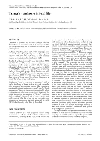
  • 1. Ultrasound Obstet Gynecol 2003; 22: 264–267 Published online 9 June 2003 in Wiley InterScience (www.interscience.wiley.com). DOI: 10.1002/uog.151 Turner’s syndrome in fetal life E. SURERUS, I. C. HUGGON and L. D. ALLAN Fetal Cardiology Unit, Harris Birthright Research Centre for Fetal Medicine, King’s College, London, UK KEYWORDS: cardiac defects; echocardiography; fetus; first trimester; karyotype; Turner’s syndrome ABSTRACT Objective To compare the incidence and type of heart disease found in association with 45X karyotype in fetal life with postnatal life and to examine the outcome after fetal diagnosis. Methods Fifty-three fetuses with a 45X karyotype were examined echocardiographically over a 4-year period between 1999 and 2002. Of these, 47 were referred because of increased nuchal translucency (NT). Results A cardiac abnormality was detected in 33/53 (62.2%) fetuses. The most common diagnosis was coarctation of the aorta in 24/53 (45.3%) fetuses, followed by the hypoplastic left heart syndrome (HLHS) in 7/53 (13.2%) fetuses. The mean NT was significantly higher in fetuses with a heart defect than in those with normal echocardiography. Termination of pregnancy was carried out in 45/53 (84.9%) fetuses and intrauterine death occurred in six cases. Two of four fetuses with a mosaic karyotype are currently alive. Conclusion Turner’s syndrome is associated with a higher incidence of heart defects detected prenatally when compared to postnatal reports. The commonest associated heart defects detected prenatally are HLHS and coarctation of the aorta, in contrast to postnatal life where a bicuspid aortic valve is the most common diagnosis. The typical intrauterine presentation of Turner’s syndrome with a markedly increased NT or with hydrops and with a typical 45X karyotype has an extremely poor prognosis for intrauterine survival. Copyright  2003 ISUOG. Published by John Wiley & Sons, Ltd. INTRODUCTION Turner’s syndrome is a well-recognized clinical entity in children and adults, which includes cardiovascular abnormalities, webbing of the neck, short stature and ovarian dysfunction. It is characteristically associated with monosomy of the X chromosome on karyotyping, but nearly half of the cases recognized postnatally have other X chromosome anomalies, such as mosaicism, ring formation or deletions1,2 . Structural heart disease is a feature of Turner’s syndrome, occurring in 15–30% of those presenting in postnatal life2–5. The most frequent cardiac abnormalities are a bicuspid aortic valve, which occurs in 14–19% of cases, or coarctation of the aorta in around 4–10% of cases. More severe left heart disease, including the hypoplastic left heart syndrome (HLHS), is also reported6. The prognosis for girls presenting with Turner’s syndrome beyond the neonatal period is generally good with appropriate treatment. In particular, intellectual development is usually normal. The endocrine deficiencies can usually be overcome by supplementation7 . In fetal life there is a typical group of obstetric ultrasound findings associated with Turner’s syndrome, including cystic hygroma and fetal hydrops, which are most commonly found up to 21 weeks’ gestation8–10. In addition, a 45X karyotype is commonly identified at chorionic villus sampling (CVS) after the detection of increased nuchal translucency (NT) at the 11–14- week scan. Usually, the NT in Turner’s syndrome is markedly increased above the normal range11, and may be associated with additional features of fluid retention. The prognosis following presentation in this fashion appears to be very much worse than that associated with postnatal presentation of Turner’s syndrome. There is a known high rate of spontaneous fetal loss in utero, with a suggested incidence of at least 80% of fetuses with Turner’s syndrome dying between 10 weeks’ gestation and term12, with some authors reporting a mortality of 99% from the first trimester8 . Presentation with hydrops may itself predict fetal loss10,13 . The objective of this study was to compare the incidence and type of heart disease found in association with 45X karyotype in fetal life with that reported in postnatal life and to document the outcome after fetal diagnosis. Correspondence to: Prof. L. D. Allan, Harris Birthright Research Centre, 3rd Floor Jubilee Wing, King’s College Hospital, Denmark Hill, London SE5 9RS, UK (e-mail: la48@columbia.edu) Accepted: 27 March 2003 Copyright  2003 ISUOG. Published by John Wiley & Sons, Ltd. ORIGINAL PAPER
  • 2. Fetal Turner’s syndrome 265 METHODS A retrospective search of our database between February 1999 and July 2002 identified 78 cases of Turner’s syndrome after karyotyping at the Harris Birthright Research Centre for Fetal Medicine. Of these, 61 cases had been examined echocardiographically by abdominal ultrasound. In eight cases the echocardiographic images were not diagnostic, leaving 53 cases to be analyzed in this study. Fifty-two patients had fetal echocardiography prior to invasive testing for fetal karyotype and one after a 45X karyotype had been detected. The ultrasound examination was performed using an Acuson Aspen (Acuson, Mountain View, CA, USA) with a C7 7-MHz probe. Of these 53 cases, 47 were referred because of increased NT > 4 mm, which is our arbitrary cut-off for early specialized fetal cardiac evaluation. The gestational age of this group ranged between 11 and 14 (mean, 12) weeks. A further five cases were referred because of fetal hydrops between 15 and 20 (mean, 18) weeks, and in the remaining case echocardiography was performed in a fetus in which the NT was 1.5 mm but a mosaic 45X karyotype was detected at CVS for a previous history of trisomy 18. RESULTS The diagnosis of cardiac normality or abnormality was made at the first visit in 51 fetuses. In two fetuses, diag- nostic images were not achieved at 11 and 12 weeks but were obtained at 13 and 15 weeks, respectively. Of the 53 cases, 49 were 45X karyotype and four had mosaicism for 45X. The gestational age at diagnosis was < 14 weeks in 47/53 cases. A cardiac abnormality was detected in 33/53 (62.3%) cases (Figure 1). The most common diagnosis was disproportion between the two ventricles and between the aorta and pulmonary trunk suggestive of coarctation of the aorta 24/53 (45.3%) cases, followed by the HLHS in 7/53 (13.2%) cases (Figures 2 and 3). In addition, there was one case of an atrioventricular septal defect (AVSD) and one of isolated tricuspid regurgitation. Of the four mosaic karyotypes, all had a normal heart scan. Of the group of 53 pregnancies, termination of preg- nancy (TOP) took place in 45 (84.9%) pregnancies, and spontaneous intrauterine death occurred in six cases at gestations ranging between 16 and 33 weeks (Table 1). In five of those six patients, later echocardiography con- firmed the earlier findings. Of those four cases with a mosaic karyotype, two patients chose pregnancy inter- ruption without further testing, and two continued the pregnancy without amniocentesis, which was offered. Both newborns are alive and well, with normal hearts as predicted. Of these, one had a normal postnatal karyotype and the remaining case showed no obvious signs of Turner’s syndrome at birth but postnatal kary- otyping revealed 45X in all cells analyzed. Postmortem Turner’s syndrome n = 53 Normal echocardiography n = 20 (37.7%) Abnormal echocardiography n = 33 (62.3%) Coarctation n = 24 (45.3%) HLHS n = 7 (13.2%) Other n = 2 (3.7%) Figure 1 Echocardiographic findings in the fetuses with Turner syndrome. HLHS, hypoplastic left heart syndrome; Other, atrioventricular septal defect and tricuspid regurgitation. RV LV LV RV S S Figure 2 (a) Normal four-chamber view of the heart at 12 weeks and (b) hypoplastic left heart at 12 weeks in a four-chamber view. There was no flow into the left ventricle on color flow mapping and reverse flow in the arch. LV, left ventricle; RV, right ventricle; S, spine. Copyright  2003 ISUOG. Published by John Wiley & Sons, Ltd. Ultrasound Obstet Gynecol 2003; 22: 264–267.
  • 3. 266 Surerus et al. LV RV S * Figure 3 Ventricular disproportion at 16 weeks suggestive of coarctation of the aorta. *, pleural effusion; LV, left ventricle; RV, right ventricle; S, spine. Table 1 Outcome of pregnancy according to fetal karyotype 45X karyotype Mosaic karyotype Total TOP 43 2 45 IUD 6 0 6 LB 0 2 2 Total 49 4 53 IUD, intrauterine death; LB, live birth; TOP, termination of pregnancy. examination confirmed the echocardiographic findings in five cases, two of which were predicted to have coarcta- tion, two with HLHS and one with an AVSD. The mean NT in the group of 47 fetuses with increased NT (over the 95th centile for the crown–rump length) was 9.8 (range, 4.1–17.1) mm. The mean NT was significantly higher in those cases with heart disease detected prenatally (10.9 mm), than in those without cardiac abnormalities (8.0 mm) (F statistic = 24.04, P = 0.0001). In addition, the NT was higher in those cases with monosomy X than in those with mosaicism (10.0 vs. 4.8 mm) (F statistic = 13.4, P = 0.000671). Student’s t-test was used in the statistical analysis. DISCUSSION The relatively benign course for a case of Turner’s syndrome detected postnatally appears to be in sharp contrast to the prognosis of those cases observed in fetal life7,14 . The group of Turner’s syndrome detected by increased NT between 11 and 14 weeks or with hydrops at later gestation are known to have a high rate of spontaneous fetal loss10,13 and this was confirmed in our series. Only one of the cases in our series was found at invasive testing by chance because of a previous history of trisomy 18. In contrast, Saenger states in his review14 that most prenatal diagnosis of Turner’s syndrome occurs by chance after routine amniocentesis for maternal age. Such cases may have quite a different prognosis from the group seen by us at a referral center with increased NT or hydrops. Thus, our findings may not be applicable to cases found in different clinical settings. In our study, a higher incidence of congenital heart defects was found compared to those cases identified postnatally (62% vs. 20%). Coarctation of the aorta was found in 45% of cases and the HLHS in 13% prenatally, in contrast to 4–7% and 1–2%, respectively, reported postnatally15,16 . A limitation of our study is the small number of postmortem examinations after TOP or miscarriage to confirm the prenatal diagnosis of heart defects, but in 10 cases with either postmortem or later echocardiography, the findings were confirmed. The low rate of postmortem examination was because most patients are referred to us from other hospitals for CVS with the TOP taking place locally after the result has been obtained. In addition, as most terminations took place prior to 14 weeks the mode of termination was surgical, which did not provide an intact fetus for examination. In a series of four fetuses seen prior to 14 weeks’ gestation with increased NT and Turner’s syndrome, reported by Haak et al., the HLHS was found in 3/4 cases17 . Although their numbers are small, fetal echocardiography in the first trimester successfully identified this abnormality, which they confirmed at postmortem examination. Gembruch et al. described a hypoplastic aortic arch and hypoplastic left ventricle as the commonest cardiac anomalies associated with Turner’s syndrome in their prenatal series of five cases18 . Isolated bicuspid aortic valve constitutes the majority (12–34%) of heart defects identified in postnatal life3,5,15,19 but this lesion cannot generally be detected during prenatal ultrasound and was not seen in our series. The X ring karyotype was related to a prevalence of 33.3% of cardiac defects in postnatal life by Prandstraller et al. but the most common abnormality found in association with this karyotype pattern was a bicuspid aortic valve3,15,20 . There is also a difference in the karyotype found prenatally from that found postnatally. In our series, almost all cases were of monosomy X fetuses (92.5%), with only a small proportion of mosaics (7.5%), and no cases of more minor X chromosome defects were seen. It is, of course, possible that some of the cases of mosaicism were not truly fetal mosaicism but confined to the placenta. In the two continuing pregnancies with mosaicism the parents decided against amniocentesis. In one a postnatal karyotype proved normal and the other was a true case of 45X. Similar to our findings, Monney et al. report rates of 84.5% and 13.5% for 45X and mosaic karyotype, respectively, in their prenatal series8 . In contrast, in postnatal series, Gotzsche et al. reported 58% 45X karyotypes, 35% mosaics and 7% of structural abnormalities and Douchin et al. reported 50% 45X and 50% mosaics or structural abnormalities2,21 . This Copyright  2003 ISUOG. Published by John Wiley & Sons, Ltd. Ultrasound Obstet Gynecol 2003; 22: 264–267.
  • 4. Fetal Turner’s syndrome 267 suggests that most of the children with mosaicism or more minor X anomalies, who comprise about half the postnatal series, may appear normal at the ultrasound examination in intrauterine life and also that intrauterine lethality for 45X karyotype is higher than in mosaics. Gravholt et al. observed in their series of 100 fetuses that the probability of a mosaic karyotype fetus to reach term was significantly higher than a 45X fetus surviving12 . In our series, none of the six fetuses with a 45X karyotype, where there was expectant management, survived to term, although there was a wide range in the timing of spontaneous loss (16–33 weeks). Conversely, two mosaic pregnancies continued and delivered live births. It is of interest that of our four mosaics, although three were detected for increased NT, the increase in measurement was more modest in these cases than in the group with monosomy X at 4.3, 6.7 and 6.8 mm, respectively. In postnatal series, the nature of the karyotype is also strongly associated with the severity of the cardiac abnormality identified. Fetuses with 45X karyotype have been reported to be more likely to have a heart defect, and to have a more severe form of cardiac disease, such as coarctation of the aorta, than the mosaics (29–30% vs. 16.6–24.3%)3,15 . Structural abnormalities in the X chromosome are generally associated with milder abnormalities such as bicuspid aortic valve and cardiac dysfunction3 . Gotzsche et al. report a series of 179 patients where there was a cardiovascular malformation in 38% with 45X karyotypes as against 11% with a mosaic karyotype. None of their patients with structural abnormalities of the X chromosome had cardiovascular malformations2 . In conclusion, there is a higher incidence of congenital heart disease diagnosed in cases of Turner’s syndrome where prenatal identification occurs as a result of abnormal ultrasound findings. These fetuses usually present with more severe forms of heart defects and this is associated with a specific fetal karyotype of monosomy X. In addition, the typical intrauterine presentation of Turner’s syndrome with a markedly increased NT or with hydrops has an extremely poor prognosis for survival. ACKNOWLEDGMENT This study was supported by a grant from The Fetal Medicine Foundation (Registered Charity 1037116). REFERENCES 1. Hook EB, Warburton D. The distribution of chromosomal genotypes associated with Turner’s syndrome: livebirth preva- lence rates and evidence for diminished fetal mortality and severity in genotypes associated with structural abnormalities or mosaicism. Hum Genet 1983; 64: 24–27. 2. Gotzsche CO, Krag-Olsen B, Nielsen J, Sorensen KE, Kris- tensen BO. Prevalence of cardiovascular malformations and associations with karyotypes in Turner’s syndrome. Arch Dis Child 1994; 71: 433–436. 3. Mazzanti L, Cacciari E. Congenital heart disease in patients with Turner’s syndrome. J Pediatr 1998; 133: 688–692. 4. Landin-Wilhelmsen K, Bryman I, Wilhelmsen L. Cardiac mal- formations and hypertension, but not metabolic risk factor, are common in Turner syndrome. J Clin Endocrinol Metab 2001; 86: 4166–4170. 5. Hou JW, Hwu WL, Tsai WY, Lee JS, Wang TR, Lue HC. Cardiovascular disorders in Turner’s syndrome and its correlation to karyotype. J Formos Med Assoc 1993; 92: 188–189. 6. Sybert VP. Cardiovascular malformations and complications in Turner syndrome. Pediatrics 1998; 101: E11. 7. Sas TC, de Muinck Keizer-Schrama SM. Turner’s syndrome: a pediatric perspective. Horm Res 2001; 56: 38–43. 8. Monney C, Pescia G, Addor M-C. Le syndrome de Turner. Schweiz Med Wochenschr 2000; 130: 1339–1343. 9. Twining P, Zuccollo J. The ultrasound markers of chromosomal disease: a retrospective study. Br J Radiol 1993; 66: 408–414. 10. Brown BS. The ultrasonographic features of nonimmune hydrops fetalis: a study of 30 successive patients. Can Assoc Radiol J 1986; 37: 164–168. 11. Nicolaides KH, Azar G, Snijders RJ, Gosden CM. Fetal nuchal oedema: associated malformations and chromosomal defects. Fetal Diagn Ther 1992; 7: 123–131. 12. Gravholt CH, Juul S, Naeraa RW, Hansen J. Prenatal and postnatal prevalence of Turner’s syndrome: a registry study. BMJ 1996; 312: 16–21. 13. MacLeod AM, McHugo JM. Prenatal diagnosis of nuchal cystic hygroma. Br J Radiol 1991; 64: 802–807. 14. Saenger P. Turner’s syndrome. N Engl J Med 1996; 335: 1749–1754. 15. Prandstraller D, Mazzanti L, Picchio FM, Magnani C, Berga- maschi R, Perri A, Tsingos E, Cacciari E. Turner’s syndrome: cardiologic profile according to the different chromosomal pat- terns and long-term clinical follow up of 136 nonpreselected patients. Pediatr Cardiol 1999; 20: 108–112. 16. Van Egmond H, Orye E, Praet M, Coppens M, Devloo- Blancquaert A. Hypoplastic left heart syndrome and 45X karyotype. Br Heart J 1988; 60: 69–71. 17. Haak MC, Bartelings MM, Gittrnberger-de Groot AC, Van Vugt JM. Cardiac malformations in first-trimester fetuses with increased nuchal translucency: ultrasound diagnosis and postmortem morphology. Ultrasound Obstet Gynecol 2002; 20: 14–21. 18. Gembruch U, Baschat AA, Knopfle G, Hansmann M. Results of chromosomal analysis in fetuses with cardiac anomalies as diag- nosed by first- and early second-trimester echocardiography. Ultrasound Obstet Gynecol 1997; 10: 391–396. 19. Miller MJ, Geffner ME, Lippe BM, Itami RM, Kaplan AS, DiSessa TG, Isabel-Jones JB, Friedman WF. Echocardiography reveals a high incidence of bicuspid aortic valve in Turner syndrome. J Pediatr 1983; 102: 47–50. 20. Mazzanti L, Prandstraller D, Tassinari D, Rubino I, Santucci S, Picchio FM, Forabosco A, Cacciari E. Heart disease in Turner’s syndrome. Helv Paediatr Acta 1988; 43: 25–31. 21. Douchin S, Rossignol AM, Klein SK, Siche JP, Baguet JP, Bost M. Heart malformations and vascular complications with Turner’s syndrome. Prospective study of 26 patients. Arch Mal Coeur Vaiss 2000; 93: 565–570. Copyright  2003 ISUOG. Published by John Wiley & Sons, Ltd. Ultrasound Obstet Gynecol 2003; 22: 264–267.