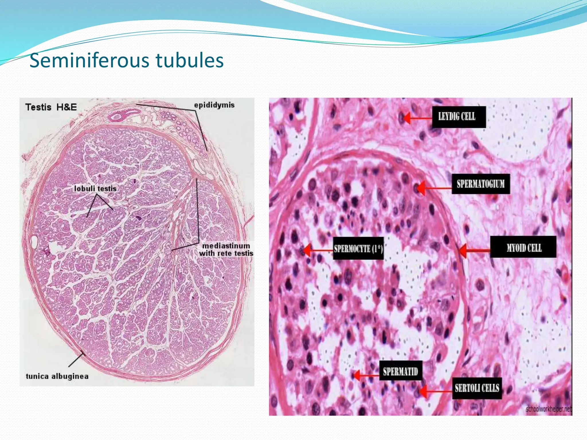
By: Lilian B. Rosario, MD, FPPS
good morning !!
Objective: Identify structures, components, functions
DIAGRAM:
EXTERNAL GENITAL ORGANS
Penis:
3 cylindrical Cavernous bodies
(erectile tissue)
- 2 Corpora Cavernosa
Penis- dorsum area
- 1 Corpus spongiosum’:
median, ventral
- enlarged portion form
GLANS of Penis-
covered bv PREPUCE
- Its central portion
occupied by Spongy
Urethra (Penile;
Cavernous Urethra)
Penis:
 Tunica albuginea- dense collagenous CT, supplied by elastic elements;
binds the 3 cavernous bodies, forms
A capsule/body
 Flaccid Penis: T. Albuginea of corpora cavernous Penis
is 2 mm thick; But
Erect Penis: T. albuginea is 0.5 mm thin
External to T. albuginea: Hypodermis of the skin-
envelopes the penis; richly supplied with blood
vessels; but devoid of Fat cells
Skin of Penis: no hair follicles; limited sweat glands
Glands of Tyson: undersurface of the prepuce; proximal portion
of Glans of penis
- atypical sebaceous glands ( not associated with hair follicles)
- the secretions forms cheesy material ‘SMEGMA’
SCROTUM:
Internal Genital Organs: TESTES
- Produce male gametes:
spermatozoa
- Pair of oval bodies
lodged in the scrotum
- Each is 4-5 cm long, 3 cm
wide, weighs: 14 g
Testes:
 T.Albuginea: tough fibrous
capsule; dense irregular
CT; enclosed testes
 Mediastinum testis:
thickened T. albuginea;
blood& lymphatic v
enter/exit
 Ductuli efferentes: leave
the testis
 Septulae testis: CT septa
from T. albuginea,
incomplete division of
testis into” Lobuli testis”
 Seminiferous tubules:
highly coiled tubes; “
spermatogenesi”
Seminiferous tubules
Seminiferous tubules: SPERMATOGENESIS : 3 stages
SPERMIOGENESIS: 3 Phases
 Golgi Phase: golgi complex =
proacrosomal granules fused=
acrosomal vesicle; nucleus
condense; centrioles at the
opposite of a.v; then a.v
attached to one pole of nucleus
 Acrosomal phase: A.V forms
acrosome; flagellum formation;
cytoplasm migrates taking
mitochondria to form Middle
piece
 Maturation phase:
transfromation of spermatid to
spaermatozoon. Residual
cytoplasm is shed and
phagocytized by Sertoli cells
 ENDS : Spermiation- release of
spermatozoon into the fluid
filled lumen of Seminiferous
tubules.
Testes:
Spermatozoon: Head – elongated highly condensed
nucleus covered by acrosomal cap.
Acrosome: hydrolytic enzymes that during fertization,
enable spermatozoa to disperse the cells of Corona
radiata and digest Zona pellucida of mature oocyte.
Tail: neck; middle /pricipal/ end piece
Testicular lobules: division of testis by connective tissue
septa into 250 pyramidal compartments which contains
seminiferous tubules ( supported by CT stroma- highly
vascular reticular CT: scattered LEYDIG cells, reticular
cells, mesenchymal, macrophages
Interstitial Cells( of Leydig): produce TESTOSTERONE,
regulated by LH secreted by pituitary gland.
Large ovoid cells w/ distinct nucleus;
Crystal (of Reinke) in cytoplasm
Testes:
Seminiferous tubules: terminates at
mediastinus testis by becoming
cont w/ tubuli recti; outer fibrous
sheath w/ basal lamina :
Myoid Cells- resemble sm m cells,
contracts to propel spermatozoa
into the duct.
Inner strat epith basal lamina of
wall: has Gametes in various
stages; supporting cells: Sertoli
cells
Sertoli cells: tall cells w/ elongated
nucleus; near bases forms tight
junctions at interstitial lumen:
Blood- Testes Barrier.
- nutritional support, protection
for gametes; prod of fluids for
transport of spermatozoa
- prod INHBIN ; (-) feedback
effect to reduce FSH( increased
activity of SC) secretion
DUCTS of the Testis
Intratesticular Ducts:
Tubuli Recti
Rete Testis
Ductuli Efferentes
Extratesticular Ducts:
Epididymis & Ductus
epididymis
Ductus Deferens ( Vas
deferens)
Ejaculatory duct
Urethra
Intratesticular genital ducts:
Tubuli Recti (straight tubules)
- immediate cont. seminife
rous tubules; initial
segment: Sertoli cells w/o
gametes.
Rete Testis: network of
- anastomosing canals w/
irreg lumen at mediasti-
num testis where T.Recti
empty.
- canals lined: single layer
squamous/ cuboidal cells
on basal lamina
Ductuli Efferentes: 12- 20 fine tubules
penetrate T. albuginea: where R.Testis
drain
- leaving the testis becomes highly
totuos and merge to form single tube
Ductus epididymis
Extratesticular ducts: EPIDIDYMIS & DUCTUS EPIDIDYMIS
-C shaped structure, 7.5 cm
length, superior/ post of
testes
- Parts:
- Head- superior pole w/
- thin myoepithelium (distal portion
- ductuli efferentes + initial portion of
- ductus epididymis)
- - receives spermatozoa via
- efferent ducts; diluted
- sperm
- Body- posterior surface
- Tail- inferior pole of testis;
- - thicker myoepithelium
- - conc of sperm increased,
-contains blood v that supply the
ducts & CT that binds the coils of
ducts
-Ductus Epididymis: main component of Epi
didymis
Epididymis / Ductus epididymis
-Covered : 2 layered Pseudostrat
epith, separated by BM
surrounded by CT w/ sm. m
cells
-2 types of cells:
- PC: tall columnar cells at the
head w/c decreased in size along
ductus (cuboidal); apical
surfaces: + Stereocilia:
- Absorbs 90% of fluids from
Seminiferous t.
- BC: small pyramidal cells at basal
lamina between bases and PC=
precursors of PC (?)
Spermatozoa: 2-6 days in Ductus E;
leaves ductus w/ fully motile tail
but still has to undergo
CAPACITATION before it can
fertilize the ovum.
Extratesticular ducts: DUCTUS DEFERENS (Vas deferens)
* Fibromuscular tube; 45 cm long;
embedded in the Spermatic cord
frm ductus E- deep inguinal ring-
abdomino pelvic cavity
* 3 layers:
- inner Mucosa; epith (pseudostrat
w/ stereocilia)+
lamina propia (elastic f.)
- middle Muscularis: sm m fibers in
3 distinct layers
- Adventitia: outer fibrous has:
Blood v, lymphatic v and nerves
that supply the duct
• Ampulla: distal segment of Vas D
* Store & Carries sperm from
Epididymis to Ejaculatory duct
Extratesticular ducts: EJACULATORY Duct & URETHRA
Ejaculatory Duct: formed
by union of Ductus
Deferens w/ Duct of
Seminal Vesicle
- penetrates Prostate
gland to empty into post
part of Prostatic Urethra
Urethra: last segment of
the duct system both
reproductive and
urrinary systems
EJACULATORY DUCT
Simple columnar: lining
epithelium
Has NO muscular layer
Mucosa: surrounded by Fibro-
muscular tissue ( part of stroma
of prostate gland)
URETHRA
- 20 cms length:
- Prostatic urethra:
transitional epith; glands
of Littre ( tubo alveolar
mucus secreting )
- Membranous urethra:
pseudostrat columnar
- Spongy urethra:
pseudostrat col to non-
- Keratinized strat sq.
ACCESSORY GLANDS
Prostate gland
Seminal vesicles (paired)
Bulbourethral glands “Glands
of Cowper”
-combined secretions of the 3
glands: most volume of male
ejaculate (semen)
Prostate: 30% seminal volume
Seminal v: 60% volume
Bulbourethal: insignificant
amount ( drop or so)
-Semen= 2-5ml/ ejaculation
- = 20 t0 250 million spermatozoa
- Per ml ( 10% of the ejaculate)
Prostate Gland
- Largest; chest nut shaped gland
- Stroma: dense CT w/ rich
supply of Sm m fibers
“ fibromuscular organ”
- forms a capsule, a septa
divides into poorly
defined lobules
- Parenchyma: 3 groups of
Tubuloalveolar glands:
- main prostatic glands- bulk
- submucosal glands- inner 3rd
- mucosal glands- small,
surrounds urethra
Prostate gland: cont
Lining of the alveoli: mostly
simple columnar ; least simple
squamous, cuboidal, pseudo-
strat col
Lumen: lamellated concretions
“Corpora Amylacea”- has
glycoproteins & other subs,
deposited on cell fragments,
inc in size/ number: calcify
with age.
Secretions: alkaline fluid-
enzymes, fibrinolysin, prosta-
glandins, compound w/
antibiotic properties.
SEMINAL VESICLES
Paired saclike structures,
above prostate gland w/in
pelvis
Has capsulated highly coiled
narrow tube
Wall: Mucosa- branched
folds”maze”
Muscularis- sm.m f
circularly, longitudinally
arranged
Adventitia- CT
Secretions: Fructose ( energy
for spermatozoa), Pg,
fibrinogen.
Seminal vesicles:
BULBOURETHRAL GLANDS ( of COWPER)
Paired Pea sized yellowish
organs embedded in the
sphincter urethral m.,
posterolateral to membranous
urethra
Secretory units: capsulated with
CT
Tubulo-alveolar mucus glands
Lining of main secretory duct:
simple columnar
epithelium
Secretions: clear, viscous fluid
discharged shortly before
ejaculation
- helps lubricate the urethra for
easier passage of the
spermatozoa.
Thank You…..

The Male Reproductive system lecture.pptx

  • 1.
    By: Lilian B.Rosario, MD, FPPS
  • 2.
  • 3.
    Objective: Identify structures,components, functions
  • 4.
  • 5.
  • 6.
    Penis: 3 cylindrical Cavernousbodies (erectile tissue) - 2 Corpora Cavernosa Penis- dorsum area - 1 Corpus spongiosum’: median, ventral - enlarged portion form GLANS of Penis- covered bv PREPUCE - Its central portion occupied by Spongy Urethra (Penile; Cavernous Urethra)
  • 7.
    Penis:  Tunica albuginea-dense collagenous CT, supplied by elastic elements; binds the 3 cavernous bodies, forms A capsule/body  Flaccid Penis: T. Albuginea of corpora cavernous Penis is 2 mm thick; But Erect Penis: T. albuginea is 0.5 mm thin External to T. albuginea: Hypodermis of the skin- envelopes the penis; richly supplied with blood vessels; but devoid of Fat cells Skin of Penis: no hair follicles; limited sweat glands Glands of Tyson: undersurface of the prepuce; proximal portion of Glans of penis - atypical sebaceous glands ( not associated with hair follicles) - the secretions forms cheesy material ‘SMEGMA’
  • 8.
  • 10.
    Internal Genital Organs:TESTES - Produce male gametes: spermatozoa - Pair of oval bodies lodged in the scrotum - Each is 4-5 cm long, 3 cm wide, weighs: 14 g
  • 11.
    Testes:  T.Albuginea: toughfibrous capsule; dense irregular CT; enclosed testes  Mediastinum testis: thickened T. albuginea; blood& lymphatic v enter/exit  Ductuli efferentes: leave the testis  Septulae testis: CT septa from T. albuginea, incomplete division of testis into” Lobuli testis”  Seminiferous tubules: highly coiled tubes; “ spermatogenesi”
  • 12.
  • 13.
  • 15.
    SPERMIOGENESIS: 3 Phases Golgi Phase: golgi complex = proacrosomal granules fused= acrosomal vesicle; nucleus condense; centrioles at the opposite of a.v; then a.v attached to one pole of nucleus  Acrosomal phase: A.V forms acrosome; flagellum formation; cytoplasm migrates taking mitochondria to form Middle piece  Maturation phase: transfromation of spermatid to spaermatozoon. Residual cytoplasm is shed and phagocytized by Sertoli cells  ENDS : Spermiation- release of spermatozoon into the fluid filled lumen of Seminiferous tubules.
  • 16.
    Testes: Spermatozoon: Head –elongated highly condensed nucleus covered by acrosomal cap. Acrosome: hydrolytic enzymes that during fertization, enable spermatozoa to disperse the cells of Corona radiata and digest Zona pellucida of mature oocyte. Tail: neck; middle /pricipal/ end piece Testicular lobules: division of testis by connective tissue septa into 250 pyramidal compartments which contains seminiferous tubules ( supported by CT stroma- highly vascular reticular CT: scattered LEYDIG cells, reticular cells, mesenchymal, macrophages Interstitial Cells( of Leydig): produce TESTOSTERONE, regulated by LH secreted by pituitary gland. Large ovoid cells w/ distinct nucleus; Crystal (of Reinke) in cytoplasm
  • 17.
    Testes: Seminiferous tubules: terminatesat mediastinus testis by becoming cont w/ tubuli recti; outer fibrous sheath w/ basal lamina : Myoid Cells- resemble sm m cells, contracts to propel spermatozoa into the duct. Inner strat epith basal lamina of wall: has Gametes in various stages; supporting cells: Sertoli cells Sertoli cells: tall cells w/ elongated nucleus; near bases forms tight junctions at interstitial lumen: Blood- Testes Barrier. - nutritional support, protection for gametes; prod of fluids for transport of spermatozoa - prod INHBIN ; (-) feedback effect to reduce FSH( increased activity of SC) secretion
  • 19.
    DUCTS of theTestis Intratesticular Ducts: Tubuli Recti Rete Testis Ductuli Efferentes Extratesticular Ducts: Epididymis & Ductus epididymis Ductus Deferens ( Vas deferens) Ejaculatory duct Urethra
  • 21.
    Intratesticular genital ducts: TubuliRecti (straight tubules) - immediate cont. seminife rous tubules; initial segment: Sertoli cells w/o gametes. Rete Testis: network of - anastomosing canals w/ irreg lumen at mediasti- num testis where T.Recti empty. - canals lined: single layer squamous/ cuboidal cells on basal lamina Ductuli Efferentes: 12- 20 fine tubules penetrate T. albuginea: where R.Testis drain - leaving the testis becomes highly totuos and merge to form single tube Ductus epididymis
  • 22.
    Extratesticular ducts: EPIDIDYMIS& DUCTUS EPIDIDYMIS -C shaped structure, 7.5 cm length, superior/ post of testes - Parts: - Head- superior pole w/ - thin myoepithelium (distal portion - ductuli efferentes + initial portion of - ductus epididymis) - - receives spermatozoa via - efferent ducts; diluted - sperm - Body- posterior surface - Tail- inferior pole of testis; - - thicker myoepithelium - - conc of sperm increased, -contains blood v that supply the ducts & CT that binds the coils of ducts -Ductus Epididymis: main component of Epi didymis
  • 23.
    Epididymis / Ductusepididymis -Covered : 2 layered Pseudostrat epith, separated by BM surrounded by CT w/ sm. m cells -2 types of cells: - PC: tall columnar cells at the head w/c decreased in size along ductus (cuboidal); apical surfaces: + Stereocilia: - Absorbs 90% of fluids from Seminiferous t. - BC: small pyramidal cells at basal lamina between bases and PC= precursors of PC (?) Spermatozoa: 2-6 days in Ductus E; leaves ductus w/ fully motile tail but still has to undergo CAPACITATION before it can fertilize the ovum.
  • 24.
    Extratesticular ducts: DUCTUSDEFERENS (Vas deferens) * Fibromuscular tube; 45 cm long; embedded in the Spermatic cord frm ductus E- deep inguinal ring- abdomino pelvic cavity * 3 layers: - inner Mucosa; epith (pseudostrat w/ stereocilia)+ lamina propia (elastic f.) - middle Muscularis: sm m fibers in 3 distinct layers - Adventitia: outer fibrous has: Blood v, lymphatic v and nerves that supply the duct • Ampulla: distal segment of Vas D * Store & Carries sperm from Epididymis to Ejaculatory duct
  • 25.
    Extratesticular ducts: EJACULATORYDuct & URETHRA Ejaculatory Duct: formed by union of Ductus Deferens w/ Duct of Seminal Vesicle - penetrates Prostate gland to empty into post part of Prostatic Urethra Urethra: last segment of the duct system both reproductive and urrinary systems
  • 26.
    EJACULATORY DUCT Simple columnar:lining epithelium Has NO muscular layer Mucosa: surrounded by Fibro- muscular tissue ( part of stroma of prostate gland)
  • 27.
    URETHRA - 20 cmslength: - Prostatic urethra: transitional epith; glands of Littre ( tubo alveolar mucus secreting ) - Membranous urethra: pseudostrat columnar - Spongy urethra: pseudostrat col to non- - Keratinized strat sq.
  • 29.
    ACCESSORY GLANDS Prostate gland Seminalvesicles (paired) Bulbourethral glands “Glands of Cowper” -combined secretions of the 3 glands: most volume of male ejaculate (semen) Prostate: 30% seminal volume Seminal v: 60% volume Bulbourethal: insignificant amount ( drop or so) -Semen= 2-5ml/ ejaculation - = 20 t0 250 million spermatozoa - Per ml ( 10% of the ejaculate)
  • 30.
    Prostate Gland - Largest;chest nut shaped gland - Stroma: dense CT w/ rich supply of Sm m fibers “ fibromuscular organ” - forms a capsule, a septa divides into poorly defined lobules - Parenchyma: 3 groups of Tubuloalveolar glands: - main prostatic glands- bulk - submucosal glands- inner 3rd - mucosal glands- small, surrounds urethra
  • 31.
    Prostate gland: cont Liningof the alveoli: mostly simple columnar ; least simple squamous, cuboidal, pseudo- strat col Lumen: lamellated concretions “Corpora Amylacea”- has glycoproteins & other subs, deposited on cell fragments, inc in size/ number: calcify with age. Secretions: alkaline fluid- enzymes, fibrinolysin, prosta- glandins, compound w/ antibiotic properties.
  • 32.
    SEMINAL VESICLES Paired saclikestructures, above prostate gland w/in pelvis Has capsulated highly coiled narrow tube Wall: Mucosa- branched folds”maze” Muscularis- sm.m f circularly, longitudinally arranged Adventitia- CT Secretions: Fructose ( energy for spermatozoa), Pg, fibrinogen.
  • 33.
  • 34.
    BULBOURETHRAL GLANDS (of COWPER) Paired Pea sized yellowish organs embedded in the sphincter urethral m., posterolateral to membranous urethra Secretory units: capsulated with CT Tubulo-alveolar mucus glands Lining of main secretory duct: simple columnar epithelium Secretions: clear, viscous fluid discharged shortly before ejaculation - helps lubricate the urethra for easier passage of the spermatozoa.
  • 35.