This document presents a new approach for ECG feature extraction and heartbeat classification using mathematical morphology. The key points are:
1) Mathematical morphology filtering is used to preprocess the ECG signal before feature extraction in order to normalize variations between heartbeats.
2) Features are extracted from the preprocessed ECG signal and fed into a Kohonen self-organizing map for clustering.
3) A Learning Vector Quantization classifier is then trained on the clustered features to classify heartbeats as normal or abnormal.
4) The method is evaluated on the MIT-BIH Arrhythmia Database and shows improved classification performance over using the original unprocessed ECG signal.
![Mathematical morphology based
ECG feature extraction
for the purpose of heartbeat classification
Pawel Tadejko, Waldemar Rakowski
Technical University of Bialystok
Faculty of Computer Science
15-351 Bialystok, Wiejska 45A, Poland
{ptad@ii, W.Rakowski@}.pb.bialystok.pl
Abstract
The paper presents the classification performance of an
automatic classifier of the electrocardiogram (ECG) for
the detection abnormal beats with new concept of feature
extraction stage. Feature sets were based on ECG mor-
phology and RR-intervals. Configuration adopted a Ko-
honen self-organizing maps (SOM) for analysis of signal
features and clustering. In this study, a classifier was devel-
oped with SOM and Learning Vector Quantization (LVQ)
algorithms using the data from the records recommended
by ANSI/AAMI EC57 standard. This paper compares two
strategies for classification of annotated QRS complexes:
based on orginal ECG morphology features and proposed
new apporach - based on preprocessed ECG morphology
features. The mathematical morphology filtering is used for
the preprocessing of ECG signal. The problem of choosing
an appropriate structuring element of mathematical mor-
phology filtering for ECG signal processing was studied.
The performance of the algorithm is evaluated on the MIT-
BIH Arrhythmia Database following the AAMI recommen-
dations. Using this method the results of recognition beats
either as normal or arrhythmias was improved.
Index-terms
ECG, preprocesing, mathematical morphology, ECG fil-
tering, feature extraction, heartbeat classification
1. Introduction
The analysis of heart beat cycles in ECG signal is very
important for long-term monitoring of heart patients. How-
ever, it is very costly for the medical expert to analyze the
ECG recording beat by beat since the ECG records may last
for hours. Therefore, it is justified to develop a computer-
assisted technique to examine and annotate the ECG record-
ing to facilitate review by medical experts. This computer
annotation will assist doctors to select only the abnormal
beats for further analysis.
1.1. Arrhtyhmias classification
Automatic classification of cardiac rhythms still remains
a vital problem in clinical cardiology, especially when it is
performed in real time. Several researchers have addressed
the problem of automatic classification of cardiac arrhyth-
mias [2, 3, 4, 5, 16, 19, 22]. Annotation of ECG recording
requires the detection of various types of heartbeats. This
is a pattern recognition task. Very often, a classifier is to be
trained to recognize different types of beats. The training set
of the classifier is usually a large database, which consists
of the ECG beats from a large pool of patients. However,
these classifiers suffer from the problem of poor generaliza-
tion because there are usually some variations in the ”nor-
mal” range among human beings. Even doctors may expe-
rience difficulty in assessing abnormal ECG beats if only
considering the reference values based on the general pa-
tient population.
1.2. Heartbeat classifier
There are two general approaches to the training process:
building general heartbeat classifier [2, 5, 16, 19, 22] and
patient-adapting classifier [3, 4].
However, most of the approaches proposed in the liter-
ature deal with a limited number of arrhythmic types and
process the entire ECG signal extracting several features
from it, such as the P wave, which is an extremely time-
consuming process and sometimes difficult due to the pres-
ence of noise. Other researches suggested that there is a
6th International Conference on Computer Information
Systems and Industrial Management Applications (CISIM'07)
0-7695-2894-5/07 $20.00 © 2007](https://image.slidesharecdn.com/tadejko2007-230320145531-9596b016/85/tadejko2007-pdf-1-320.jpg)
![Mathematical morphology based
ECG feature extraction
for the purpose of heartbeat classification
Pawel Tadejko, Waldemar Rakowski
Technical University of Bialystok
Faculty of Computer Science
15-351 Bialystok, Wiejska 45A, Poland
{ptad@ii, W.Rakowski@}.pb.bialystok.pl
Abstract
The paper presents the classification performance of an
automatic classifier of the electrocardiogram (ECG) for
the detection abnormal beats with new concept of feature
extraction stage. Feature sets were based on ECG mor-
phology and RR-intervals. Configuration adopted a Ko-
honen self-organizing maps (SOM) for analysis of signal
features and clustering. In this study, a classifier was devel-
oped with SOM and Learning Vector Quantization (LVQ)
algorithms using the data from the records recommended
by ANSI/AAMI EC57 standard. This paper compares two
strategies for classification of annotated QRS complexes:
based on orginal ECG morphology features and proposed
new apporach - based on preprocessed ECG morphology
features. The mathematical morphology filtering is used for
the preprocessing of ECG signal. The problem of choosing
an appropriate structuring element of mathematical mor-
phology filtering for ECG signal processing was studied.
The performance of the algorithm is evaluated on the MIT-
BIH Arrhythmia Database following the AAMI recommen-
dations. Using this method the results of recognition beats
either as normal or arrhythmias was improved.
Index-terms
ECG, preprocesing, mathematical morphology, ECG fil-
tering, feature extraction, heartbeat classification
1. Introduction
The analysis of heart beat cycles in ECG signal is very
important for long-term monitoring of heart patients. How-
ever, it is very costly for the medical expert to analyze the
ECG recording beat by beat since the ECG records may last
for hours. Therefore, it is justified to develop a computer-
assisted technique to examine and annotate the ECG record-
ing to facilitate review by medical experts. This computer
annotation will assist doctors to select only the abnormal
beats for further analysis.
1.1. Arrhtyhmias classification
Automatic classification of cardiac rhythms still remains
a vital problem in clinical cardiology, especially when it is
performed in real time. Several researchers have addressed
the problem of automatic classification of cardiac arrhyth-
mias [2, 3, 4, 5, 16, 19, 22]. Annotation of ECG recording
requires the detection of various types of heartbeats. This
is a pattern recognition task. Very often, a classifier is to be
trained to recognize different types of beats. The training set
of the classifier is usually a large database, which consists
of the ECG beats from a large pool of patients. However,
these classifiers suffer from the problem of poor generaliza-
tion because there are usually some variations in the ”nor-
mal” range among human beings. Even doctors may expe-
rience difficulty in assessing abnormal ECG beats if only
considering the reference values based on the general pa-
tient population.
1.2. Heartbeat classifier
There are two general approaches to the training process:
building general heartbeat classifier [2, 5, 16, 19, 22] and
patient-adapting classifier [3, 4].
However, most of the approaches proposed in the liter-
ature deal with a limited number of arrhythmic types and
process the entire ECG signal extracting several features
from it, such as the P wave, which is an extremely time-
consuming process and sometimes difficult due to the pres-
ence of noise. Other researches suggested that there is a
6th International Conference on Computer Information
Systems and Industrial Management Applications (CISIM'07)
0-7695-2894-5/07 $20.00 © 2007](https://image.slidesharecdn.com/tadejko2007-230320145531-9596b016/75/tadejko2007-pdf-1-2048.jpg)
![need to incorporate local information of a specific patient
to improve the recognition of abnormal ECG beats and thus
help to improve the generalization.
This work proposes a method for the normalization of
variations in ECG beats, based on mathematical morphol-
ogy and resampling model, which can be easily applied to
the ECG signal.
2. Mathematical morphology
By ”morphological signal processing” we mean a broad
and coherent collection of theoretical concepts, mathemat-
ical tools for signal analysis [18]. Originally MM was
applied to analyzing images from geological or biologi-
cal specimens. However, its rich theoretical framework,
algorithmic efficiency, easy implementability on special
hardware, and suitability for many shape-oriented prob-
lems have propelled its widespread diffusion and adoption
by many academic and industry groups in many countries
as one among the dominant image analysis methodologies
[1, 11, 12, 13].
As a result, MM nowadays offers many theoretical and
algorithmic tools to and inspires new directions in many re-
search areas from the fields of signal processing, image pro-
cessing and machine vision, and pattern recognition.
2.1. Mathematical morphology transforma-
tions
Morphological filters are nonlinear signal transforma-
tions that locally modify geometric features of signals.
They stem from the basic operations of a set-theoretical
method for signal analysis, called mathematical morphol-
ogy, which was introduced by Serra [18].
In morphological filtering [1, 11, 12, 13], each signal is
viewed as a set, and its geometrical features are modified
by morphologically convolving the signal with a structuring
element (SE), which is another set of simple shape and size.
By varying the structuring element we can extract different
types of information from the signal.
A structuring element is characterized by its shape,
width, and height. The values of the structuring element
determine the shape of the output waveform.
2.2. Elementary mathematical morphology
operators
In the sequel we use definitions of grey-level morphol-
ogy basic operators in the same form as in [18]. Let us re-
call that erosion of a function f : R → R by a structuring
element b : R → R can be defined as
(f b)(s) = min
x
{f(s+x)−b(x) : ∗(s+x) ∈ Df ∧x ∈ Db}
(1)
where Df sup f, Db sup b. In a similar way, dilation ⊕ is
an operator given by
(f⊕b)(s) = max
x
{f(s−x)+b(x) : (s−x) ∈ Df ∧x ∈ Db}
(2)
Two other operators: closing • and opening ◦ are defined
with help of (2) and (3), i.e.
f • b = (f ⊕ b) b, f ◦ b = (f b) ⊕ b (3)
3. Self-Organization Map and Learning Vector
Quantization
The Self-organizing Map (SOM) is an artificial neural
network architecture based on unsupervised, competitive
learning [8]. It provides a topology preserving, smooth
mapping from a high-dimensional input space to the map
units usually arranged as a two-dimensional lattice of neu-
rons (nodes). Thus, the SOM can serve as a tool for cluster
analysis of complex, high-dimensional data.
A parametric reference vector m, is associated with every
node. A data vector x is compared to all reference vectors
in any metric and the best matching node is defined, e.g.,
by the smallest Euclidean distance between the data vec-
tor and any of the reference vectors. During learning, those
nodes that are topographically close in the array up to a cer-
tain distance will activate each other to learn from the same
input:
mj(t + 1) = mj(t) + hcj(t)[x(t) − mj(t)] (4)
where t is an integer representing time, and hcj is the so-
called neighbourhood kernel describing the neighbourhood
that is updated around the best-matching node in response
to the present feature vector x(t). Initially, the neighbor-
hood is large. The size reduces as clustering converges, un-
til no neighboring neurons will get updated. Several suit-
able kernels can be used, e.g. a so-called bubble kernel or
a gaussian kernel, relating to different ways of determining
the activating cells. The kernel also includes the learning
rate parameter α(t).
With time, the size of the neighbourhood and the learn-
ing rate are diminished. The described learning process
leads to a smoothing effect on the weight vectors in the
neighbourhood and by continued learning to global order-
ing of the nodes [8, 9].
Learning Vector Quantization (LVQ) [8, 10] is a super-
vised, clustering-based classification technique which clas-
sifies a feature vector x(t) according to the label of the clus-
ter prototype (code word) into which x(t) is clustered. Clas-
sification error occurs when the feature vectors within the
same cluster (hence, assigned to the same class label) are
actually drawn from different classes. To minimize clas-
sification error, the LVQ algorithm fine tunes the cluster-
ing boundary between clusters of different class labels by
6th International Conference on Computer Information
Systems and Industrial Management Applications (CISIM'07)
0-7695-2894-5/07 $20.00 © 2007](https://image.slidesharecdn.com/tadejko2007-230320145531-9596b016/85/tadejko2007-pdf-2-320.jpg)
![modifying the position of the clustering center (prototype or
code word). This method is called “learning vector quanti-
zation” because this clustering based classification method
is similar to the “vector quantization” method used for sig-
nal compression in the areas of communication and signal
processing.
4. Evaluation Method
The proposed method consists of three steps (Figure 1):
(a) preprocesssing (feature extraction), (b) feature vector
preparation (feature selection), (c) arrhythmic episode clas-
sification. The MIT-BIH arrhythmia database [15] is used
for evaluation of the method.
We evaluate various combinations of morphological fil-
ters and conduct experiments for different structuring ele-
ments [17, 20]. It turns out that results are strong depends
on shape and size of structuring element. Since the opening
and closing operations are intended to remove impulses, the
structuring element must be designed so that the waves in
the ECG signal are not removed by the process.
4.1. Datasets
To evaluate the performance of our approaches, we used
the 48 tapes of the MIT/BIH arrhytmia database, which
comes along with a very detailed annotation for each beat.
The Association for the Advancment of Medical Instrumen-
tation (AAMI) has summarized those detailed classes to
four classes of clinical relevance, as shown in Table 1 [14].
Four records (102, 104, 107, and 217), including paced
beats, are excluded from the study in compliance with the
standards recommended for reporting performance results
of cardiac rhythms by the AAMI.
The original signals in the MIT/BIH arrhythmia database
are two-leads, sampled at 360 Hz. The ECG signal of Lead
1 is used in this study.
4.2. Denoising and baseline wander elimi-
nation
The electrocardiogram (ECG) signal is the electrical in-
terpretation of the heart activity; it consists of a set of, well
defined, successive waves denoted: P, Q, R, S, and T waves
[7]. However, as the major part of real signals; the real
picked-up ECG signal is corrupted by several sources of
noise: EMG (electromyogram) signal (a high frequency sig-
nal related to muscle activity), the BLW (the baseline wan-
dering: a low frequency signal caused mainly by the breath-
ing action), the electrode motion (usually represented by a
sharp variation of the baseline).
Background normalization is performed by estimating
the drift in the background and subtracting it from the in-
coming data. Processing the data through a sequence of
opening and closing operations performs impulsive noise
suppression.
Let us take two possibly different structuring elements
b1 (for opening) and b2 (for closing) of the type considered
so far. In this way we obtain as follows:
bgn(f) = f − 1/2[(f ◦ b1) • b2 + (f • b2) ◦ b1] (5)
The ECG signal, as well as any baseline drift, is estimated
by processing the data using an opening operation followed
by a closing operation. Processing the data using a closing
operation followed by an opening operation forms a second
estimate of the signal. The result from this step is the aver-
age of the two estimates. For opening operation structuring
element b1 has size of L and for closing b2 size of 2L. The
size of the first SE L should be longer than QRS interval.
4.3. Feature vector
Here we investigate the use of raw amplitude of the time
domain ECG signals after noise suppression and baseline
drift removal as feature vectors to represent the ECG beats.
After the R-peak is located, the ECG signal in a window of
550 ms is taken as an ECG beat. The lengths of the sig-
nal before and after the R-peak in each beat are 140 ms and
410 ms, respectively, such that the window covers most of
the characterization of the ECG beat. The signal in each
window is then resampled to form a feature vector of 20-
dimensions. The R-R interval (the interval between two
consecutive R-peaks) is also used in this study by appending
it to the 20-dimensional feature vector.
4.4. Additional preprocessing
It has been proven that as belonging to nonlinear filtering
techniques - morphological dilation and erosion satisfy the
causality and the additive semigroup property required by
multiscale analysis for signals of any dimension with local
maxima and local minima as singular points [1, 12, 13].
Many experiments have been done to test the perfor-
mance of morphological filters used in ECG signal prepro-
cessing [17, 20]. The experiments show that non-standard
filter block construction, especially combination of elemen-
tary morphology operators (as sequence operations), has
very big impact for characteristic of output signal.
We propose a feature-preserved transformation for sig-
nal processing of ECG data, based on mathematical mor-
phology filtering. For extraction and normalization of the
ECG features, the length of the SE should be less than QRS
interval.
Therefore, we chose 10, 20, 30 points as the length (L) of
the SE. Here, we take the flat structuring element (SEXXF),
triangle SE (SEXXT, y = −abs(x) + L), line SE (SEXXL,
6th International Conference on Computer Information
Systems and Industrial Management Applications (CISIM'07)
0-7695-2894-5/07 $20.00 © 2007](https://image.slidesharecdn.com/tadejko2007-230320145531-9596b016/85/tadejko2007-pdf-3-320.jpg)
![y = (x + L)/2) and scaling function of symlet wavelet
(SESXX, name ”sym10”) to illustrate preprocessing effect.
All function was centered on the origins and XX means 10,
20 and 30 points length.
4.5. SOM clustering and LVQ training
The issue of SOM quality is a complicated one [6]. Typi-
cally two evaluation criterias are used: resolution and topol-
ogy preservation. There are many ways to measure them.
The ones used here were chosen for their simplicity:
• qe (quantization error) - Average distance between
each data vector and its BMUs (best matching units).
Measures map resolution,
• te (topographic error) - the proportion of all data vec-
tors for which first and second BMUs are not adjacent
units. Measures topology preservation.
The average quantization error and the topographic error
are measures used for this purpose. In order to study the be-
haviour of these factors we chose hexagonal topology map
with automatic determination of map size.
According to Kohonen, there are three different LVQ al-
gorithms, called LVQ1, LVQ2, and LVQ3 developed at sub-
sequent stages to handle classification problems with differ-
ent natures. In this study, the learning-rate LVQ1 algorithm
was used for the training and fine-tuning of the code book
respectively. This stage is based on applying the training
and accuracy classification using the MATLAB SOM Tool-
box [21] and LVQ PAK [10].
5. Results
The method allocates manually detected heartbeats (us-
ing MIT/BIT database annotations) to one of the classes
showed in Table 1 (MIT-BIH heartbeat types). The labels in
the annotation files of MIT/BIH database made by cardiolo-
gists are used as the ground truth in evaluating the classifier.
Figure 2 shows the quantization error for all MIT/BIH
datasets. Average distance between each data vector and its
BMUs is smaller for almost all SE except SES10, SES20.
Classifier performance has been estimated using two
destination classification types in this study on the dataset
comprises data from recordings 100, 103, 105, 113, 117,
121, 123, 200, 202, 210, 212, 213, 214, 219, 221, 222, 228,
231, 232, 233, 234. In the first eveluation, we used four
destination classes according to AAMI recommendations,
in second one - MIT-BIH heartbeat types showed in Table
1. The signals from 21 records used as training data and
then each of them was used as testing set.
To determine the clustering performance of our method,
on next step each record was processed as testing data.
Figure 2. SOM clustering quantization error
(qe) for each recording of the test set for dif-
ferent structuring element: without prepro-
cessing (black line) vs. preprocessing with
MM filtering stage (different SE - gray lines)
The better results were obtained for classification of MIT-
BIH heartbeat types. Some interesting trends emerge from
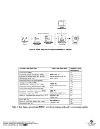
these results. The results show (Table 2) that the classifica-
tion performance for PV (premature ventricular contraction,
PVC) and FS (fusion of ventricular and normal beat) pre-
processed by MM filter are notably higher than the same re-
sulting without any preprocessing stage. Specifically, accu-
rate detection of premature ventricular contractions (PVCs)
is imperative to prepare for the possible onset of lifethreat-
ening arrhythmias. According to AAMI recommendations
classes, we see that also aggregate classification perfor-
mance for normal beats (NL, LB, RB, AE, NE) from any
others heartbeat may be improved with mathematical mor-
phology preprocessing.
6. Conclusions
However, the parameters of morphological operators for
the ECG signal preprocessing intended to extract feature
where tested on a limited number of subjects. Preliminary
results showed that the proposed algorithm leads to an im-
provement in the heartbeat classification using MIT/BIH
database.
The future research will be oriented on the improvement
of the performance of the presented alghorithm.
7. Acknowledgement
Research for this paper was supported in part by grant
from Technical University of Bialystok, no. W/WI/13/07.
6th International Conference on Computer Information
Systems and Industrial Management Applications (CISIM'07)
0-7695-2894-5/07 $20.00 © 2007](https://image.slidesharecdn.com/tadejko2007-230320145531-9596b016/85/tadejko2007-pdf-5-320.jpg)
![MIT-BIH heartbeat types
NL AP NQ PV PB LB AA FS VE RB AE NE NP
total beats 9175 432 306 788 3 1021 8 106 1 881 35 43 12
w/o. preproc. 95,99 86,34 92,81 80,71 0,00 49,56 0,00 56,60 0,00 87,63 97,14 58,14 16,67
w. MM SE10F 95,79 86,34 92,81 81,09 0,00 51,22 0,00 65,09 0,00 87,29 97,14 41,86 0,00
w. MM SES10 93,65 82,87 90,85 81,47 0,00 51,32 0,00 70,75 0,00 94,10 97,14 39,53 0,00
w. MM SES20 94,19 84,26 93,14 87,56 0,00 51,22 0,00 68,87 0,00 86,83 97,14 46,51 0,00
Table 2. Classification performance [%] of MIT-BIH heartbeat type each recording of dataset using
the AAMI recommended performance measures: without preprocessing vs. preprocessing with MM
filtering stage
References
[1] C. Chu-Song, W. Ja-Ling, and H. Yi-Ping. Theoretical as-
pects of vertically invariant gray-level morphological oper-
ators and their application on adaptive signal and image fil-
tering. IEEE Trans. on Signal Processing, 47(4), 1999.
[2] P. de Chazal, M. O’Dwyer, and R. Reilly. Automatic clas-
sification of heartbeats using ecg morphology and heartbeat
interval features. IEEE Transactions on Biomedical Engi-
neering, 51(7):1196–1206, 2004.
[3] P. de Chazal and R. Reilly. A patient-adapting heart-
beat classifier using ecg morphology and heartbeat interval
features. IEEE Transactions on Biomedical Engineering,
53(12):2535–2543, 2006.
[4] Y. Hu, S. Palreddy, and W. Tompkins. A patient-adaptable
ecg beat classifier using a mixture of experts approach. IEEE
Transactions on Biomedical Engineering, 44(9):891–900,
1997.
[5] L. Khadra, A. Al-Fahoum, and S. Binajjaj. A quantitative
analysis approach for cardiac arrhythmia classification us-
ing higher order spectral techniques. IEEE Transactions on
Biomedical Engineering, 52(11):1840–1845, 2005.
[6] K. Kivimoto. Topology preservation in som. Proc. of Inter-
national Conference on Neural NetWorks. Washington DC,
1:294–300, 1996.
[7] B.-U. Kohler, C. Hennig, and R. Orglmeister. The principles
of software qrs detection. IEEE Engineering in Medicine
and Biology, 2002.
[8] T. Kohonen. Self-organizing maps. Springer-Verlag, 1995.
[9] T. Kohonen, J. Hynninen, J. Kangas, and J. Laaksonen.
Som-pak - the self-organizing map program package. Re-
port A31, Helsinki University of Technology, Laboratory of
Computer and Information Science.
[10] T. Kohonen, J. Kangas, J. Laaksonen, and K. Torkkola. Lvq-
pak: A program package for the correct application of learn-
ing vector quantization algorithms. In Proceedings of the
International Joint Conference on Neural Networks, Balti-
more, IEEE, 1:725–730, 1992.
[11] P. Maragos. Morphological signal and image processing.
CRC Press, 2000.
[12] P. Maragos and R. Schafer. Morphological filters-part i:
Their set-theoretic analysis and relations to linear shift-
invariant filters. IEEE Trans. on Acoustic, Speech and Signal
Processing, ASSP-35(8), 1987.
[13] P. Maragos and R. Schafer. Morphological systems for mul-
tidimensional signal processing. Proceedings of the IEEE,
78(4), 1990.
[14] R. Mark and R. Wallen. Aami-recommended practice: Test-
ing and reporting performance results of ventricular arrhyth-
mia detection algorithms. Association for the Advancement
of Medical Instrumentation, Arrhythmia Monitoring Sub-
committee, AAMI ECAR, 1987.
[15] M.I.T. Harvard-mit division of health sciences and
technology, mit/bih arrhythmia database cd-rom.
http://www.physionet.org/physiobank/database/mitdb/,
1992.
[16] S. Mitra, M. Mitra, and B. Chaudhuri. A rough-set-based in-
ference engine for ecg classification. IEEE Transactions on
Instrumentation and Measurement, 55(6):2198–2206, 2006.
[17] K. Piekarski, P. Tadejko, and W. Rakowski. Properties of
morphological operators applied to analysis of ecg signals.
Biometrics, Computer Security Systems and Artificial Intel-
ligence Applications, Springer US, pages 279–288, 2006.
[18] I. Serra. Image analysis and mathematical morphology. New
York Academic, 1982.
[19] L. Shyu, Y. Wu, and W. Hu. Using wavelet trans-
form and fuzzy neural network for vpc detection from the
holter ecg. IEEE Transactions on Biomedical Engineering,
51(7):1269–1273, 2004.
[20] P. Tadejko and W. Rakowski. Matlab simulink in modeling
morphological filters for electrocardiogram signal process-
ing. Simulation in Research and Expansion, XIII Science
Workshop, PTSK, 2005.
[21] J. Vesanto, J. Himberg, E. Alhoniemi, and J. Parhankan-
gas. Som toolbox for matlab. Som Toolbox
Team, Helsinki University of Technology, Finland,
http://www.cis.hut.fi/projects/somtoolbox/, ver. 2, 2007.
[22] Z. Zhang, H. Jiang, D. Ge, and X. Xiang. Pattern recognition
of cardiac arrhythmias using scalar autoregressive modeling.
Fifth World Congress on Intelligent Control and Automation
WCICA, 6:5545–5548, 2004.
6th International Conference on Computer Information
Systems and Industrial Management Applications (CISIM'07)
0-7695-2894-5/07 $20.00 © 2007](https://image.slidesharecdn.com/tadejko2007-230320145531-9596b016/85/tadejko2007-pdf-6-320.jpg)