Download to read offline


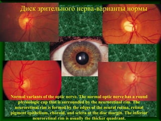
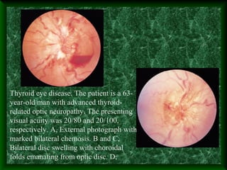
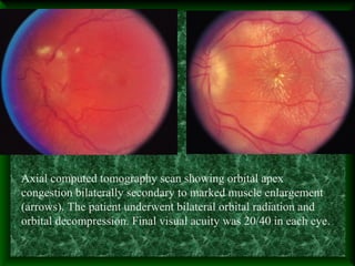
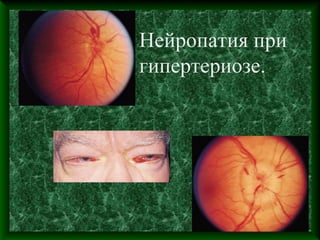
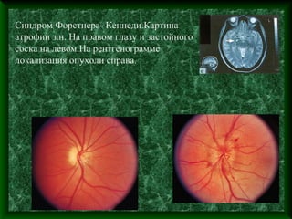
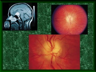
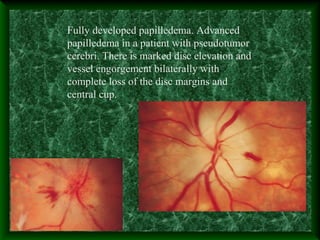

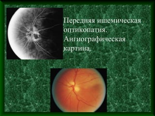
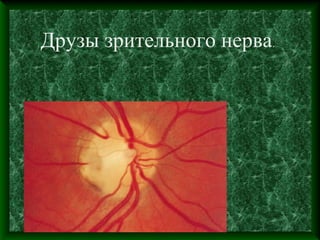
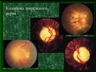

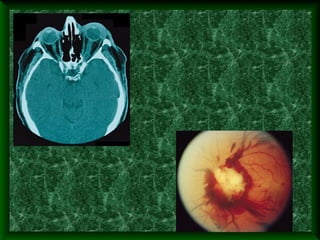

Документ описывает изменения зрительного нерва при различных патологиях, включая воспаление, гипертензию, сосудистые и врожденные нарушения. Приводятся клинические примеры, такие как болезнь глаз при гипертиреозе и передняя ишемическая оптикопатия с соответствующими визуальными и ангиографическими данными. В заключение подчеркивается необходимость оформления будущих выступлений в соответствии с представленным образцом.