SlideShare a Scribd company logo
1 of 10
Download to read offline
Horizontal Gene Transfer of Antibiotic Resistance Genes in the Human Gut
Sarah Maciorowski
Abstract
Horizontal gene transfer (HGT) of antibiotic resistance genes is of increasing interest
because of its impact on the evolution of bacteria. There are millions of genes and hundreds of
bacterial species in the human gut, so HGT likely occurs frequently in the gut microbiota. To test
HGT through conjugation, specific obligate and facultative anaerobes were co-cultured with E.
coli in anaerobic conditions then selected on antibiotic plates. To test HGT by transformation,
bacterial strains commonly found in the human gut microbiota were studied by pooling genomic
DNA and culturing recipient A. baylyi strains with the pooled DNA. After co-culturing antibiotic
sensitive E. coli with antibiotic resistant B. uniformis, E. coli gained the ability to grow in the
presence of the antibiotic Chloramphenicol. E. coli also grew in the presence of
Chloramphenicol when co-cultured with E. fergusonii. A. baylyi was similar in its growth in the
presence of Chloramphenicol when transformed with Bacteroidetes DNA. By studying and
experimenting with gut microbiota conditions, we can further understand the driving factors in
HGT of antibiotic resistance genes.
Introduction
In the human body, the ratio of bacterial cells to human cells and bacterial genes to
human genes is 10:1 and 100:1, respectively (Gill et al., 2006). The majority of the microbes that
are found in the human body are located in the gut (Liu et al., 2012). There are approximately
2.5 million bacterial genes making it a prime location to study horizontal gene transfer (HGT).
HGT is the exchange of genes between bacteria, independent of reproduction (Syvanen,
1994). The main mechanisms of HGT include transformation, transduction, and conjugation.
Transformation occurs when the bacterium intakes exogenous DNA from its surroundings
(Kaznowski and Wlodarczak, 1991). Transduction occurs when a virus injects DNA into the cell,
while conjugation is the exchange of genes between donor and recipient bacteria through direct
contact (Lederberg and Tatum, 1946). One particularly interesting class of genes that can be
exchanged through HGT is antibiotic resistance genes. While antibiotics target a certain area of
the cell to kill the bacteria or prevent it from replicating, antibiotic resistance genes encode
proteins allowing the bacteria to grow in the presence of an antibiotic (Bush, 1988).
Antibiotic resistance genes have become of increasing interest in the health field because
of their ability to alter the genome sequence of a bacterium and complicate treatment for
bacterial infections. Any bacteria that can collect resistance genes can become resistant to one or
more antibiotics. Bacteria acquire antibiotic resistance genes either through mutations or from
another bacterium through vertical or horizontal gene transfer. Vertical transfer happens within a
generation as the resistant bacteria reproduce, while horizontal transfer occurs across the
Bacteria phylogeny (Freeman, 1951). Over time, bacteria can collect multiple resistance genes
and become multidrug-resistant—resistant to many different antibiotic classes (Amábile-Cuevas,
 
2006). If bacteria in the gut transfer antibiotic resistance genes to pathogens, it becomes much
more difficult to treat bacterial infections. Over 70% of the bacteria that cause infections are
resistant to at least one commonly used drug (Stone, 2010). Antibiotic resistant pathogens cause
patients to undergo more difficult treatments and purchase costly antibiotics. Antibiotic resistant
bacterial infections cost the United States healthcare industry an estimated $28 to $45 billion per
year in extra care (Stone, 2010). The inability to treat infectious pathogens in patients has caused
the death rate from bacterial infections to increase by 675% over the past 20 years (NIAID,
2012). Treating infectious diseases has become more difficult with the increase of antibiotic
resistant genes in bacteria.
Two recent studies examined HGT in the human microbiome (Smillie, 2011; Liu, 2012).
Smillie et al. focused on the HGT between human, non-human, and environmental sites, while
Liu et al. focused on HGT between different sites in the human body. Although these two studies
concentrated on HGT, HGT among bacteria at the same body habitat has not been closely
studied. The experimental or computational conditions in these studies tend to create ideal
conditions for detection of HGT, so the driving factors of transfer are still unknown.
In order for HGT to occur, bacteria must incubate in their intended growing conditions.
Growth conditions include temperature, moisture, pH, etc. When it comes to oxygen, some
bacteria thrive in its presence, while others find it a toxic substance. Obligate aerobes require
oxygen for growth, while obligate anaerobes have to grow in the absence of oxygen. Facultative
anaerobes are organisms that can switch between aerobic and anaerobic. These organisms grow
by fermentation or respiration depending on the oxygen condition they find themselves. Similar
to facultative anaerobes, aerotolerant anaerobes can grow with or without oxygen (Fox, 2011).
Since the conditions in the human gut are anaerobic, we chose bacteria strains that could live
anaerobically to mimic the gut.
We aimed to test and understand the factors of HGT without forcing ideal conditions for
conjugation or transformation. By using genetically un-manipulated bacteria and standard culture
conditions, we were able to replicate the kind of conditions for horizontal gene transfer seen in
the gut and test if antibiotic-resistance arose. If the originally antibiotic-sensitive recipient
bacterium was resistant to an antibiotic, then HGT occurred and an antibiotic resistance gene was
passed along. We used E. coli and A. baylyi as antibiotic-sensitive bacteria, hypothesizing HGT
would occur (Rouxel et al., 1991). After examining both conjugation and transformation,
Chloramphenicol resistance seems to be the most readily transferred type of resistance gene.
Materials and Methods
Bacteria and Antibiotics
Each donor bacterial strain was originally isolated from the human gut (Table 1). Strains
were grown statically in Mega Media (Washington University, St. Louis) at 37° C for 24 hours.
The recipient strains came from BioBricks and ATCC and were frozen in 15% glycerol (Table
2). Each antibiotic came from Sigma in St. Louis. Penicillin and Tetracycline were stored at
room temperature, while Chloramphenicol and Trimethoprim were stored at 4°C. Stock
 
solutions of each antibiotic were made at the following concentrations: 100 mg/mL Penicillin, 5
mg/mL Tetracycline, 50 mg/mL Chloramphenicol, and 50 mg/mL Trimethoprim.
Validating Donor Bacteria through Sequencing
The identities of the bacterial cultures used were confirmed through the sequence of the
culture’s 16S rRNA gene. For each strain used, colony PCR was performed using commercially
available PCR mix (Thermo Scientific, ReddyMix) and the following primers: forward primer
(Bac_27F: 10um; 5’-AGAGTTTGATCATGGCTCAG-3’) and reverse primer (Bac_1391R:
10um; 5’-GACGGGCGGTGTGTGCA-3’). PCR products were confirmed on an agarose gel
before purification with QIAquick PCR Purification Kit (QIAGEN). Sanger sequencing was
performed by GeneWiz, Inc. Sequences from GeneWiz were analyzed using the Ribosomal
Database Project classifier (http://rdp.cme.msu.edu/classifier/classifier.jsp).
Validating Recipient Bacteria through Fluorescence
To verify that our stock of E. coli was CFP-positive, we diluted the culture to get
individual colonies on Luria-Bertani (LB) plates (US Biological). Single colonies were picked
and added to the wells of a 96-well plate, each containing 200 uL of LB Broth (US Biological).
Fluorescence was read every 15 minutes for 24 hours on a fluorescence reader kept at 37°C, and
recorded using the Gen5 Secure software package (BioTek Instruments, 1.11.5).
Plates Containing Antibiotics
Standard LB agar (LB Agar Lennox, US Biological) plates were used to grow CFP-
positive E. coli with or without antibiotics at one of the following concentrations: 128 ug/mL
Penicillin, 8 ug/mL Tetracycline, 8 ug/mL Chloramphenicol, or 8 ug/mL Trimethoprim (Sigma,
St. Louis). Standard LB agar plates were used to grow A. baylyi with or without antibiotics at one
of the following concentrations: 128 ug/mL Penicillin, 8 ug/mL Tetracycline, 12 ug/mL
Chloramphenicol, or 50 ug/mL Trimethoprim.
Plates for Coy Chamber
To prepare plates for the obligate-anaerobe bacteria, components for part 1 (Table 3)
were sterilized by autoclave, while the components for part 2 were combined and filter-sterilized
(FS). FS-part 2 was added to autoclaved part 1, and the solution was mixed until combined.
Plates were then poured and left to solidify. Plates were covered in aluminum foil due to the light
sensitivity of Vitamin K, Histidine-Hematin, and Resazurin. Once plates were solid, they were
moved into the anaerobic chamber (Coy Laboratory Products) to become anaerobic.
Conditions for Conjugation of Obligate Anaerobic Donor
In an anaerobic chamber, a 1:1 volume of donor and recipient bacterial strains were co-
cultured together to create a 650 uL culture. The co-culture was grown at 37 °C in the anaerobic
 
chamber for 48 hours or 5 days. 100 uL of co-culture was spread on a LB agar plate containing
one of the antibiotics. The bacteria were incubated at 37 °C for 24 hours.
Conditions for Conjugation of Facultative Anaerobic Donor
A 1:1 ratio of donor and recipient strains of bacteria were co-cultured together to create a
650 uL culture. The co-culture was grown at 37°C for 5 days. 100 uL of co-culture was spread
on a LB agar plate containing one of the antibiotics. The bacteria were incubated at 37 °C for 24
hours. To quantify fluorescent colonies, CFP was read at 480 nm using DPController and
DPManager programs (Olympus Optical Co. 1.2.1.108).
Conditions for Transformation of Pooled DNA
To test the transformation between bacterial DNA and A. baylyi, four pools of genomic
DNA were collected (Table 4). 50 uL of A. baylyi was placed on standard BHI plates in a single
drop. 5 uL of pooled DNA from different bacterial phyla was added to the drop. Once all the
liquid was absorbed into the agar, plates were incubated for 24 hours at 37°C. 750 uL of LB
broth was added to the plates, and cells were scraped. To assay for the potentially transformed
colonies, 100 uL of the suspended cells were spread on LB plates inoculated with antibiotics.
Plates were incubated at 37°C for 24 hours.
Validating Resistance
To validate the presence of a resistance gene in the colony, half of the colony was added
to LB broth with the antibiotic concentration that corresponded to the plate on which the colonies
grew. Colonies grew overnight in the shaker at 37°C.
Results
Colony Growth between B. uniformis and E. coli
We used anaerobic conditions to test the horizontal gene transfer between B. uniformis
and E. coli because B. uniformis is an obligate anaerobe. After 48 hours, E. coli co-cultured with
B. uniformis grew on standard LB plates but did not grow in the presence of any antibiotic
(Figure 1). After 5 days, E. coli co-cultured with B. uniformis was able to grow in the presence of
Chloramphenicol.
Colony Growth between E. fergusonii and E. coli
We tested the horizontal gene transfer between E. fergusonii and E. coli through
conjugation by using conditions for facultative anaerobes. After co-culturing E. coli with E.
fergusonii for five days, we saw growth on Penicillin, Tetracycline, and Chloramphenicol plates
(Figure 2). Because E. fergusonii and E. coli grow in the presence of oxygen, we looked for the
fluorescent property of CFP to determine if E. coli grew in the presence of any antibiotics. To
identify CFP-positive colonies, we first looked at the colonies through white light and then at
 
480 nm to determine if the colonies contained CFP (Figure 3). After looking for CFP, E. coli
only grew in the presence of Chloramphenicol when co-cultured with E. fergusonii (Figure 4).
Colony Growth between A. baylyi and DNA
We tested transformation of HGT by using A. baylyi combined with genomic DNA pools
from one of the following four phyla: Firmicutes, Actinobacteria, Proteobacteria, and
Bacteroidetes (Table 4). After incubation, A. baylyi was present on each of the standard control
plates but was unable to grow in the presence of most antibiotics. However, A. baylyi combined
with Bacteroidetes DNA was able to grow in the presence of Chloramphenicol (Figure 5).
Discussion
Our results highly suggest horizontal gene transfer between donor and recipient strains of
bacteria without forcing ideal conditions for such gene transfer. The growth of E. coli co-
cultured with B. uniformis in the presence of Chloramphenicol strongly implies that horizontal
gene transfer of antibiotic resistance genes occurred between bacteria that are members of the
human gut microbiota, which supports the HGT mechanism of conjugation. E. coli also grew in
the presence of Chloramphenicol after being co-cultured with a facultative anaerobe. Because
antibiotic-sensitive E. coli grew in the presence of Chloramphenicol after co-culturing with E.
fergusonii, HGT between the donor and recipient bacteria strain is strongly suggested. Through
the HGT mechanism of transformation, we were able to test transfers between A. baylyi and
bacterial DNA from different phyla. A. baylyi combined with Bacteroidetes DNA growing in the
presence of Chloramphenicol suggests that HGT occurred in the transformation experiment.
After testing HGT transformation and conjugation, it seems that Chloramphenicol resistance
genes are most readily transfer from donor and recipient bacterial strain.
Although we expected more antibiotic resistance genes to transfer from donor to recipient
strain, there were multiple variables that could have played a part in the abundance of
unexpected colony growth. Co-culturing B. uniformis and E. coli together in the Coy Chamber
could have contaminated B. uniformis. The sterile technique used in the Coy Chamber is UV
light, which might have not been strong enough to prevent contamination in pure B. uniformis
cultures, which allowed it to grow in aerobic conditions. The growth of E. coli co-cultured with
Chloramphenicol in the presence of Chloramphenicol could have been due to adaptation of E.
coli to Chloramphenicol. Also, the lack of growth on antibiotic plates in the conjugation and
transformation experiments could be due to the fact that horizontal gene transfer did not occur,
and the two strains did not exchange DNA.
In order to further confirm the presence of an antibiotic resistance gene in the recipient
strain of bacteria, PCR amplification of the putative antibiotic resistance gene will be performed.
Antibiotic resistant colonies will be grown up in an antibiotic liquid culture to select for
antibiotic resistance genes again. The antibiotic resistance gene that was passed from B.
uniformis or E. fergusonii and is now present in the E. coli will be amplified through PCR. The
resistance gene that was transferred from the Bacteroidetes phyla to A. baylyi would also be
 
tested with PCR and sequenced. If the gene is confirmed through sequencing, then the horizontal
gene transfer mechanism did indeed occur.
After testing two main methods of horizontal gene transfer, Chloramphenicol resistance
genes most readily transfer from donor to recipient strains of bacteria in the conditions studied.
Chloramphenicol and possibly Amphenicol antibiotics could quickly become ineffective to treat
certain infections since Chloramphenicol resistance genes most readily transferred out of the four
antibiotics we tested. Since Chloramphenicol resistance genes readily transfer, it is likely that
other Amphenicol antibiotics transfer just as easily creating bacteria that are multi-drug resistant.
Multi-drug resistant bacteria will make treating infectious diseases increasingly difficult
especially because antibiotic resistance from HGT events can occur in as little as five days. It is
possible that HGT can occur even more frequently in the human gut because of the 2.5 million
bacterial genes and high bacterial density of the gut. Now that we have demonstrated that
horizontal gene transfer can occur under more realistic conditions between donor and recipient
bacterial strains, more antibiotics along with various donor strains can be tested to increase our
knowledge about HGT of antibiotic resistance genes in the human gut.
Resources
1. Amábile-Cuevas, Carlos et al. Antimicrobial Resistance in Bacteria. Horizon Bioscience
(2006).
2. Bush. Beta-lactamase inhibitors from laboratory to clinic. Clinical Microbiology Reviews
1, 109-123 (1988).
3. Fox, Alvin. Microbiology and Immunology On-line. University of South Carolina
Available online at: http://pathmicro.med.sc.edu/fox/nutrition.htm (2011).
4. Freeman, Victor J. Studies on the Virulence of Bacteriophage-infected strains of
Corynebacterium Diphtheriae. Journal of Bacteriology 61, 675-688 (1951).
5. Gill, Steven R. et al. Metagenomic Analysis of the Human Distal Gut Microbiome.
Science 312, 1355-1359 (2006).
6. Kaznowski, A and Wlodarczak K. Susceptibilities of motile Aeromonas sp. to
antimicrobial agents. Zentralbl Bakteriol 275, 85-93 (1991).
7. Lederberg, Joshua and Tatum, Edward. Gene recombination in Escherichia-
coli Nature 158, 558 (1946).
8. Liu, Li. et al. The human microbiome: A hot spot of microbial horizontal gene transfer.
Elsevier 100, 265-270 (2012).
9. NIAID. Antimicrobial (Drug) Resistance: Quick Facts. National Institutes of Health
Available online at: http://www.niaid.nih.gov/topics/antimicrobialResistance/
Understanding/ Pages/quickFacts.aspx (2012).
10. Rouxel, T. et al. Evidence for horizontal gene transfer in Escherichia coli speciation.
Elsevier 222, 851-856 (1991).
11. Smillie, Chris S. et al. Ecology drives a global network of gene exchange connecting the
human microbiome Nature 480, 241-244 (2011).
 
12. Stone, Patricia W. Economic burden of healthcare-associated infections: an American
perspective. Expert Rev Pharmacoecon Outcomes Res.9, 417-422 (2010).
13. Syvanen, M. Horizontal gene transfer: evidence and possible consequences. Annu. Rev.
Genet. 28, 237-261 (1994).
Acknowledgments
The author is grateful for the opportunity that Dr. Gautam Dantas provided. The utmost
thanks goes out to her mentor Molly Gibson and tutor Vitas Wagner as well as everyone
involved in the Young Scientist Program at Washington University.
Table 1. Donor Strains Used.
Table 2. Recipient Strains and Antibiotics Used.
Donor Strains
Strain Phyla Anticipated Growth Conditions
Escherichia fergusonii Proteobacteria Facultative Anaerobe
Bacteroides uniformis Bacteroidetes Obligate Anaerobe
Recipient Strains Antibiotics
Strain Phyla Antibiotic Antibiotic Class
Escherichia coli (MG1655) Proteobacteria Penicillin (PE) Beta lactam
Acinetobacter baylyi (ADP1) Proteobacteria Tetracycline (TE) Tetracycline
Chloramphenicol (CH) Amphenicol
Trimethoprim (TR) Pyrimidine derivative
 
Table 3. Components for Anaerobic Plates.
Components for Part 1 Amount Components for Part 2 Amount
Tryptone Peptone (US Biological, MA) 5 g D-(+)-Glucose (Sigma) 1 g
Yeast Extract (US Biological, MA) 2.5 g Vitamin K Solution (Sigma) 500 uL
L-Cysteine HCl (Sigma) 0.25 g Distilled H2O 10 uL
1 M Potassium Phosphate Buffer, pH 7.2
(1M KH2PO4 and 1M K2HPO4)
50 mL Resazurin (Sigma) 2 mL
TYG Salts Solution (MgSO4·7H2O,
NaHCO3, NaCl, Distilled H2O)
20 mL Histidine-Hematin (0.2 M histidine,
pH 8.0 and hematin (Sigma))
500 uL
0.8% CaCl2 500 uL
FeSO4 ·7H2O (0.4 mg/ml) 500 uL
Agar (US Biological, MA) 7 g
Distilled H2O 416 mL
Total: 487 mL Total: 13 mL
Figure 2. Quantified Colony Growth between B. uniformis and E. coli. A combination of B. uniformis, E. coli,
and Penicillin, Tetracycline, Trimethoprim, or Chloramphenicol were co-cultured and plated on LB and LB with the
corresponding antibiotic. The quantity of colonies is shown for all combinations of B. uniformis, E. coli, and an
antibiotic. *The colonies that grew in the presence of an antibiotic were validated to have resistance genes.
*	
  
Figure 1. Colony Growth between E. fergusonii and E. coli. A combination of E. fergusonii, E. coli, and
Penicillin, Tetracycline, Trimethoprim, or Chloramphenicol were co-cultured and plated on LB and LB with the
corresponding antibiotic. The quantity of colonies is shown for all combinations of E. fergusonii, E. coli, and an
antibiotic. *The colonies that grew in the presence of an antibiotic were validated to have resistance genes.
*	
  
 
Figure 3. CFP Fluorescent Colonies. To measure E. coli growth between E. coli and E.
fergusonii, the CFP of the colonies was read. A1, B1, and C1 are colonies that can be seen in white
light (1a). Colonies A2 and B2 can be seen at 480 nm, while colony C1 is nonexistent (1b).
1a	
   1b	
  
Figure 5. Quantified Colony Growth between A. baylyi and DNA. A.
baylyi combined with bacteria DNA from Firmicutes, Actinobacteria,
Proteobacteria, or Bacteroidetes phyla were spread on antibiotic plates.
*The colonies that grew in the presence of an antibiotic were validated to
have resistance genes.
*	
  
Figure 4. Quantified E. coli Growth between E. fergusonii and E. coli. All colonies that grew from
co-cultures containing combinations of E. fergusonii, E. coli, and an antibiotic were analyzed for E. coli.
After looking at the CFP fluorescent colonies, we quantified E. coli. *The colonies that grew in the
presence of an antibiotic were validated to have resistance genes.
*	
  
 
Table 4. Pooled Bacteria DNA.

More Related Content

What's hot

Antimicrobial resistance as an emerging food-borne infectious disease
Antimicrobial resistance as an emerging food-borne infectious diseaseAntimicrobial resistance as an emerging food-borne infectious disease
Antimicrobial resistance as an emerging food-borne infectious diseaseJean Jacques Bernatas
 
Variability in plant pathogens
Variability in plant pathogensVariability in plant pathogens
Variability in plant pathogensVigneshVikki10
 
Environmental Transmission of Antimicrobial Resistance
Environmental Transmission of Antimicrobial ResistanceEnvironmental Transmission of Antimicrobial Resistance
Environmental Transmission of Antimicrobial ResistancePranab Chatterjee
 
The Role of the Skin Microbiome in Atopic Dermatitis (Eczema)
The Role of the Skin Microbiome in Atopic Dermatitis (Eczema)The Role of the Skin Microbiome in Atopic Dermatitis (Eczema)
The Role of the Skin Microbiome in Atopic Dermatitis (Eczema)Laura Berry
 
The environmental dimensions of antibiotic resistance
The environmental dimensions of antibiotic resistanceThe environmental dimensions of antibiotic resistance
The environmental dimensions of antibiotic resistanceSIANI
 
Edible vaccines-A new approach to oral immunization
Edible vaccines-A new approach to oral immunizationEdible vaccines-A new approach to oral immunization
Edible vaccines-A new approach to oral immunizationJagadabhi Ravinder Raju
 
Anti-microbial Resistance
Anti-microbial ResistanceAnti-microbial Resistance
Anti-microbial ResistanceRudresh Rudri
 
Seminário 6 horner-devineetal 2003_bacteria (1)
Seminário 6 horner-devineetal 2003_bacteria (1)Seminário 6 horner-devineetal 2003_bacteria (1)
Seminário 6 horner-devineetal 2003_bacteria (1)Carlos Alberto Monteiro
 
The gut microbiota — masters of host development and physiology
The gut microbiota — masters of host development and physiologyThe gut microbiota — masters of host development and physiology
The gut microbiota — masters of host development and physiologyAlfonso Enrique Islas Rodríguez
 
Quorum sensing in Archaea
Quorum sensing in ArchaeaQuorum sensing in Archaea
Quorum sensing in ArchaeaZahra Naz
 
The antibiotic free movement - Enhancing the nutritional value of feed
The antibiotic free movement - Enhancing the nutritional value of feedThe antibiotic free movement - Enhancing the nutritional value of feed
The antibiotic free movement - Enhancing the nutritional value of feedMilling and Grain magazine
 
Pieces of the phytobiome: multitrophic and environmental influences on plant ...
Pieces of the phytobiome: multitrophic and environmental influences on plant ...Pieces of the phytobiome: multitrophic and environmental influences on plant ...
Pieces of the phytobiome: multitrophic and environmental influences on plant ...CIAT
 
Breeding Approaches Towards Disease Resistance In Livestocks
Breeding Approaches Towards Disease Resistance In LivestocksBreeding Approaches Towards Disease Resistance In Livestocks
Breeding Approaches Towards Disease Resistance In LivestocksSharadindu Shil
 
Antimicrobial resistance (AMR) in N. gonorrhoeae (GC) - global problem but v...
Antimicrobial resistance (AMR) in N. gonorrhoeae (GC) - global problem but v...Antimicrobial resistance (AMR) in N. gonorrhoeae (GC) - global problem but v...
Antimicrobial resistance (AMR) in N. gonorrhoeae (GC) - global problem but v...Игорь Шадеркин
 
genetic Resistance against gastrointestinal nematodes in sheep: Association w...
genetic Resistance against gastrointestinal nematodes in sheep: Association w...genetic Resistance against gastrointestinal nematodes in sheep: Association w...
genetic Resistance against gastrointestinal nematodes in sheep: Association w...Ishfaq Maqbool
 
Transgenic strategies for improving rice disease resistance
Transgenic strategies for improving rice disease resistanceTransgenic strategies for improving rice disease resistance
Transgenic strategies for improving rice disease resistanceKiranKumarN24
 
Maintenance of antibiotic resistance traits during relaxed selection in a lon...
Maintenance of antibiotic resistance traits during relaxed selection in a lon...Maintenance of antibiotic resistance traits during relaxed selection in a lon...
Maintenance of antibiotic resistance traits during relaxed selection in a lon...Kyle Card
 

What's hot (19)

Antimicrobial resistance as an emerging food-borne infectious disease
Antimicrobial resistance as an emerging food-borne infectious diseaseAntimicrobial resistance as an emerging food-borne infectious disease
Antimicrobial resistance as an emerging food-borne infectious disease
 
Variability in plant pathogens
Variability in plant pathogensVariability in plant pathogens
Variability in plant pathogens
 
Environmental Transmission of Antimicrobial Resistance
Environmental Transmission of Antimicrobial ResistanceEnvironmental Transmission of Antimicrobial Resistance
Environmental Transmission of Antimicrobial Resistance
 
The Role of the Skin Microbiome in Atopic Dermatitis (Eczema)
The Role of the Skin Microbiome in Atopic Dermatitis (Eczema)The Role of the Skin Microbiome in Atopic Dermatitis (Eczema)
The Role of the Skin Microbiome in Atopic Dermatitis (Eczema)
 
The environmental dimensions of antibiotic resistance
The environmental dimensions of antibiotic resistanceThe environmental dimensions of antibiotic resistance
The environmental dimensions of antibiotic resistance
 
Edible vaccines-A new approach to oral immunization
Edible vaccines-A new approach to oral immunizationEdible vaccines-A new approach to oral immunization
Edible vaccines-A new approach to oral immunization
 
Anti-microbial Resistance
Anti-microbial ResistanceAnti-microbial Resistance
Anti-microbial Resistance
 
Seminário 6 horner-devineetal 2003_bacteria (1)
Seminário 6 horner-devineetal 2003_bacteria (1)Seminário 6 horner-devineetal 2003_bacteria (1)
Seminário 6 horner-devineetal 2003_bacteria (1)
 
The gut microbiota — masters of host development and physiology
The gut microbiota — masters of host development and physiologyThe gut microbiota — masters of host development and physiology
The gut microbiota — masters of host development and physiology
 
Quorum sensing in Archaea
Quorum sensing in ArchaeaQuorum sensing in Archaea
Quorum sensing in Archaea
 
The antibiotic free movement - Enhancing the nutritional value of feed
The antibiotic free movement - Enhancing the nutritional value of feedThe antibiotic free movement - Enhancing the nutritional value of feed
The antibiotic free movement - Enhancing the nutritional value of feed
 
Pieces of the phytobiome: multitrophic and environmental influences on plant ...
Pieces of the phytobiome: multitrophic and environmental influences on plant ...Pieces of the phytobiome: multitrophic and environmental influences on plant ...
Pieces of the phytobiome: multitrophic and environmental influences on plant ...
 
Poster
PosterPoster
Poster
 
Breeding Approaches Towards Disease Resistance In Livestocks
Breeding Approaches Towards Disease Resistance In LivestocksBreeding Approaches Towards Disease Resistance In Livestocks
Breeding Approaches Towards Disease Resistance In Livestocks
 
Antimicrobial resistance (AMR) in N. gonorrhoeae (GC) - global problem but v...
Antimicrobial resistance (AMR) in N. gonorrhoeae (GC) - global problem but v...Antimicrobial resistance (AMR) in N. gonorrhoeae (GC) - global problem but v...
Antimicrobial resistance (AMR) in N. gonorrhoeae (GC) - global problem but v...
 
genetic Resistance against gastrointestinal nematodes in sheep: Association w...
genetic Resistance against gastrointestinal nematodes in sheep: Association w...genetic Resistance against gastrointestinal nematodes in sheep: Association w...
genetic Resistance against gastrointestinal nematodes in sheep: Association w...
 
Transgenic strategies for improving rice disease resistance
Transgenic strategies for improving rice disease resistanceTransgenic strategies for improving rice disease resistance
Transgenic strategies for improving rice disease resistance
 
Maintenance of antibiotic resistance traits during relaxed selection in a lon...
Maintenance of antibiotic resistance traits during relaxed selection in a lon...Maintenance of antibiotic resistance traits during relaxed selection in a lon...
Maintenance of antibiotic resistance traits during relaxed selection in a lon...
 
FOOD AS VACCINE
FOOD AS VACCINE FOOD AS VACCINE
FOOD AS VACCINE
 

Viewers also liked

Importance of gene transfer in antibiotic resistance
Importance of gene transfer in antibiotic resistanceImportance of gene transfer in antibiotic resistance
Importance of gene transfer in antibiotic resistanceNadhirah Rosman
 
Antibiotic resistance in bacteria 1
Antibiotic resistance in bacteria 1Antibiotic resistance in bacteria 1
Antibiotic resistance in bacteria 1anusil
 
Antibiotic resistance
Antibiotic resistance Antibiotic resistance
Antibiotic resistance Naser Tadvi
 

Viewers also liked (8)

Importance of gene transfer in antibiotic resistance
Importance of gene transfer in antibiotic resistanceImportance of gene transfer in antibiotic resistance
Importance of gene transfer in antibiotic resistance
 
Drug Resistance!
Drug Resistance!Drug Resistance!
Drug Resistance!
 
Antibiotic resistance
Antibiotic resistanceAntibiotic resistance
Antibiotic resistance
 
Direct Gene Transfer Methods
Direct Gene Transfer MethodsDirect Gene Transfer Methods
Direct Gene Transfer Methods
 
Antibiotic resistance in bacteria 1
Antibiotic resistance in bacteria 1Antibiotic resistance in bacteria 1
Antibiotic resistance in bacteria 1
 
Antibiotic resistance
Antibiotic resistanceAntibiotic resistance
Antibiotic resistance
 
Antibiotic resistance
Antibiotic resistance Antibiotic resistance
Antibiotic resistance
 
Build Features, Not Apps
Build Features, Not AppsBuild Features, Not Apps
Build Features, Not Apps
 

Similar to Horizontal Gene Transfer of Antibiotic Resistance in the Gut

Antibiotic Resistant Bacteria
Antibiotic Resistant BacteriaAntibiotic Resistant Bacteria
Antibiotic Resistant BacteriaKevin B Hugins
 
Activation-of-human-immunodeficiency-virus-type-1-expression-by-Gardnerella-v...
Activation-of-human-immunodeficiency-virus-type-1-expression-by-Gardnerella-v...Activation-of-human-immunodeficiency-virus-type-1-expression-by-Gardnerella-v...
Activation-of-human-immunodeficiency-virus-type-1-expression-by-Gardnerella-v...Farhad B. Hashemi, PhD
 
articulo descripcion y analisis del articulo
articulo descripcion y analisis del articuloarticulo descripcion y analisis del articulo
articulo descripcion y analisis del articuloyessica756439
 
Bacteriophage therapy for antimicrobial resistant and biofilm forming [Autosa...
Bacteriophage therapy for antimicrobial resistant and biofilm forming [Autosa...Bacteriophage therapy for antimicrobial resistant and biofilm forming [Autosa...
Bacteriophage therapy for antimicrobial resistant and biofilm forming [Autosa...kamal shrestha
 
Syngulon - Breakout session Synthetic Biology June 10, 2022.pdf
Syngulon - Breakout session Synthetic Biology June 10, 2022.pdfSyngulon - Breakout session Synthetic Biology June 10, 2022.pdf
Syngulon - Breakout session Synthetic Biology June 10, 2022.pdfSyngulon
 
Antibiotic resistome by Dr Namita shukla
Antibiotic resistome by Dr Namita shuklaAntibiotic resistome by Dr Namita shukla
Antibiotic resistome by Dr Namita shuklaDrNamitashukla
 
Fungal Contamination
Fungal ContaminationFungal Contamination
Fungal ContaminationAshley Lott
 
Isolation and characterization of bacteria from tropical soils
Isolation and characterization of bacteria from tropical soilsIsolation and characterization of bacteria from tropical soils
Isolation and characterization of bacteria from tropical soilsJessica Cristina
 
Fernando Vaquero-El impacto de las ciencias ómicas en la medicina, la nutrici...
Fernando Vaquero-El impacto de las ciencias ómicas en la medicina, la nutrici...Fernando Vaquero-El impacto de las ciencias ómicas en la medicina, la nutrici...
Fernando Vaquero-El impacto de las ciencias ómicas en la medicina, la nutrici...Fundación Ramón Areces
 
How Much Do You Know about Bacteriocin?
How Much Do You Know about Bacteriocin?How Much Do You Know about Bacteriocin?
How Much Do You Know about Bacteriocin?Creative BioMart
 

Similar to Horizontal Gene Transfer of Antibiotic Resistance in the Gut (20)

Antibiotic Resistant Bacteria
Antibiotic Resistant BacteriaAntibiotic Resistant Bacteria
Antibiotic Resistant Bacteria
 
edible vaccine
edible vaccineedible vaccine
edible vaccine
 
Bruno pot
Bruno pot Bruno pot
Bruno pot
 
Activation-of-human-immunodeficiency-virus-type-1-expression-by-Gardnerella-v...
Activation-of-human-immunodeficiency-virus-type-1-expression-by-Gardnerella-v...Activation-of-human-immunodeficiency-virus-type-1-expression-by-Gardnerella-v...
Activation-of-human-immunodeficiency-virus-type-1-expression-by-Gardnerella-v...
 
Final Proposal
Final ProposalFinal Proposal
Final Proposal
 
Antibiotics and their Resistance Gene Path to Humans
Antibiotics and their Resistance Gene Path to Humans Antibiotics and their Resistance Gene Path to Humans
Antibiotics and their Resistance Gene Path to Humans
 
Ph02 edible vaccines
Ph02 edible vaccinesPh02 edible vaccines
Ph02 edible vaccines
 
articulo descripcion y analisis del articulo
articulo descripcion y analisis del articuloarticulo descripcion y analisis del articulo
articulo descripcion y analisis del articulo
 
Bacteriophage therapy for antimicrobial resistant and biofilm forming [Autosa...
Bacteriophage therapy for antimicrobial resistant and biofilm forming [Autosa...Bacteriophage therapy for antimicrobial resistant and biofilm forming [Autosa...
Bacteriophage therapy for antimicrobial resistant and biofilm forming [Autosa...
 
Bacteriophage therapy
Bacteriophage therapyBacteriophage therapy
Bacteriophage therapy
 
FimH ADHESIN
FimH ADHESINFimH ADHESIN
FimH ADHESIN
 
Syngulon - Breakout session Synthetic Biology June 10, 2022.pdf
Syngulon - Breakout session Synthetic Biology June 10, 2022.pdfSyngulon - Breakout session Synthetic Biology June 10, 2022.pdf
Syngulon - Breakout session Synthetic Biology June 10, 2022.pdf
 
Antibiotic resistome by Dr Namita shukla
Antibiotic resistome by Dr Namita shuklaAntibiotic resistome by Dr Namita shukla
Antibiotic resistome by Dr Namita shukla
 
Fungal Contamination
Fungal ContaminationFungal Contamination
Fungal Contamination
 
Isolation and characterization of bacteria from tropical soils
Isolation and characterization of bacteria from tropical soilsIsolation and characterization of bacteria from tropical soils
Isolation and characterization of bacteria from tropical soils
 
Dr d p rajani
Dr d p rajaniDr d p rajani
Dr d p rajani
 
Fernando Vaquero-El impacto de las ciencias ómicas en la medicina, la nutrici...
Fernando Vaquero-El impacto de las ciencias ómicas en la medicina, la nutrici...Fernando Vaquero-El impacto de las ciencias ómicas en la medicina, la nutrici...
Fernando Vaquero-El impacto de las ciencias ómicas en la medicina, la nutrici...
 
Biofilms
BiofilmsBiofilms
Biofilms
 
How Much Do You Know about Bacteriocin?
How Much Do You Know about Bacteriocin?How Much Do You Know about Bacteriocin?
How Much Do You Know about Bacteriocin?
 
Edible vaccines
Edible vaccinesEdible vaccines
Edible vaccines
 

Horizontal Gene Transfer of Antibiotic Resistance in the Gut

  • 1. Horizontal Gene Transfer of Antibiotic Resistance Genes in the Human Gut Sarah Maciorowski Abstract Horizontal gene transfer (HGT) of antibiotic resistance genes is of increasing interest because of its impact on the evolution of bacteria. There are millions of genes and hundreds of bacterial species in the human gut, so HGT likely occurs frequently in the gut microbiota. To test HGT through conjugation, specific obligate and facultative anaerobes were co-cultured with E. coli in anaerobic conditions then selected on antibiotic plates. To test HGT by transformation, bacterial strains commonly found in the human gut microbiota were studied by pooling genomic DNA and culturing recipient A. baylyi strains with the pooled DNA. After co-culturing antibiotic sensitive E. coli with antibiotic resistant B. uniformis, E. coli gained the ability to grow in the presence of the antibiotic Chloramphenicol. E. coli also grew in the presence of Chloramphenicol when co-cultured with E. fergusonii. A. baylyi was similar in its growth in the presence of Chloramphenicol when transformed with Bacteroidetes DNA. By studying and experimenting with gut microbiota conditions, we can further understand the driving factors in HGT of antibiotic resistance genes. Introduction In the human body, the ratio of bacterial cells to human cells and bacterial genes to human genes is 10:1 and 100:1, respectively (Gill et al., 2006). The majority of the microbes that are found in the human body are located in the gut (Liu et al., 2012). There are approximately 2.5 million bacterial genes making it a prime location to study horizontal gene transfer (HGT). HGT is the exchange of genes between bacteria, independent of reproduction (Syvanen, 1994). The main mechanisms of HGT include transformation, transduction, and conjugation. Transformation occurs when the bacterium intakes exogenous DNA from its surroundings (Kaznowski and Wlodarczak, 1991). Transduction occurs when a virus injects DNA into the cell, while conjugation is the exchange of genes between donor and recipient bacteria through direct contact (Lederberg and Tatum, 1946). One particularly interesting class of genes that can be exchanged through HGT is antibiotic resistance genes. While antibiotics target a certain area of the cell to kill the bacteria or prevent it from replicating, antibiotic resistance genes encode proteins allowing the bacteria to grow in the presence of an antibiotic (Bush, 1988). Antibiotic resistance genes have become of increasing interest in the health field because of their ability to alter the genome sequence of a bacterium and complicate treatment for bacterial infections. Any bacteria that can collect resistance genes can become resistant to one or more antibiotics. Bacteria acquire antibiotic resistance genes either through mutations or from another bacterium through vertical or horizontal gene transfer. Vertical transfer happens within a generation as the resistant bacteria reproduce, while horizontal transfer occurs across the Bacteria phylogeny (Freeman, 1951). Over time, bacteria can collect multiple resistance genes and become multidrug-resistant—resistant to many different antibiotic classes (Amábile-Cuevas,
  • 2.   2006). If bacteria in the gut transfer antibiotic resistance genes to pathogens, it becomes much more difficult to treat bacterial infections. Over 70% of the bacteria that cause infections are resistant to at least one commonly used drug (Stone, 2010). Antibiotic resistant pathogens cause patients to undergo more difficult treatments and purchase costly antibiotics. Antibiotic resistant bacterial infections cost the United States healthcare industry an estimated $28 to $45 billion per year in extra care (Stone, 2010). The inability to treat infectious pathogens in patients has caused the death rate from bacterial infections to increase by 675% over the past 20 years (NIAID, 2012). Treating infectious diseases has become more difficult with the increase of antibiotic resistant genes in bacteria. Two recent studies examined HGT in the human microbiome (Smillie, 2011; Liu, 2012). Smillie et al. focused on the HGT between human, non-human, and environmental sites, while Liu et al. focused on HGT between different sites in the human body. Although these two studies concentrated on HGT, HGT among bacteria at the same body habitat has not been closely studied. The experimental or computational conditions in these studies tend to create ideal conditions for detection of HGT, so the driving factors of transfer are still unknown. In order for HGT to occur, bacteria must incubate in their intended growing conditions. Growth conditions include temperature, moisture, pH, etc. When it comes to oxygen, some bacteria thrive in its presence, while others find it a toxic substance. Obligate aerobes require oxygen for growth, while obligate anaerobes have to grow in the absence of oxygen. Facultative anaerobes are organisms that can switch between aerobic and anaerobic. These organisms grow by fermentation or respiration depending on the oxygen condition they find themselves. Similar to facultative anaerobes, aerotolerant anaerobes can grow with or without oxygen (Fox, 2011). Since the conditions in the human gut are anaerobic, we chose bacteria strains that could live anaerobically to mimic the gut. We aimed to test and understand the factors of HGT without forcing ideal conditions for conjugation or transformation. By using genetically un-manipulated bacteria and standard culture conditions, we were able to replicate the kind of conditions for horizontal gene transfer seen in the gut and test if antibiotic-resistance arose. If the originally antibiotic-sensitive recipient bacterium was resistant to an antibiotic, then HGT occurred and an antibiotic resistance gene was passed along. We used E. coli and A. baylyi as antibiotic-sensitive bacteria, hypothesizing HGT would occur (Rouxel et al., 1991). After examining both conjugation and transformation, Chloramphenicol resistance seems to be the most readily transferred type of resistance gene. Materials and Methods Bacteria and Antibiotics Each donor bacterial strain was originally isolated from the human gut (Table 1). Strains were grown statically in Mega Media (Washington University, St. Louis) at 37° C for 24 hours. The recipient strains came from BioBricks and ATCC and were frozen in 15% glycerol (Table 2). Each antibiotic came from Sigma in St. Louis. Penicillin and Tetracycline were stored at room temperature, while Chloramphenicol and Trimethoprim were stored at 4°C. Stock
  • 3.   solutions of each antibiotic were made at the following concentrations: 100 mg/mL Penicillin, 5 mg/mL Tetracycline, 50 mg/mL Chloramphenicol, and 50 mg/mL Trimethoprim. Validating Donor Bacteria through Sequencing The identities of the bacterial cultures used were confirmed through the sequence of the culture’s 16S rRNA gene. For each strain used, colony PCR was performed using commercially available PCR mix (Thermo Scientific, ReddyMix) and the following primers: forward primer (Bac_27F: 10um; 5’-AGAGTTTGATCATGGCTCAG-3’) and reverse primer (Bac_1391R: 10um; 5’-GACGGGCGGTGTGTGCA-3’). PCR products were confirmed on an agarose gel before purification with QIAquick PCR Purification Kit (QIAGEN). Sanger sequencing was performed by GeneWiz, Inc. Sequences from GeneWiz were analyzed using the Ribosomal Database Project classifier (http://rdp.cme.msu.edu/classifier/classifier.jsp). Validating Recipient Bacteria through Fluorescence To verify that our stock of E. coli was CFP-positive, we diluted the culture to get individual colonies on Luria-Bertani (LB) plates (US Biological). Single colonies were picked and added to the wells of a 96-well plate, each containing 200 uL of LB Broth (US Biological). Fluorescence was read every 15 minutes for 24 hours on a fluorescence reader kept at 37°C, and recorded using the Gen5 Secure software package (BioTek Instruments, 1.11.5). Plates Containing Antibiotics Standard LB agar (LB Agar Lennox, US Biological) plates were used to grow CFP- positive E. coli with or without antibiotics at one of the following concentrations: 128 ug/mL Penicillin, 8 ug/mL Tetracycline, 8 ug/mL Chloramphenicol, or 8 ug/mL Trimethoprim (Sigma, St. Louis). Standard LB agar plates were used to grow A. baylyi with or without antibiotics at one of the following concentrations: 128 ug/mL Penicillin, 8 ug/mL Tetracycline, 12 ug/mL Chloramphenicol, or 50 ug/mL Trimethoprim. Plates for Coy Chamber To prepare plates for the obligate-anaerobe bacteria, components for part 1 (Table 3) were sterilized by autoclave, while the components for part 2 were combined and filter-sterilized (FS). FS-part 2 was added to autoclaved part 1, and the solution was mixed until combined. Plates were then poured and left to solidify. Plates were covered in aluminum foil due to the light sensitivity of Vitamin K, Histidine-Hematin, and Resazurin. Once plates were solid, they were moved into the anaerobic chamber (Coy Laboratory Products) to become anaerobic. Conditions for Conjugation of Obligate Anaerobic Donor In an anaerobic chamber, a 1:1 volume of donor and recipient bacterial strains were co- cultured together to create a 650 uL culture. The co-culture was grown at 37 °C in the anaerobic
  • 4.   chamber for 48 hours or 5 days. 100 uL of co-culture was spread on a LB agar plate containing one of the antibiotics. The bacteria were incubated at 37 °C for 24 hours. Conditions for Conjugation of Facultative Anaerobic Donor A 1:1 ratio of donor and recipient strains of bacteria were co-cultured together to create a 650 uL culture. The co-culture was grown at 37°C for 5 days. 100 uL of co-culture was spread on a LB agar plate containing one of the antibiotics. The bacteria were incubated at 37 °C for 24 hours. To quantify fluorescent colonies, CFP was read at 480 nm using DPController and DPManager programs (Olympus Optical Co. 1.2.1.108). Conditions for Transformation of Pooled DNA To test the transformation between bacterial DNA and A. baylyi, four pools of genomic DNA were collected (Table 4). 50 uL of A. baylyi was placed on standard BHI plates in a single drop. 5 uL of pooled DNA from different bacterial phyla was added to the drop. Once all the liquid was absorbed into the agar, plates were incubated for 24 hours at 37°C. 750 uL of LB broth was added to the plates, and cells were scraped. To assay for the potentially transformed colonies, 100 uL of the suspended cells were spread on LB plates inoculated with antibiotics. Plates were incubated at 37°C for 24 hours. Validating Resistance To validate the presence of a resistance gene in the colony, half of the colony was added to LB broth with the antibiotic concentration that corresponded to the plate on which the colonies grew. Colonies grew overnight in the shaker at 37°C. Results Colony Growth between B. uniformis and E. coli We used anaerobic conditions to test the horizontal gene transfer between B. uniformis and E. coli because B. uniformis is an obligate anaerobe. After 48 hours, E. coli co-cultured with B. uniformis grew on standard LB plates but did not grow in the presence of any antibiotic (Figure 1). After 5 days, E. coli co-cultured with B. uniformis was able to grow in the presence of Chloramphenicol. Colony Growth between E. fergusonii and E. coli We tested the horizontal gene transfer between E. fergusonii and E. coli through conjugation by using conditions for facultative anaerobes. After co-culturing E. coli with E. fergusonii for five days, we saw growth on Penicillin, Tetracycline, and Chloramphenicol plates (Figure 2). Because E. fergusonii and E. coli grow in the presence of oxygen, we looked for the fluorescent property of CFP to determine if E. coli grew in the presence of any antibiotics. To identify CFP-positive colonies, we first looked at the colonies through white light and then at
  • 5.   480 nm to determine if the colonies contained CFP (Figure 3). After looking for CFP, E. coli only grew in the presence of Chloramphenicol when co-cultured with E. fergusonii (Figure 4). Colony Growth between A. baylyi and DNA We tested transformation of HGT by using A. baylyi combined with genomic DNA pools from one of the following four phyla: Firmicutes, Actinobacteria, Proteobacteria, and Bacteroidetes (Table 4). After incubation, A. baylyi was present on each of the standard control plates but was unable to grow in the presence of most antibiotics. However, A. baylyi combined with Bacteroidetes DNA was able to grow in the presence of Chloramphenicol (Figure 5). Discussion Our results highly suggest horizontal gene transfer between donor and recipient strains of bacteria without forcing ideal conditions for such gene transfer. The growth of E. coli co- cultured with B. uniformis in the presence of Chloramphenicol strongly implies that horizontal gene transfer of antibiotic resistance genes occurred between bacteria that are members of the human gut microbiota, which supports the HGT mechanism of conjugation. E. coli also grew in the presence of Chloramphenicol after being co-cultured with a facultative anaerobe. Because antibiotic-sensitive E. coli grew in the presence of Chloramphenicol after co-culturing with E. fergusonii, HGT between the donor and recipient bacteria strain is strongly suggested. Through the HGT mechanism of transformation, we were able to test transfers between A. baylyi and bacterial DNA from different phyla. A. baylyi combined with Bacteroidetes DNA growing in the presence of Chloramphenicol suggests that HGT occurred in the transformation experiment. After testing HGT transformation and conjugation, it seems that Chloramphenicol resistance genes are most readily transfer from donor and recipient bacterial strain. Although we expected more antibiotic resistance genes to transfer from donor to recipient strain, there were multiple variables that could have played a part in the abundance of unexpected colony growth. Co-culturing B. uniformis and E. coli together in the Coy Chamber could have contaminated B. uniformis. The sterile technique used in the Coy Chamber is UV light, which might have not been strong enough to prevent contamination in pure B. uniformis cultures, which allowed it to grow in aerobic conditions. The growth of E. coli co-cultured with Chloramphenicol in the presence of Chloramphenicol could have been due to adaptation of E. coli to Chloramphenicol. Also, the lack of growth on antibiotic plates in the conjugation and transformation experiments could be due to the fact that horizontal gene transfer did not occur, and the two strains did not exchange DNA. In order to further confirm the presence of an antibiotic resistance gene in the recipient strain of bacteria, PCR amplification of the putative antibiotic resistance gene will be performed. Antibiotic resistant colonies will be grown up in an antibiotic liquid culture to select for antibiotic resistance genes again. The antibiotic resistance gene that was passed from B. uniformis or E. fergusonii and is now present in the E. coli will be amplified through PCR. The resistance gene that was transferred from the Bacteroidetes phyla to A. baylyi would also be
  • 6.   tested with PCR and sequenced. If the gene is confirmed through sequencing, then the horizontal gene transfer mechanism did indeed occur. After testing two main methods of horizontal gene transfer, Chloramphenicol resistance genes most readily transfer from donor to recipient strains of bacteria in the conditions studied. Chloramphenicol and possibly Amphenicol antibiotics could quickly become ineffective to treat certain infections since Chloramphenicol resistance genes most readily transferred out of the four antibiotics we tested. Since Chloramphenicol resistance genes readily transfer, it is likely that other Amphenicol antibiotics transfer just as easily creating bacteria that are multi-drug resistant. Multi-drug resistant bacteria will make treating infectious diseases increasingly difficult especially because antibiotic resistance from HGT events can occur in as little as five days. It is possible that HGT can occur even more frequently in the human gut because of the 2.5 million bacterial genes and high bacterial density of the gut. Now that we have demonstrated that horizontal gene transfer can occur under more realistic conditions between donor and recipient bacterial strains, more antibiotics along with various donor strains can be tested to increase our knowledge about HGT of antibiotic resistance genes in the human gut. Resources 1. Amábile-Cuevas, Carlos et al. Antimicrobial Resistance in Bacteria. Horizon Bioscience (2006). 2. Bush. Beta-lactamase inhibitors from laboratory to clinic. Clinical Microbiology Reviews 1, 109-123 (1988). 3. Fox, Alvin. Microbiology and Immunology On-line. University of South Carolina Available online at: http://pathmicro.med.sc.edu/fox/nutrition.htm (2011). 4. Freeman, Victor J. Studies on the Virulence of Bacteriophage-infected strains of Corynebacterium Diphtheriae. Journal of Bacteriology 61, 675-688 (1951). 5. Gill, Steven R. et al. Metagenomic Analysis of the Human Distal Gut Microbiome. Science 312, 1355-1359 (2006). 6. Kaznowski, A and Wlodarczak K. Susceptibilities of motile Aeromonas sp. to antimicrobial agents. Zentralbl Bakteriol 275, 85-93 (1991). 7. Lederberg, Joshua and Tatum, Edward. Gene recombination in Escherichia- coli Nature 158, 558 (1946). 8. Liu, Li. et al. The human microbiome: A hot spot of microbial horizontal gene transfer. Elsevier 100, 265-270 (2012). 9. NIAID. Antimicrobial (Drug) Resistance: Quick Facts. National Institutes of Health Available online at: http://www.niaid.nih.gov/topics/antimicrobialResistance/ Understanding/ Pages/quickFacts.aspx (2012). 10. Rouxel, T. et al. Evidence for horizontal gene transfer in Escherichia coli speciation. Elsevier 222, 851-856 (1991). 11. Smillie, Chris S. et al. Ecology drives a global network of gene exchange connecting the human microbiome Nature 480, 241-244 (2011).
  • 7.   12. Stone, Patricia W. Economic burden of healthcare-associated infections: an American perspective. Expert Rev Pharmacoecon Outcomes Res.9, 417-422 (2010). 13. Syvanen, M. Horizontal gene transfer: evidence and possible consequences. Annu. Rev. Genet. 28, 237-261 (1994). Acknowledgments The author is grateful for the opportunity that Dr. Gautam Dantas provided. The utmost thanks goes out to her mentor Molly Gibson and tutor Vitas Wagner as well as everyone involved in the Young Scientist Program at Washington University. Table 1. Donor Strains Used. Table 2. Recipient Strains and Antibiotics Used. Donor Strains Strain Phyla Anticipated Growth Conditions Escherichia fergusonii Proteobacteria Facultative Anaerobe Bacteroides uniformis Bacteroidetes Obligate Anaerobe Recipient Strains Antibiotics Strain Phyla Antibiotic Antibiotic Class Escherichia coli (MG1655) Proteobacteria Penicillin (PE) Beta lactam Acinetobacter baylyi (ADP1) Proteobacteria Tetracycline (TE) Tetracycline Chloramphenicol (CH) Amphenicol Trimethoprim (TR) Pyrimidine derivative
  • 8.   Table 3. Components for Anaerobic Plates. Components for Part 1 Amount Components for Part 2 Amount Tryptone Peptone (US Biological, MA) 5 g D-(+)-Glucose (Sigma) 1 g Yeast Extract (US Biological, MA) 2.5 g Vitamin K Solution (Sigma) 500 uL L-Cysteine HCl (Sigma) 0.25 g Distilled H2O 10 uL 1 M Potassium Phosphate Buffer, pH 7.2 (1M KH2PO4 and 1M K2HPO4) 50 mL Resazurin (Sigma) 2 mL TYG Salts Solution (MgSO4·7H2O, NaHCO3, NaCl, Distilled H2O) 20 mL Histidine-Hematin (0.2 M histidine, pH 8.0 and hematin (Sigma)) 500 uL 0.8% CaCl2 500 uL FeSO4 ·7H2O (0.4 mg/ml) 500 uL Agar (US Biological, MA) 7 g Distilled H2O 416 mL Total: 487 mL Total: 13 mL Figure 2. Quantified Colony Growth between B. uniformis and E. coli. A combination of B. uniformis, E. coli, and Penicillin, Tetracycline, Trimethoprim, or Chloramphenicol were co-cultured and plated on LB and LB with the corresponding antibiotic. The quantity of colonies is shown for all combinations of B. uniformis, E. coli, and an antibiotic. *The colonies that grew in the presence of an antibiotic were validated to have resistance genes. *   Figure 1. Colony Growth between E. fergusonii and E. coli. A combination of E. fergusonii, E. coli, and Penicillin, Tetracycline, Trimethoprim, or Chloramphenicol were co-cultured and plated on LB and LB with the corresponding antibiotic. The quantity of colonies is shown for all combinations of E. fergusonii, E. coli, and an antibiotic. *The colonies that grew in the presence of an antibiotic were validated to have resistance genes. *  
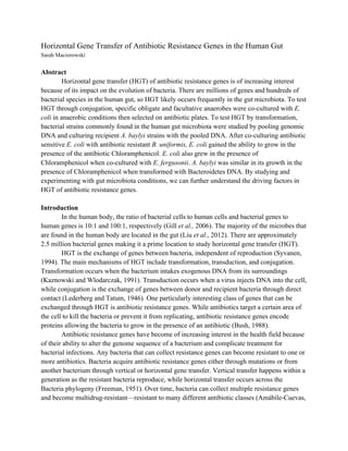
  • 9.   Figure 3. CFP Fluorescent Colonies. To measure E. coli growth between E. coli and E. fergusonii, the CFP of the colonies was read. A1, B1, and C1 are colonies that can be seen in white light (1a). Colonies A2 and B2 can be seen at 480 nm, while colony C1 is nonexistent (1b). 1a   1b   Figure 5. Quantified Colony Growth between A. baylyi and DNA. A. baylyi combined with bacteria DNA from Firmicutes, Actinobacteria, Proteobacteria, or Bacteroidetes phyla were spread on antibiotic plates. *The colonies that grew in the presence of an antibiotic were validated to have resistance genes. *   Figure 4. Quantified E. coli Growth between E. fergusonii and E. coli. All colonies that grew from co-cultures containing combinations of E. fergusonii, E. coli, and an antibiotic were analyzed for E. coli. After looking at the CFP fluorescent colonies, we quantified E. coli. *The colonies that grew in the presence of an antibiotic were validated to have resistance genes. *  
  • 10.   Table 4. Pooled Bacteria DNA.