More Related Content
More from Mohammed Hanif (A.Ag.)
More from Mohammed Hanif (A.Ag.) (20)
Biofilms
- 1. MINIREVIEW
Microbial biofilms and gastrointestinal diseases
Erik C. von Rosenvinge1,2
, Graeme A. O’May3
, Sandra Macfarlane4
, George T. Macfarlane4
& Mark E. Shirtliff3
1 Department of Gastroenterology and Hepatology, University of Maryland School of Medicine, Baltimore, MD, USA
2 Department of Veterans Affairs, VA Maryland Health Care System, Baltimore, MD, USA
3 Department of Microbial Pathogenesis, University of Maryland School of Dentistry, Baltimore, MD, USA
4 Microbiology and Gut Biology Group, University of Dundee, Ninewells Hospital Medical School, Dundee, UK
This timely review on the significance of microbial biofilms and gastrointestinal disease will stimulate research in this field.
Keywords
biofilm; microbiota; gastrointestinal disease;
gastrointestinal tract.
Correspondence
Mark E. Shirtliff, Department of Microbial
Pathogenesis, University of Maryland School
of Dentistry, Baltimore, MD 21201, USA.
Tel.: +1 410 706 2263
fax: 1 410 706 0193
e-mail: mshirtliff@umaryland.edu
Received: 9 September 2012; revised 12
December 2012; accepted 12 December
2012. Final version published online 29
January 2013.
doi:10.1111/2049-632X.12020
Editor: Ake Forsberg
Abstract
The majority of bacteria live not planktonically, but as residents of sessile biofilm
communities. Such populations have been defined as ‘matrix-enclosed microbial
accretions, which adhere to both biological and nonbiological surfaces’. Bacterial
formation of biofilm is implicated in many chronic disease states. Growth in this
mode promotes survival by increasing community recalcitrance to clearance by
host immune effectors and therapeutic antimicrobials. The human gastrointestinal
(GI) tract encompasses a plethora of nutritional and physicochemical environ-
ments, many of which are ideal for biofilm formation and survival. However, little is
known of the nature, function, and clinical relevance of these communities. This
review summarizes current knowledge of the composition and association with
health and disease of biofilm communities in the GI tract.
Introduction
The human gastrointestinal (GI) tract extends from the
esophagus through the stomach, small intestine, and
large intestine (colon) and terminates in the rectum (Fig. 1).
The small intestine is divided proximally-to-distally into the
duodenum, jejunum, and ileum. This collection of intercon-
nected organs harbors a diversity of microhabitats that are
colonized by microorganisms to varying degrees, depending
on local environmental conditions. For the purposes of this
article, the oral and nasal cavities will not be regarded as
being part of the GI tract, although these anatomical spaces
also contain great microbiological complexity (Ledder et al.,
2007).
There exists in the GI tract a gradient of colonization, from
the relatively sparsely populated esophagus and stomach to
the much more heavily colonized colon, which can contain
up to 1012
culturable bacteria per gram luminal contents
(Hopkins et al., 2002). Evolution has dictated that the GI
tract possess a large surface area to facilitate efficient
nutrient uptake, its primary physiological role in the body.
This coupled to high nutrient availability and a constant
influx of microorganisms, together with stable autochtho-
nous populations, makes the GI tract an ideal site for the
development of sessile microbial biofilm communities. The
microbiome of the gut has recently been determined in 124
subjects, and the microbial diversity indicates that the entire
cohort harbors only between 1000 and 1150 prevalent
bacterial species and each individual at least 160 such
species (Qin et al., 2010). In addition, there were common
microbial flora in subjects tested with 75 species common to
> 50% of individuals and 57 species common to > 90%.
Those microorganisms in closest proximity to host tissues
have the most opportunity for interaction with host
physiology, immunity, and metabolism; thus, mucosal
populations are arguably the most important component of
any host–microbiota interaction, whether beneficial or det-
rimental. The GI tract microbiota has been implicated in
disease states such as inflammatory bowel disease (IBD;
Macpherson et al., 1996), colon cancer (Horie et al.,
1999a, b), gastric cancer (Bj€orkholm et al., 2003), and
irritable bowel syndrome (IBS; Swidsinski et al., 2005). In
Pathogens and Disease (2013), 67, 25–38, © 2012 Federation of European Microbiological Societies. Published by Blackwell Publishing Ltd. All rights reserved 25
Pathogens and Disease ISSN 2049-632X
- 2. addition, recent microbiome studies have uncovered a
relationship between diet, microbiota, and health status,
particularly in older subjects (Claesson et al., 2012).
The GI tract is anatomically divided into ‘upper’ and ‘lower’
sections by the ligament of Treitz; however, from a microbial
perspective, this division applies to the GI tract poorly. The
colonization gradient in the GI tract, and particularly the
large and rapid (relative to the length of the GI tract)
increase in microbial population density from the terminal
ileum to the cecum, renders possible a convenient – if
somewhat artificial given their connectedness – microbial
distinction between the ‘upper’ and ‘lower’ GI tracts at the
level of the ileocecal valve. We will consider first the nature
and influence of microbial biofilms in the upper GI tract, that
is to say the esophagus, stomach and small intestine.
Following this, we shall venture forth into the lower GI tract.
The upper GI tract
In quantitative terms, the esophagus and stomach carry the
lightest bacterial load in the entire digestive system. In
comparison with the lower GI tract, comparatively few
microbiological investigations have been made on this part
of the gut; this is due in part to difficulties in obtaining
representative samples. In contradistinction, fecal effluent
provides a ready supply of material for investigations of
lower gut microbiology. Studies of the upper GI tract that
have been carried out indicate that it is sparsely colonized in
terms of microbial population density, but exhibits consider-
able diversity. Culturable bacteria in the healthy esophagus
are mainly Gram-positive facultatively anaerobic species
such as lactobacilli and streptococci. These are thought to
originate primarily in the oral cavity (Macfarlane & Dillon,
2007). While traditionally the stomach has been considered
inhospitable for bacteria due to its acidity, using sensitive
molecular techniques Bik et al. (2006) identified a surpris-
ingly diverse bacterial population in gastric mucosal biop-
sies.
Barrett’s esophagus
Barrett’s esophagus (BE) arises in individuals suffering from
long-term gastroesophageal reflux disease. In this condition,
squamous epithelial cells lining the distal esophagus
undergo metaplastic changes, forming a columnar mucosa
(Winters et al., 1987). Estimates of BE prevalence vary
markedly; indeed, the two largest recent studies gave
prevalences of 1.6% and 6.8%, in the general community
(Ronkainen et al., 2005) and individuals undergoing endo-
scopic examination (Rex et al., 2003), respectively. Patients
diagnosed with BE have a markedly higher risk of esoph-
ageal dysplasia and subsequent adenocarcinoma (Spechler
et al., 2001).
To date, there have been three investigations of esoph-
ageal mucosal bacterial populations in BE patients. One
such retrospective analysis of stored esophageal tissue
(Osias et al., 2004) reported increased microbial coloniza-
tion (mainly Gram-positive cocci) in patients with BE.
However, no significant difference was found when aerobic
cultures of fresh esophageal biopsy specimens were ana-
lyzed. In another investigation, a molecular cloning, and thus
nonquantitative, approach was used to identify the bacteria
on a mucosal sample from a single BE patient. Twenty-one
bacterial species were detected, of which circa 50% were
categorized as ‘unidentified’ rumen and oral isolates (Pei
et al., 2005).
The third, and more detailed, study by Macfarlane et al.
(2007) involved analysis of esophageal biopsy and aspirate
specimens taken from (1) seven individuals with confirmed
BE; and (2) seven controls. Controls, for the purposes of this
study, were defined as those persons attending the GI clinic
for upper GI tract endoscopy procedures, but who had no
evidence of BE by either endoscopic or histologic examina-
tion. Each specimen was subjected to analysis by culturing
techniques on a variety of solid media under aerobic,
anaerobic, and microaerophilic conditions, and bacterial
isolates were identified by 16S rRNA gene sequencing. The
spatial location of bacterial biofilms on mucosal samples
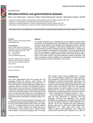
was determined by fluorescence microscopy. A total of 46
bacterial species were detected; interestingly, high levels of
Campylobacter concisus and Campylobacter rectus were
detected in four of the seven (57.1%) patients with BE, but
none of those without. Examination of biopsy material using
fluorescence microscopy revealed distinct microcolonies
existing within the mucosal layer (Fig. 2).
Nitrate in the human body is concentrated in the saliva.
Some is reduced by bacterial nitrate reductase in the mouth,
but the rest is washed into the esophagus and stomach. The
finding that the esophagus in some Barrett’s patients was
Fig. 1 The human gastrointestinal tract.
Pathogens and Disease (2013), 67, 25–38, © 2012 Federation of European Microbiological Societies. Published by Blackwell Publishing Ltd. All rights reserved26
Biofilms and GI diseases E.C. von Rosenvinge et al.
- 3. colonized heavily by nitrate-reducing campylobacters raises
the possibility that some of the cellular damage observed in
the esophagi of BE patients is caused by nitrate and nitric
oxide formation. Under low pH conditions, chemical reduc-
tion of nitrate can lead to the generation of carcinogenic
N-nitroso compounds and nitric oxide (Suzuki et al., 2005).
Nitric oxide is capable of inhibiting DNA repair enzymes and
can also be mutagenic at high concentrations (Liu et al.,
2002). Interestingly, the principal area of nitrite production
has been shown to occur at the gastroesophageal junction
(Iijima et al., 2002), lending support to the notion of bacterial
involvement in mutagenic events associated with BE.
Increased numbers of nitrate-reducing veillonellas were
also found in patients with BE (Macfarlane et al., 2007)
compared with control subjects, and these organisms have
been reported to be present in higher levels in oral
squamous cell carcinomas (Nagy et al., 1998).
Thus, the role of microorganisms and specifically sessile
biofilm bacteria in the pathogenesis of BE is intriguing.
However, more work is needed to ascertain what, if any,
affect the unique bacterial communities identified in BE
patients exert on the host.
The stomach
Historically, the stomach was thought to be a sterile
environment; the discovery of Helicobacter pylori coloniza-
tion dramatically altered this belief. More recently, sensitive
molecular techniques have identified the presence of a
diverse population of bacteria, including 128 phylotypes
from eight bacterial phyla in a study of gastric mucosal
biopsies taken from 23 adult subjects (Bik et al., 2006). Not
surprisingly, 67% of the identified phylotypes had previously
been identified in oral specimens. Sampling contamination
or passage of transient microorganisms, either from
ingested food or from swallowed oropharyngeal bacteria
that are not resident in the stomach, is certainly also
present, but their importance is unknown.
Helicobacter pylori
In a significant proportion of the population, the gastric
mucosa is colonized by H. pylori (Lehours & Yilmaz, 2007),
a phenomenon associated with peptic ulcer disease,
achlorhydria (Graham et al., 1988), corpus-predominant
gastritis (Harford et al., 2000), and gastric (Peek & Blaser,
2002), and possibly also esophageal (Ye et al., 2004),
adenocarcinomas.
Biofilm formation by H. pylori has been observed in vitro
at air/liquid interfaces in media with a high carbon/nitrogen
ratio (Stark et al., 1999). The capacity to form biofilm does
not appear related to cell surface hydrophobicity, motility, or
auto-aggregation (Yonezawa et al., 2010), but is strain-
dependent (Yonezawa et al., 2009). Furthermore, attach-
ment of H. pylori to glass surfaces and biofilm formation has
been reported (Cole et al., 2004). Surface properties
affected H. pylori morphology; the highly infectious spiral
form was associated with attachment to nonpolymeric
substances. Presence of serum in the medium inhibits
attachment (Williams et al., 2008). Interestingly, addition of
mucin (10% w/v type III porcine) resulted in an increase in
planktonic, but not biofilm, H. pylori numbers; thus, the
proportion of adherent cells declined upon addition of mucin
(Cole et al., 2004). This may be due to mucin-mediated
inhibition of H. pylori binding (Simon et al., 1997). However,
the significance of this finding is uncertain as the actual
number of adherent H. pylori cells remained unchanged.
Helicobacter pylori strain TK1402 was able to produce
biofilms with greater biomass than other strains; such
biofilms contained abundant outer membrane vesicles
(Yonezawa et al., 2009).
Helicobacter pylori biofilms have also been directly
visualized within the gastric mucosa (Carron et al., 2006;
Coticchia et al., 2006; Cellini et al., 2008; Cammarota et al.,
2010). Indeed, in subjects with peptic ulcer disease, biofilm
covered c. 97% of the surface of urease-positive biopsies
compared to c. 1.5% of urease-negative controls (Coticchia
et al., 2006). Within 3 days of initial colonization of the
gastric mucosa, H. pylori induces profound hypochlorhydria
and activates pro-inflammatory pathways that are involved
in further development of mucosal pathology (Zavros et al.,
2005). Although the precise mechanism of pathogenesis
remains unclear, production of IL-1beta by monocytes and
neutrophils, themselves recruited through H. pylori-induced
IL-8 production by mucosal epithelial cells (Bimczok et al.,
2010), inhibits H+
, K+
-ATPase (proton pump) a-subunit
expression (G€o~oz et al., 2000; Saha et al., 2007). In
addition, these infections often demonstrate in vitro and in
vivo recalcitrance to even quadruple antimicrobial therapy
using antibiotics to which the strains are supposedly
sensitive (Megraud et al., 1991; Gisbert, 2008; Cammarota
et al., 2010).
Helicobacter pylori possesses a number of virulence
factors that assist in gastric mucosal colonization and
persistence. Recent evidence has suggested that H. pylori
heat shock protein 60 (Hsp60) may be involved in angio-
genesis (Lin et al., 2010), itself vital for tumor development.
Helicobacter pylori vacuolating toxin (VacA) disrupts actin
interaction with parietal cell apical membranes, preventing
recruitment and fusion of H, K-ATPase-containing tubulove-
sicles and causing hypochlorhydria (Wang et al., 2008).
Perhaps the best-known H. pylori virulence factor is urease
(Mobley et al., 1988), which assists colonization and per-
sistence by modulating the highly acidic conditions in the
immediate environment of H. pylori cells. Urease may act
(a) (b)
Fig. 2 Fluorescence microscopy image of mucosal biopsies from BE
patients showing distinct microcolonies existing within the mucosal
layer. Original magnification, 9 60 (Macfarlane et al., 2007).
Pathogens and Disease (2013), 67, 25–38, © 2012 Federation of European Microbiological Societies. Published by Blackwell Publishing Ltd. All rights reserved 27
E.C. von Rosenvinge et al. Biofilms and GI diseases
- 4. either within the bacterial cytoplasm (Weeks et al., 2000),
on the cell surface (Baik et al., 2004), or extracellularly
(Gobert et al., 2002). Urease-mediated increases in gastric
pH may be useful not only for survival of H. pylori; recent
evidence suggests that the viscoelasticity of gastric mucus
increases as pH rises, facilitating movement of H. pylori
through the mucus layer (Celli et al., 2009).
Recently, a study of the biofilm-disrupting compound
N-acetylcysteine (NAC) has demonstrated the importance
of the biofilm phenotype in human H. pylori infection
(Cammarota et al., 2010). In this study of 40 patients, all
with a history of multiple failed attempts at H. pylori
eradication, SEM documented biofilm in all patients
(100%). Patients were randomized to receive 1-week
treatment with NAC or placebo prior to culture-guided
antibiotic therapy. Thirteen of the 20 patients (65%) who
received NAC cleared their infection while only four of the
20 patients (20%) who received placebo did so (P < 0.01).
Ten of those who successfully eradicated their H. pylori
infection agreed to a follow-up upper endoscopy, and in
these patients, SEM showed disappearance of biofilm in all.
While these exciting findings should be confirmed in larger
studies, they suggest that the biofilm phenotype plays an
important role in human GI infection and provides the first
evidence that biofilm-directed therapy can be successful for
GI diseases.
The small intestine
After being expelled from the stomach through the pyloric
sphincter, digestive material is in a highly liquid state due to
the addition of gastric juices in the stomach, bile, mucus,
and other secretions present in the duodenum itself. The
end result is a high flow rate through the small intestine, with
average transit times being in the region of 2–4 h. This
washing-through of gut contents contributes to the low
bacterial load of the duodenum, jejunum, and ileum; bacteria
passing through these organs have little opportunity to
attach to the mucosa and form biofilm. Bacterial population
density increases along the length of the small intestine until
a colonic-like community structure is established in the
vicinity of the ileo-cecal valve, where numbers of micro-
organisms present can reach 108
–109
CFU per gram
contents. A variety of disease states can result in larger
numbers of bacteria in the small bowel, for example,
achlorhydria (Williams & McColl, 2006).
Enteral nutrition
Patients who are unable to masticate or swallow normally,
typically due to cerebrovascular disease, oropharyngeal or
esophageal carcinoma, or craniofacial trauma, require
nutritional support via an enteral tube. Enteral nutrition
(EN) is typically preferred to parenteral nutrition as both
animal and human studies have shown it to be safer and
more physiological in that it preserves gut barrier and
absorptive functions, and immune mechanisms. The 2011
American Society for Gastrointestinal Endoscopy
guidelines on the role of endoscopy in enteral feeding
recommends nasoenteric feeding as the preferred
approach to feeding patients who are expected to resume
peroral nutrition within 30 days (Jain et al., 2011). In
patients not predicted to resume peroral nutrition within
30 days, they suggest that nutrition be provided by a
percutaneous endoscopic gastrostomy (PEG) feeding
tube, after first addressing factors such as patient prefer-
ences, quality of life, and overall prognosis with the
patient and their family. Alternatives to PEG include
surgically placed or interventional radiology–placed gas-
trostomy tubes. Patients with severe gastroesophageal
reflux, delayed gastric emptying, or repeated tube feeding-
related aspiration pneumonia may benefit from direct or
trans-gastric jejunal feeding.
Low gastric pH is generally considered to be a major
factor suppressing microbial colonization of the stomach;
however, some enteric bacteria possess one or more acid
resistance mechanism(s) (Castanie-Cornet et al., 1999) that
can confer protection from the bactericidal effects of acid
during passage through the stomach. Many innate defense
mechanisms break down in EN patients, where a lack of
sensory stimuli associated with food intake inhibits saliva
production and peristalsis, while reduced swallowing may
result in lower gastric acid production and reduce nitrite
concentrations. The net effect is greater susceptibility to
microbial overgrowth in the stomach and small intestine, at
times resulting in diarrhea, although more serious compli-
cations such as malabsorption and sepsis also occur (Cabre
Gassull, 1993). The formation of microbial biofilms on EN
tubes is an unavoidable consequence of bacterial over-
growth. These structures are difficult to eradicate with
antimicrobial agents (Walters et al., 2003) and can harbor
pathogens (Bauer et al., 2002) and/or microorganisms
carrying antibiotic resistance genes (Ohlsen Lorenz,
2010).
Nasogastric feeding. During passage through the nasal
cavity and esophagus, the NG tube is exposed to nasopha-
ryngeal and esophageal microbiotas. Additionally, the exte-
rior environment and the feeding formula itself, which may
be contaminated (Mathus-Vliegen et al., 2006), are other
sources of tube contamination. The location of NG tubes in
the nasopharynx, esophagus, and stomach ensures a
regular supply of nutrients, together with the presence of
large numbers of bacteria. Under such conditions, biofilm
formation is inevitable. It should also be noted that the NG
tube passes close to the larynx, raising the possibility of
respiratory tract colonization.
Marrie et al. (1990) undertook microbiological assess-
ments of the external surfaces of the gastric portion of NG
tubes recovered from hospitalized patients. They reported
that the majority of such tubes were covered in an
amorphous biofilm, composed primarily of microcolonies
within which bacterial cells were enclosed by an extracel-
lular matrix. These microcolonies were composed both of
bacteria of varying morphotypes and yeast cells. Interest-
ingly, a proportion of the observed microcolonies were found
to be composed of dead cells and empty cell walls. NG
tubes that had been in situ for as little as 24 h were
colonized extensively.
Pathogens and Disease (2013), 67, 25–38, © 2012 Federation of European Microbiological Societies. Published by Blackwell Publishing Ltd. All rights reserved28
Biofilms and GI diseases E.C. von Rosenvinge et al.
- 5. A further study evaluated colonization of the oropharynx of
elderly patients by Pseudomonas aeruginosa (Leibovitz
et al., 2003). Pseudomonas aeruginosa was detected in 18
of 53 (34%) patients receiving NG and none of the controls,
while other Gram-negative bacteria were detected in 34
(64%) of NG patients and four (8%) of the controls. Addition-
ally, SEM revealed P. aeruginosa biofilm on tube surfaces.
Pulsed-field gel electrophoresis analysis suggested that the
oropharynx was the source of tube contamination.
A further study used first-ever introduced NG tubes that
had been self-removed by patients between one and 7 days
after placement; these tubes were examined by SEM and
confocal laser scanning microscopy (Leibovitz et al., 2005).
The surfaces of the majority of tubes were covered by
biofilm. No quantitative data on the extent, morphology, or
composition of NG biofilm was provided in this study.
Segal et al. (2006) investigated the microbiological com-
position of gastric juices and the nasal cavities of 107
subjects undergoing NG feeding. Potentially pathogenic
microorganisms (defined in this study as Gram-negative
bacteria or Staphylococcus aureus) were isolated from 74%
and 69% of gastric and nasopharyngeal samples, respec-
tively. The most common organisms isolated from gastric
juice were Proteus spp. (26%) and Escherichia coli (22%),
while Proteus spp. (24%) and Pseudomonas spp. (21%)
were the species isolated most frequently from the orophar-
ynx. This study also noted high gastric pH (4.57 Æ 0.65 after
3 h NG feeding, and 4.2 Æ 0.9 after 12 h). High pH
correlated strongly with isolation of pathogenic bacteria,
underlining the importance of gastric acid in host defense.
The authors hypothesized that the colonized stomach may
act as a reservoir of pathogens, leading to aspiration
pneumonia in some cases.
Due to the presence of this array of pathogenic biofilm
populations on NG tubes, it is not surprising that they can
act as a microbial reservoir for a number of diseases
associated with NG tubes including nasogastric tube
syndrome, microbial pneumonia, sinusitis, middle ear effu-
sion, acute necrotizing esophagitis, and even death (Gold-
enberg et al., 1990; Le Moal et al.,1999; Apostolakis et al.,
2001; Bullock et al., 2004; Lin et al., 2006). As with all
mature biofilms forming on indwelling medical devices, the
NG tube should be removed and antimicrobial chemother-
apy applied to resolve the infection.
Gastrostomy feeding. PEG has the advantage of reduced
nasal and oropharyngeal irritation and is typically easier to
manage in the home or other community setting, and PEG
insertion can facilitate discharge from hospital. PEG tubes
can be left in situ for extended periods, but often they require
replacement due to either deterioration of the PEG tube
itself or its accidental removal by patients.
Candida spp. readily colonize PEG tubes, a phenomenon
that may lead to tube deterioration (Gottlieb et al., 1992).
Dautle et al. undertook a comprehensive analysis of PEG
tube microbiotas using molecular techniques. Random
amplified polymorphic DNA (RAPD) analysis was used on
material obtained from biofilms that had formed on 18
gastrostomy devices taken from pediatric patients whose
age ranged from 6 months to 17 years. These devices had
remained in place for a mean time of 20 months (range,
3–47 months). Data indicated that PEG tube biofilms in
pediatric patients were compositionally diverse, containing
enterococci, staphylococci, E. coli, lactobacilli, candidas,
pseudomonads, and bacilli (Dautle et al., 2003).
The gastric and duodenal microbiotas of PEG patients
and populations on PEG tube surfaces themselves were
evaluated by culturing methods. Interestingly, those individ-
uals who received antibiotics prior to PEG tube placement
had both an increased prevalence of some types of infection
and decreased mortality rates. The organisms isolated were
mainly candidas, enterobacteria, streptococci, staphylo-
cocci, and lactobacilli (Table 1; O’May et al., 2005a, b).
Data suggested that gastric pH had no significant effect on
the density of colonization in the stomachs and duodena of
EN patients, although it did affect microbiota composition:
Bifidobacterium, Klebsiella, and Staphylococcus spp. were
detected only in aspirates with a pH of greater than three.
Significantly, E. coli, staphylococci, and candidas were
detected only in aspirates from patients who had received
antibiotic treatment during their stay in hospital. This was
supported by the work of Smith et al. (2011) who used real-
time PCR and FISH to investigate microbial colonization of
the gastric mucosa of eight PEG patients. Mean levels of
enterobacteria and staphylococci were significantly higher
in PEG patients than in controls; however, levels of the
pro-inflammatory cytokines IL-1a, IL-6, and TNF-a were
lower in PEG patients. As with NG tubes, PEG tubes
contaminated with a variety of pathogenic microbial biofilms
can produce a number of infections, most importantly
peristomal infection and the potential for sepsis (Blomberg
et al., 2012). Resolution of infection, and prevention of
Table 1 Characterization of microorganisms detected in gastric and
duodenal aspirates obtained from patients undergoing a PEG placement
procedure (O’May et al., 2005a, b)
Genus
Population size*
Gastric aspirates Duodenal aspirates
Streptococcus 5.2 Æ 0.6 (5) 4.8 Æ 0.5 (11)
Staphylococcus 5.8 Æ 0.7 (4) 4.7 Æ 0.8 (6)
Proprionibacterium 3.8 Æ 0.4 (3) ND
Peptostreptococcus 3.8 Æ 0.4 (3) 5.7 Æ 0.9 (4)
Lactobacillus 4.0 Æ 0.2 (6) 4.0 Æ 0.3 (6)
Klebsiella ND 4.7 Æ 0.6 (5)
Gemella 3.7 (1) 4.5 Æ 1.2 (2)
Eubacterium 3.6 Æ 0.1 (3) 4.6 Æ 0.4 (3)
Escherichia 5.4 Æ 0.4 (5) 4.5 Æ 0.6 (6)
Corynebacterium 4.4 Æ 1.1 (3) 4.4 Æ 0.6 (5)
Clostridium 3.5 Æ 0.4 (2) 4.7 Æ 0.4 (2)
Bifidobacterium 4.7 Æ 0.3 (3) 4.8 Æ 0.4 (6)
Actinomyces 3.9 Æ 0.1 (2) 5.5 Æ 0.6 (3)
Candida 4.6 Æ 0.5 (5) 3.7 Æ 0.2 (5)
ND, Not detected.
*Data are expressed as mean log10 CFU mlÀ1
Æ standard deviation
(N); Ntotal = 20.
Pathogens and Disease (2013), 67, 25–38, © 2012 Federation of European Microbiological Societies. Published by Blackwell Publishing Ltd. All rights reserved 29
E.C. von Rosenvinge et al. Biofilms and GI diseases
- 6. re-infection, may require removal of the PEG because
antibiotics alone will not clear biofilm pathogens from a
contaminated tube.
In general, data obtained by in vitro modeling using a
chemostat-based system mirrored those of human studies
(O’May et al., 2005a, b). Lowering of pH from six to three had
no significant effect on the density of planktonic or biofilm
communities; indeed, a significant (circa 107
CFU mlÀ1
)
microbiota was detected at pH 3. It is important to note that
because of the continuous culture methods employed in this
study, these recovery data must represent cells actively
multiplying in such low pH values. Low pH altered markedly
microbiota composition: candidas and lactobacilli were
aciduric while numbers of E. coli and Klebsiella pneumoniae
decreased concomitantly with pH. Visualization of PEG tube
surface-associated biofilm using BacLightTM
showed micro-
colonies composed of both living and dead cells; in many
cases, yeast pseudohyphae were found to be invading the
interior of microcolonies. Where this occurred, bacterial cells
surrounding the pseudohyphae were red-stained. More
recent work has established the existence of an interaction
between S. aureus and Candida albicans pseudohyphae
during biofilm growth (Peters et al., 2010). Differential in-gel
electrophoresis demonstrated differential expression of 27
proteins during co-culture biofilm growth. Variation in
expression of the virulence-related factors such as a-lactate
dehydrogenase 1 (upregulated; Richardson et al., 2008)
and CodY (downregulated by contact with C. albicans
hyphae; Levdikov et al., 2006) suggests synergistic patho-
genesis. CodY has been shown to repress polysaccharide
intercellular adhesion-dependent biofilm formation, and
production of hemolysins alpha and delta and proteins
involved in the agr-dependent quorum-sensing system, a
global regulator of virulence (Majerczyk et al., 2010). Thus,
downregulation of CodY expression may enable enhanced
toxin-mediated virulence and increased biofilm formation in
S. aureus. This phenomenon is potentially highly significant
and merits further study.
The frequent use of EN makes understanding the
mechanisms behind and consequences of microbial colo-
nization in such patients increasingly important. Biofilm
formation is inevitable when the upper GI tract becomes
overgrown, and a stable nonshedding surface, the tube
itself, is in situ for long periods. Early data suggest that the
use of antibiotics in such patients may actually increase
the probability of colonization by potentially pathogenic
microorganisms such as S. aureus and C. albicans.
Dosing with pro-, pre-, and synbiotics either before or
after tubes are placed may represent a novel method of
altering biofilm composition toward a more commensal-
type structure.
The lower GI tract
Epithelial surfaces in the GI tract are covered by a layer of
mucus, which prevents most microorganisms reaching and
persisting on the mucosal surface. This viscoelastic gel is
protective against adhesion and invasion by many patho-
genic microorganisms, bacterial toxins, end products of
metabolism, pancreatic endopeptidases, microbial antigens,
and other damaging agents present in the lumen of the
bowel. Mucus consists primarily of water (c. 95%) and
glycoproteins that give mucus its viscosity and ability to form
gel structures.
Mucins are chemically and structurally diverse molecules;
however, they always are comprised, to some extent, of
galactose and hexosamines, with smaller quantities of
fucose (Quigley Kelly, 1995). The carbohydrate groups
exist as both linear and branched oligosaccharides; these
can comprise as much as 85% of the molecule (Smith
Podolsky, 1986). Mucin oligosaccharides are attached to a
protein core via serine or threonine residues. The attach-
ment of sulfate and sialic acids to terminal mucin oligosac-
charides confers resistance to digestion by microbial
glycosidases (Corfield et al., 2001). To survive, bacteria
resident in the colon must produce a number of hydrolytic
enzymes, for example, polysaccharidases, glycosidases,
proteases, peptidases. Mucins are important sources of
carbohydrate for saccharolytic bacteria, particularly popula-
tions in the distal colon, where the supply of fermentable
carbohydrate is usually limiting (Macfarlane et al., 1992).
Some bacteria can invade the mucus layer, and many
intestinal microorganisms use these molecules as carbon,
nitrogen, and energy sources (McCormick et al., 1988). The
removal of carbohydrates and other components, such as
sulfate, from the glycoprotein compromises its protective
function (Schrager Oates, 1978), particularly when the
rate of mucus breakdown exceeds that of its synthesis and
secretion.
Pure and mixed culture studies have established that in
many gut bacteria, synthesis of degradative enzymes,
particularly b-galactosidase, N-acetyl b-glucosaminidase,
and neuraminidase, is catabolite regulated (Macfarlane
et al., 1989, 1997; Macfarlane Gibson, 1991) and
therefore dependent on local concentrations of mucin and
other carbohydrates. While some colonic microorganisms
can produce several different glycosidases (Macfarlane
et al., 1990), the majority of experimental data suggest that
the breakdown of mucin is a cooperative activity (Macfar-
lane et al., 1999). Studies on biofilm communities in the
gut have demonstrated the presence of bacterial microcol-
onies on mucosal surfaces in healthy people (Fig. 3;
Macfarlane Macfarlane, 2004). Despite its undoubted
significance, few studies have focused on mucosal bacte-
rial communities. However, there is evidence to suggest
that mucosal populations are distinct from those in the gut
lumen (Macfarlane Macfarlane, 2004), and these are
thought to play an important role in IBD (see below).
Despite this, little is known about bacterial growth in the
mucus layer, the organisms that colonize this microcosm,
or their role in disease processes.
Chemostat-based modeling studies (Macfarlane et al.,
2005) have shown differential colonization of artificial mucin
gels by fecal bacteria in a two-stage continuous culture
system, simulating the nutrient availability of the proximal
(vessel 1) and distal (vessel 2) colon. The establishment of
bacterial communities in mucin gels was investigated by
selective culture methods, SEM, and confocal laser scan-
Pathogens and Disease (2013), 67, 25–38, © 2012 Federation of European Microbiological Societies. Published by Blackwell Publishing Ltd. All rights reserved30
Biofilms and GI diseases E.C. von Rosenvinge et al.
- 7. ning microscopy, in association with fluorescently labeled
16S rRNA gene oligonucleotide probes. Mucin gels were
rapidly colonized by heterogeneous bacterial populations
(Fig. 4), particularly members of the Bacteroides fragilis
group, enterobacteria, and clostridia. Intestinal bacterial
populations growing on mucin surfaces were found to be
phylogenetically and metabolically distinct from their plank-
tonic counterparts.
Inflammatory bowel disease
The two most common forms of idiopathic IBD are UC and
Crohn’s disease (CD). It is estimated that more than one
million Americans suffer from IBD. UC affects only the
mucosal surfaces in the large intestine and rectum. CD can
occur anywhere in the digestive tract, often with inflamma-
tory lesions spreading deep into the layers of affected
tissues. UC, CD, and acute self-limited colitis (ASLC) all
cause diarrhea, with or without accompanying bleeding.
However, UC and CD are chronic inflammatory diseases, as
opposed to ASLC (mainly infectious agents) and IBS, which
is not accompanied by overt inflammation (Steed et al.,
2008).
Recent studies of the gut microbiota of patients with IBD
have in general terms found a decline in microbial flora
diversity (Frank et al., 2007) and methanogens (Scanlan
et al., 2008), and an increase in fungal diversity (Ott et al.,
2008). Furthermore, despite strenuous efforts to identify
microbial community compositions unique to IBD states,
none have as yet been elucidated (Reiff Kelly, 2010).
Frank et al. (2007) performed an rRNA sequence analysis
of diverse intestinal biopsies from both diseased and normal
tissues of patients with IBD and healthy controls. Data
suggested depletion of the commensal phyla Firmicutes and
Bacteroidetes. The authors suggest treatment of at least
some forms of IBD by targeted antimicrobial chemotherapy.
More recently, Qin et al. (2009) utilized Illumina-based
bacterial profiling to determine the microbiome differences
between the healthy individuals and those suffering from
IBD. Patients’ microbial profiles clearly separated patients
with IBD from healthy individuals and the patients with UC
from the patients with CD.
Other authors have echoed this view. Notably, Green-
berg suggested that although a cursory examination of
available clinical trials would lead to the conclusion that the
use of antibiotics in Crohn’s is – at best – ineffective, a
more in-depth examination of both clinical and laboratory
evidence may lead to the opposite conclusion (Greenberg,
2004). As it is likely that IBD represents a number of
disease states, the symptoms of which are often indistin-
guishable, it follows that microbial community composition
will be similarly diverse. Thus, any attempt at treating such
a diversity of disease states with a single strategy is likely
to fail.
(a) (b) (c)
(d) (e) (f)
(g) (h) (i)
Fig. 3 Confocal laser scanning microscopy of a bacterial microcolony on healthy rectal mucosa stained with a live/dead stain. The microcolony was
sectioned in 1.5 lm slices from the lumen (a) to the mucosal surface (i). Original magnification, 9 60 (Macfarlane Macfarlane, 2004).
Pathogens and Disease (2013), 67, 25–38, © 2012 Federation of European Microbiological Societies. Published by Blackwell Publishing Ltd. All rights reserved 31
E.C. von Rosenvinge et al. Biofilms and GI diseases
- 8. Ulcerative colitis
UC is a chronic relapsing form of IBD, and the precise
etiology of which is unknown. In UC, the inflammatory
response is located principally within the colonic
mucosa. The distal colon is always affected, and the
disease usually progresses from its initiation site in the
distal bowel toward the proximal large intestine. UC,
depending on the severity of the condition, can severely
affect the quality of life, and if medical treatments are
not effective, surgical removal of all or most of the colon
is necessary.
Involvement of commensal gut bacteria in both the
initiation and maintenance of UC has been suggested
since the early 1970s (Hill et al., 1971). Antimicrobial
agents specifically active against obligate anaerobes have
been shown to prevent ulceration in guinea pigs (Onder-
donk Bartlett, 1979), while experiments using germ-free
animals show that they only develop colitis when repop-
ulated with fecal bacteria (Sadlack et al., 1993). A variety
of species including Fusobacterium spp., Shigella spp.
(Onderdonk et al., 1983) and adhesive E. coli (Dickinson
et al., 1980) isolated from the colitic bowel have been
implicated in disease etiology; however, no specific micro-
organisms have been found in all individuals suffering from
UC, and Koch’s postulates cannot be demonstrated. The
luminal microbiota of patients with UC has been examined
extensively (Swidsinski et al., 2005, 2008a, b; Macfarlane
et al., 2009; Swidsinski et al., 2009; Ott et al., 2008; Reiff
Kelly, 2010). There is good evidence that bacteria
growing on the gut wall play an important role in UC,
because they exist in close juxtaposition to host tissues,
and can interact with the host immune and neuroendocrine
systems. This is particularly so given that FISH imaging
has suggested that mucosal bacterial populations are in
contact with the mucosal epithelium in UC and Crohn’s
patients, but not in healthy individuals (Swidsinski et al.,
2009).
Bacterial populations compositionally distinct from those
in the gut lumen are known to exist on the mucosal surface,
and in the mucus layer in the large gut (Poxton et al., 1997),
where Bacteroides and fusobacteria appear to predominate,
but other groups such as eubacteria, clostridia, and
anaerobic Gram-positive cocci are also present as either
heterogeneous populations or microcolonies (Croucher
et al., 1983). Until relatively recently, there have been
comparatively few studies on bacteria that inhabit the
colonic mucosa, largely due to two factors: Firstly, feces
and other types of material from the gut lumen are easier to
obtain than tissue samples from the gut wall, and secondly,
in most studies individuals taking part have been treated
prophylactically with antibiotics and other types of drug (e.g.
anti-inflammatory drugs and steroids), or the bowel has
been purged before colonoscopy. As a consequence, the
metabolic and health-related significance of bacteria grow-
ing as biofilms on the colonic mucosa is only now beginning
to be elucidated.
The notion that biofilm growth in the mucus layer is
important in the pathogenesis of UC is considered likely
given that (1) mucosal bacteria have been visualized
colonizing the colonic mucosa in patients with UC
(Macfarlane et al., 2004); and (2) the condition’s intracta-
bility to antibiotic treatment. Antimicrobial agents are still
used in treating patients with IBD, mostly in people with
severe disease, as in patients with fistulae or other septic-
type complications, and occasionally as a first-line therapy.
The employment of antibiotic therapy seems mainly to be
based on reported benefits observed in individual patients,
that is, on small numbers of or individual case studies
(Greenberg, 2004; Thompson-Chagoyan et al., 2005).
Also, in a recent meta-analysis, Wang et al. (2012) found
that antimicrobial therapy improved clinical outcomes of
patients with IBD. However, the long-term improvement
may be limited due to the ‘rebound effect’ following
cessation of antibiotic treatment described by Swidsinski
et al. (2008a, b). This study suggested that while mucosal
bacterial populations are suppressed during antibiotic
treatment, those communities re-establish to at least their
previous level after therapy is stopped. In this study, the
‘rebound effect’ was observed when bacterial populations
in antibiotic-treated individuals were measured 4 weeks
(a) (b)
Fig. 4 SEM image of chemostat-housed mucin gels showing rapid colonization by heterogeneous bacterial populations, particularly members of the
Bacteroides fragilis group, enterobacteria, and clostridia (Macfarlane et al., 2005).
Pathogens and Disease (2013), 67, 25–38, © 2012 Federation of European Microbiological Societies. Published by Blackwell Publishing Ltd. All rights reserved32
Biofilms and GI diseases E.C. von Rosenvinge et al.
- 9. after cessation of treatment. Bacterial numbers were circa
25 times higher than in those who had not been treated.
This rebound effect was found to diminish over time, but
was still present up to 36 weeks after cessation of
antimicrobial therapy. The ‘rebound effect’ seemed to
cause increases in the very types of bacteria that were
the targets of antibiotic therapy, for example, Bacteroides
(targeted by metronidazole) and enterobacteria (targeted
by ciprofloxacin). The data collected in this study also
suggested, although inconclusively, that the organisms
detected were less metabolically active than in nontreated
individuals. Bacteria in antibiotic-treated samples were
visualized by DAPI staining, but not by fluorescence in
situ hybridization (FISH). The authors postulate that this
may have been due to reduced rRNA levels within the
bacteria, reflecting a lower level of protein synthesis and so
reduced metabolic activity and possibly also lower viability.
Results from this study may provide some insight as to
why IBD does not seem to respond to antibiotic treatment,
despite the widely held belief that gut mucosa-associated
bacteria are involved in disease pathogenesis. The mech-
anism behind the ‘rebound effect’ remains unclear,
although it seems likely that survivor bacteria in the mucus
layer are able to utilize nutrients that are not assimilated
by microbial communities killed by the antibiotic. Further
work is needed to confirm this, however. Of wider
importance is the question of whether this ‘rebound effect’
is a general property of biofilm, either in the body or more
universally. If so, it represents a potentially important new
area of inquiry.
A promising new therapy for IBD involves the oral
administration of probiotics, prebiotics, or synbiotics. Probi-
otics are defined as live microorganisms with a demonstra-
ble health benefit when ingested by or otherwise
administered to the human host; prebiotics are food ingre-
dients that selectively stimulate the growth and/or the
activity of intestinal bacteria that have health-promoting
properties (Steed et al., 2008). At the present time, the
overwhelming preponderance of prebiotics are nondigesti-
ble oligosaccharides (NDO), of which galacto-oligosaccha-
rides (GOS), lactulose, inulins, and their fructo-
oligosaccharide (FOS) derivatives have been by far the
most extensively investigated (Macfarlane et al., 2006,
2008). It is important to note that the term nondigestible
refers only to the host; bacteria resident in the gut are
capable of utilizing prebiotic polysaccharides as energy
sources. One key difference between pro- and prebiotics is
that probiotics are allochthonous microorganisms, whereas
prebiotics can only influence those bacteria already resident
with the gut of the patient. Therefore, incoming probiotic
bacteria have to overcome the colonization resistance
offered by the bacteria in the resident microbiota who have
already established themselves within the metabolic and
spatial microenvironments close to or on the gut wall. A
synbiotic is the combination of a pro- and prebiotic in one;
the terms comes from the idea that the two, when used
together, will (1) be more likely to be able to overcome
colonization resistance; and (2) may have a synergistic
effect on the host.
Furrie et al. (2005) reported on a double-blinded random-
ized controlled trial in which a synbiotic was fed to patients
with UC for a period of 1 month. Eighteen patients took part
in this study; those selected to receive the synbiotic were
provided with six grams of synergy 1 (oligofructose-enriched
inulin) and 2 9 1011
live Bifidobacterium longum per day,
which they were asked to take twice daily. Results showed
that bifidobacterial numbers on the rectal mucosa increased
by 40-fold in those subjects who had received the
synbiotic compared with a fourfold increase in the control
group. This was accompanied by significant reductions in
mucosal pro-inflammatory cytokines (TNF-a, IL-1b) together
with inducible human b-defensins 2, 3, and 4. b-Defensins
are antimicrobial short-chain peptides produced by gut
epithelial cells during inflammation. However, unlike other
immune system mediators such as TNF-a and IL-1b,
b-defensins are not formed by immune inflammatory cells
infiltrating the mucosa. For this reason, b-defensins are
useful markers of epithelial surface healing. Histologic
assessments indicated marked, although not significant,
reductions in inflammatory cells and crypt abscesses in
patients receiving the synbiotic, together with regeneration
of normal tissue, while sigmoidoscopy scores and clinical
activity indices in these individuals also improved. This
short-term pilot study provided preliminary data supporting
the notion that synbiotic administration has the potential to
be developed into acceptable therapies for patients suffering
from active UC, but further work is needed to investigate
the long-term efficacy of synbiotics in inducing and main-
taining remission.
Crohn’s disease
Compared to UC, the evidence for sessile mucosal bacterial
involvement in the pathogenesis and maintenance of CD is
sparse. Concentrations of mucosal bacteria in patients with
CD were found to be two logs higher than in healthy controls
or patients with IBS. Of these, Bacteroides spp. predomi-
nated in patients with CD, in some individuals comprising
c. 80% of total mucosal bacteria, compared with c. 15% in
IBS (Swidsinski et al., 2005). Furthermore, these popula-
tions were found to be directly adjacent to the epithelium in
patients with CD but not healthy controls (Swidsinski et al.,
2009). The stability of bacterial diversity over time, partic-
ularly during active CD episodes and relapses, in patients
with CD is lower than that in healthy controls (Scanlan et al.,
2006). Therefore, the constantly changing microbial popu-
lations on the colonic mucosa of patients with CD may
account – at least in part – for the aberrant immune
responses characteristic of the condition. Alternatively,
these alterations in the microbiome may themselves be
caused by changes in disease activity.
In contrast, an rRNA sequence analysis of the microbial
communities of colonic biopsies from patients with CD and
healthy controls suggested depletion of normal commen-
sals, such as Bacteroides spp. Furthermore, stratification of
patients into a number of microbiota groupings suggests
that CD represents a number of disease states (Frank et al.,
2007). However, another study suggested that the dominant
mucosal-associated bacteria in inflamed and noninflamed
Pathogens and Disease (2013), 67, 25–38, © 2012 Federation of European Microbiological Societies. Published by Blackwell Publishing Ltd. All rights reserved 33
E.C. von Rosenvinge et al. Biofilms and GI diseases
- 10. tissue in patients with CD did not differ (Vasquez et al.,
2007).
Interest in a role for adherent-invasive E. coli (AIEC) in
CD (Darfeuille-Michaud, 2002) is increasing because this
microorganism is more prevalent in patients with CD than in
healthy individuals in a number of countries, for example,
the UK (Martin et al., 2004), France (Darfeuille-Michaud
et al., 2004), and the United States (Baumgart et al., 2007).
AIEC strains are adherent to and can invade colonic
epithelial cells in vitro, as well as survive and multiply inside
macrophages. Furthermore, intracellular growth of AIEC
does not induce apoptosis or tumor necrosis factor (TNF)
production. AIEC does not appear to be genetically unique,
but does possess genes associated with the virulence of
extra-intestinal pathogenic E. coli (Martinez-Medina et al.,
2009a). The biofilm-producing capacity of AIEC strains from
the colonic mucosa was compared to that of non-AIEC
strains by Martinez-Medina et al. Specific biofilm formation
indices were significantly higher among AIEC strains com-
pared to other colonic E. coli isolates (Martinez-Medina
et al., 2009b). Moreover, AIEC strains also exhibited greater
adherence and invasion indices. Biofilm-producing AIEC
strains were more frequently motile and positive for the S
fimbriae-encoding sfa/focDE virulence genes. Thus, the
extant data on the role of AIEC in CD warrants further
investigation into the nature and pathogenic mechanisms of
this bacterium.
Patients with CD have higher levels of serum IgG specific
to a number of microbial antigens. IgG levels to the ASCA
epitope of Saccharomyces cerevisiae are elevated in many
patients with CD (McKenzie et al., 1990). This is particularly
interesting given (1) the increased incidence of S. cerevisi-
ae in patients with CD has been reported (Ott et al., 2008);
and (2) that this epitope is also expressed by both
C. albicans and Mycobacterium paratuberculosis (Mpofu
et al., 2007). Levels of flagellin-specific serum IgG, for
example, CBir1, are higher in CD populations, but not in
either those suffering from UC or in healthy controls (Lodes
et al., 2004). An intestinal E. coli strain, O83:H1, has been
found to adhere to and invade colonic epithelial cells in vitro
when flagellated, but not in the absence of a flagellum
(Eaves-Pyles et al., 2008). The serum IgG response to
OmpC, gASCA, AMPCA, ALCA, and ACCA in patients with
CD has been linked to both the complicated disease
phenotype and the need for surgery (Papp et al., 2008).
However, it is also possible that the increases in serum IgG
levels reported in the aforementioned studies are merely
reflective of a more general increase in IgG levels to multiple
microbial antigens in patients with CD. Indeed, Adams and
co-authors reported that levels of IgG specific to mannan
and flagellin were no more effective for diagnosis of CD than
IgG levels against complex mixtures of antigens from gut
commensal bacteria such as Bacteroides vulgates (Adams
et al., 2008).
The link between biofilms and disease
As described in Table 2, there have been a number of
studies that have shown the simultaneous inflammation, a
disease process, and microbial biofilm communities in the
affected GI location. A set of criteria were previously
proposed by Parsek Singh (2005) to demonstrate a link
between biofilm formation and human disease. These
criteria include direct examination of an infected tissue
revealing pathogenic bacteria in communities attached to a
surface where there is a localized infection and evidence of
recalcitrance to antibiotic treatment despite the antibiotic
sensitivity demonstrated by planktonic forms.
Table 2 Evidence of microbial populations existing as biofilms in the GI tract
Biofilm location Disease process Biofilm evidence References
Esophagus mucosa
of acid reflux patients
BE FISH on biopsy samples Macfarlane et al. (2007)
Stomach Helicobacter pylori –induced
ulcers
Culture, SEM Megraud et al. (1991); Carron et al. (2006);
Coticchia et al. (2006); Cellini et al. (2008);
Gisbert (2008); Cammarota et al. (2010)
Nasogastric tubes Pseudomonas aeruginosa,
Enterobacteriaceae, biofilms
on tubes
Culture, SEM Goldenberg et al. (1990); Le Moal et al.
(1999); Apostolakis et al. (2001); Leibovitz
et al. (2003, 2005); Bullock et al. (2004);
Lin et al. (2006); Hurrell et al. (2009)
PEG Contamination of tubing with
Candida spp., lactobacilli,
E. coli and Klebsiella
pneumoniae biofilms
Culture, fluorescence
microscopy
O’May et al. (2005a, b);
Blomberg et al. (2012)
Large intestines IBD (UC and Crohn’s) FISH imaging showing
mucosal bacterial populations in
contact with the mucosal epithelium
in patients with IBD, not in healthy
individuals
Macfarlane Macfarlane (2004);
Swidsinski et al. (2009)
Large intestines Biofilms in healthy
colons with normal flora
Culture, fluorescence microscopy Macfarlane Macfarlane (2004)
Pathogens and Disease (2013), 67, 25–38, © 2012 Federation of European Microbiological Societies. Published by Blackwell Publishing Ltd. All rights reserved34
Biofilms and GI diseases E.C. von Rosenvinge et al.
- 11. GI biofilm diseases that may fulfill these criteria include
H. pylori infection, BE, IBD including Crohn’s and ulcerative
colitis (UC), and nasogastric (NG)/PEG tubes. In the case of
H. pylori biofilms in GI diseases, the causal link between
localized biofilms and host disease, as well as recalcitrance
to antimicrobial therapy, is well documented. Helicobacter
pylori biofilms have been directly visualized within the
gastric mucosa, and the resistance of these microbial
populations to eradication by antimicrobials can make
treatment difficult (Megraud et al., 1991; Carron et al.,
2006; Coticchia et al., 2006; Cellini et al., 2008; Gisbert,
2008; Cammarota et al., 2010). Another GI disease, BE, is
correlated with the local nitrate reduction demonstrated by
the biofilm communities of campylobacters and veillonellas
that may contribute to the metaplastic changes seen in the
squamous epithelial cells of the esophagus in BE patients
(Macfarlane et al., 2007). Although intriguing, designing a
prospective study to demonstrate a causal relationship
between the presence of these bacteria and progression
to BE represents a significant challenge. The microbial
communities associated with IBD have been described as
well as the positive effects on antibiotic treatment in these
diseases (Macfarlane Macfarlane, 2004; Wang et al.,
2012). However, like other biofilm diseases, once antibiotic
therapy is withdrawn, patients can suffer from a ‘rebound
effect’ in which the biofilm bacteria not eliminated by the
antimicrobial agents are able to reseed the GI tract and
restore the symptoms associated with IBD, whether Crohn’s
or UC (Swidsinski et al., 2009). Biofilms have also been well
documented in the contamination of indwelling medical
devices on neonatal and elderly nasogastric tubes and
PEGs (Goldenberg et al., 1990; Le Moal et al.,1999; Apos-
tolakis et al., 2001; Leibovitz et al., 2003; Bullock et al.,
2004; Leibovitz et al., 2005; O’May et al., 2005a, b; Lin
et al., 2006; Hurrell et al., 2009; Blomberg et al., 2012). The
microbial species includes Enterobacteriaceae, S. aureus,
lactobacilli, and Candida spp., all having well-described
recalcitrance to antimicrobial agents when grown as a
biofilm compared to their planktonic counterparts. There-
fore, in the plethora of diseases associated with these
tubes, removal of the device may be the only way to resolve
the infection.
Conclusions
The GI tract contains the highest concentration of bacteria
anywhere within the human body. It is constantly exposed to
materials originating from the external environment, which
help to maintain a constant supply of nutrients for its
resident microbiotas. A more conducive environment for
biofilm formation is difficult to imagine. Information available
at the present time suggests that microorganisms residing in
the GI tract do indeed form biofilms on any available
surface, including those introduced as part of a medical
intervention. Despite this ubiquity, the number of studies on
these unique microbial communities is small when com-
pared to other sites in the human body. These communities
will, in future, no doubt be found to be involved in the
pathogenesis of many human diseases.
References
Adams RJ, Heazlewood SP, Gilshenan KS, O’Brien M, McGuckin
MA Florin TH (2008) IgG antibodies against common gut
bacteria are more diagnostic for Crohn’s disease than IgG against
mannan or flagellin. Am J Gastroenterol 103: 386–396.
Apostolakis LW, Funk GF, Urdaneta LF, McCulloch TM Jeyapalan
MM (2001) The nasogastric tube syndrome: two case reports and
review of the literature. Head Neck 23: 59–63.
Baik SC, Kim KM, Song SM et al. (2004) Proteomic analysis of the
sarcosine-insoluble outer membrane fraction of Helicobacter
pylori strain 26695. J Bacteriol 186: 949–955.
Bauer TT, Torres A, Ferrer R, Heyer CM, Schultze-Werninghaus G
Rasche K (2002) Biofilm formation in endotracheal tubes.
Association between pneumonia and the persistence of patho-
gens. Monaldi Arch Chest Dis 57: 84–87.
Baumgart M, Dogan B, Rishniw M et al. (2007) Culture independent
analysis of ileal mucosa reveals a selective increase in invasive
Escherichia coli of novel phylogeny relative to depletion of Clostrid-
iales in Crohn’s disease involving the ileum. ISME J 1: 403–418.
Bik EM, Eckburg PB, Gill SR, Nelson KE, Purdom EA, Francois F,
Perez-Perez G, Blaser MJ Relman DA (2006) Molecular
analysis of the bacterial microbiota in the human stomach. P Natl
Acad Sci USA 103: 732–737.
Bimczok D, Clements RH, Waites KB, Novak L, Eckhoff DE,
Mannon PJ, Smith PD Smythies LE (2010) Human primary
gastric dendritic cells induce a Th1 response to H. pylori. Mucosal
Immunol 3: 260–269.
Bj€orkholm B, Falk P, Engstrand L Nyren O (2003) Helicobacter
pylori: resurrection of the cancer link. J Intern Med 253: 102–119.
Blomberg J, Lagergren J, Martin L, Mattsson F Lagergren P
(2012) Complications after percutaneous endoscopic gastrosto-
my in a prospective study. Scand J Gastroenterol 47: 737–742.
Bullock TK, Waltrip TJ, Price SA Galandiuk S (2004) A
retrospective study of nosocomial pneumonia in postoperative
patients shows a higher mortality rate in patients receiving
nasogastric tube feeding. Am Surg 70: 822–826.
Cabre E Gassull MA (1993) Complications of enteral feeding.
Nutrition 9: 1–9.
Cammarota G, Branca G, Ardito F et al. (2010) Biofilm demolition
and antibiotic treatment to eradicate resistant Helicobacter pylori:
a clinical trial. Clin Gastroenterol Hepatol 8: 817–820.e813.
Carron MA, Tran VR, Sugawa C Coticchia JM (2006) Identifica-
tion of Helicobacter pylori biofilms in human gastric mucosa. J
Gastrointest Surg. 10: 712–717.
Castanie-Cornet MP, Penfound TA, Smith D, Elliott JF Foster JW
(1999) Control of acid resistance in Escherichia coli. J Bacteriol
181: 3525–3535.
Celli JP, Turner BS, Afdhal NH, Keates S, Ghiran I, Kelly CP, Ewoldt
RH, McKinley GH, So P, Erramilli S Bansil R (2009) Helicob-
acter pylori moves through mucus by reducing mucin viscoelas-
ticity. P Natl Acad Sci USA 106: 14321–14326.
Cellini L, Grande R, Di Campli E, Traini T, Giulio MD, Lannutti
SN Lattanzio R (2008) Dynamic colonization of Helicobacter
pylori in human gastric mucosa. Scand J Gastroenterol 43:
178–185.
Claesson MJ, Jeffery IB, Conde S et al. (2012) Gut microbiota
composition correlates with diet and health in the elderly. Nature
488: 178–184.
Cole SP, Harwood J, Lee R, She R Guiney DG (2004)
Characterization of monospecies biofilm formation by Helicob-
acter pylori. J Bacteriol 186: 3124–3132.
Corfield AP, Carroll D, Myerscough N Probert CS (2001) Mucins
in the gastrointestinal tract in health and disease. Front Biosci 6:
D1321–D1357.
Pathogens and Disease (2013), 67, 25–38, © 2012 Federation of European Microbiological Societies. Published by Blackwell Publishing Ltd. All rights reserved 35
E.C. von Rosenvinge et al. Biofilms and GI diseases
- 12. Coticchia JM, Sugawa C, Tran VR, Gurrola J, Kowalski E Carron
MA (2006) Presence and density of Helicobacter pylori biofilms in
human gastric mucosa in patients with peptic ulcer disease. J
Gastrointest Surg 10: 883–889.
Croucher SC, Houston AP, Bayliss CE Turner RJ (1983) Bacterial
populations associated with different regions of the human colon
wall. Appl Environ Microbiol 45: 1025–1033.
Darfeuille-Michaud A (2002) Adherent-invasive Escherichia coli: a
putative new E. coli pathotype associated with Crohn’s disease.
Int J Med Microbiol 292: 185–193.
Darfeuille-Michaud A, Boudeau J, Bulois P, Neut C, Glasser AL,
Barnich N, Bringer MA, Swidsinski A, Beaugerie L Colombel JF
(2004) High prevalence of adherent-invasive Escherichia coli
associated with ileal mucosa in Crohn’s disease. Gastroenterol-
ogy 127: 412–421.
Dautle MP, Wilkinson TR Gauderer MW (2003) Isolation and
identification of biofilm microorganisms from silicone gastrostomy
devices. J Pediatr Surg 38: 216–220.
Dickinson RJ, Varian SA, Axon AT Cooke EM (1980) Increased
incidence of faecal coliforms with in vitro adhesive and invasive
properties in patients with ulcerative colitis. Gut 21: 787–792.
Eaves-Pyles T, Allen CA, Taormina J, Swidsinski A, Tutt CB, Jezek
GE, Islas-Islas M Torres AG (2008) Escherichia coli isolated
from a Crohn’s disease patient adheres, invades, and induces
inflammatory responses in polarized intestinal epithelial cells. Int J
Med Microbiol 298: 397–409.
Frank DN, St Amand AL, Feldman RA, Boedeker EC, Harpaz N
Pace NR (2007) Molecular-phylogenetic characterization of
microbial community imbalances in human inflammatory bowel
diseases. P Natl Acad Sci USA 104: 13780–13785.
Furrie E, Macfarlane S, Kennedy A, Cummings JH, Walsh SV,
O’neil DA Macfarlane GT (2005) Synbiotic therapy (Bifidobac-
terium longum/Synergy 1) initiates resolution of inflammation in
patients with active ulcerative colitis: a randomised controlled pilot
trial. Gut 54: 242–249.
Gisbert JP (2008) “Rescue” regimens after Helicobacter pylori
treatment failure. World J Gastroenterol 14: 5385–5402.
Gobert AP, Mersey BD, Cheng Y, Blumberg DR, Newton JC
Wilson KT (2002) Cutting edge: urease release by Helicobacter
pylori stimulates macrophage inducible nitric oxide synthase. J
Immunol 168: 6002–6006.
Goldenberg SP, Wain SL Marignani P (1990) Acute necrotizing
esophagitis. Gastroenterology 98: 493–496.
G€o~oz M, Hammond CE, Larsen K, Mukhin YV Smolka AJ (2000)
Inhibition of human gastric H(+)-K(+)-ATPase alpha-subunit gene
expression by Helicobacter pylori. Am J Physiol Gastrointest Liver
Physiol 278: G981–G991.
Gottlieb K, DeMeo M, Borton P Mobarhan S (1992) Gastrostomy
tube deterioration and fungal colonization. Am J Gastroenterol 87:
1683.
Graham DY, Alpert LC, Smith JL Yoshimura HH (1988) Iatrogenic
Campylobacter pylori infection is a cause of epidemic achlorhyd-
ria. Am J Gastroenterol 83: 974–980.
Greenberg GR (2004) Antibiotics should be used as first-line
therapy for Crohn’s disease. Inflamm Bowel Dis 10: 318–320.
Harford WV, Barnett C, Lee E, Perez-Perez G, Blaser MJ
Peterson WL (2000) Acute gastritis with hypochlorhydria: report
of 35 cases with long term follow up. Gut 47: 467–472.
Hill MJ, Drasar BS, Hawksworth G, Aries V, Crowther JS Williams
RE (1971) Bacteria and aetiology of cancer of large bowel. Lancet
1: 95–100.
Hopkins MJ, Sharp R Macfarlane GT (2002) Variation in human
intestinal microbiota with age. Dig Liver Dis 34(suppl 2): S12–
S18.
Horie H, Kanazawa K, Okada M, Narushima S, Itoh K Terada A
(1999a) Effects of intestinal bacteria on the development of colonic
neoplasm: an experimental study. Eur J Cancer Prev 8: 237–245.
Horie H, Kanazawa K, Kobayashi E, Okada M, Fujimura A,
Yamagiwa S Abo T (1999b) Effects of intestinal bacteria on
the development of colonic neoplasm II. Changes in the immu-
nological environment. Eur J Cancer Prev 8: 533–537.
Hurrell E, Kucerova E, Loughlin M, Caubilla-Barron J, Hilton A,
Armstrong R, Smith C, Grant J, Shoo S Forsythe S (2009)
Neonatal enteral feeding tubes as loci for colonisation by
members of the Enterobacteriaceae. BMC Infect Dis 9: 146.
Iijima K, Henry E, Moriya A, Wirz A, Kelman AW McColl KE (2002)
Dietary nitrate generates potentially mutagenic concentrations of
nitric oxide at the gastroesophageal junction. Gastroenterology
122: 1248–1257.
Jain R, Maple JT, Anderson MA et al. (2011) The role of endoscopy
in enteral feeding. Gastrointest Endosc 74: 7–12.
Le Moal G, Lemerre D, Grollier G, Desmont C, Klossek JM Robert
R (1999) Nosocomial sinusitis with isolation of anaerobic bacteria
in ICU patients. Intensive Care Med 25: 1066–1071.
Ledder RG, Gilbert P, Huws SA, Aarons L, Ashley MP, Hull PS
McBain AJ (2007) Molecular analysis of the subgingival micro-
biota in health and disease. Appl Environ Microbiol 73: 516–
523.
Lehours P Yilmaz O (2007) Epidemiology of Helicobacter pylori
infection. Helicobacter 12(suppl 1): 1–3.
Leibovitz A, Dan M, Zinger J, Carmeli Y, Habot B Segal R (2003)
Pseudomonas aeruginosa and the oropharyngeal ecosystem of
tube-fed patients. Emerg Infect Dis 9: 956–959.
Leibovitz A, Baumoehl Y, Steinberg D Segal R (2005) Biody-
namics of biofilm formation on nasogastric tubes in elderly
patients. Isr Med Assoc J 7: 428–430.
Levdikov VM, Blagova E, Joseph P, Sonenshein AL Wilkinson AJ
(2006) The structure of CodY, a GTP- and isoleucine-responsive
regulator of stationary phase and virulence in Gram-positive
bacteria. J Biol Chem 281: 11366–11373.
Lin CC, Lin CD, Cheng YK, Tsai MH Chang CS (2006) Middle ear
effusion in intensive care unit patients with prolonged endotra-
cheal intubation. Am J Otolaryngol 27: 109–111.
Lin CS, He PJ, Hsu WT, Wu MS, Wu CJ, Shen HW, Hwang CH, Lai
YK, Tsai NM Liao KW (2010) Helicobacter pylori-derived Heat
shock protein 60 enhances angiogenesis via a CXCR2-mediated
signaling pathway. Biochem Biophys Res Commun 397: 283–
289.
Liu L, Xu-Welliver M, Kanugula S Pegg AE (2002) Inactivation and
degradation of O(6)-alkylguanine-DNA alkyltransferase after
reaction with nitric oxide. Cancer Res 62: 3037–3043.
Lodes MJ, Cong Y, Elson CO, Mohamath R, Landers CJ, Targan
SR, Fort M Hershberg RM (2004) Bacterial flagellin is a
dominant antigen in Crohn’s disease. J Clin Invest 113: 1296–
1306.
Macfarlane S Dillon JF (2007) Microbial biofilms in the human
gastrointestinal tract. J Appl Microbiol 102: 1187–1196.
Macfarlane GT Gibson GR (1991) Formation of glycoprotein
degrading enzymes by Bacteroides fragilis. FEMS Microbiol Lett
61: 289–293.
Macfarlane S Macfarlane GT (2004) Bacterial diversity in the
human gut. Adv Appl Microbiol 54: 261–289.
Macfarlane GT, Cummings JH, Macfarlane S Gibson GR (1989)
Influence of retention time on degradation of pancreatic enzymes
by human colonic bacteria grown in a 3-stage continuous culture
system. J Appl Bacteriol 67: 520–527.
Macfarlane GT, Hay S, Macfarlane S Gibson GR (1990) Effect of
different carbohydrates on growth, polysaccharidase and glyco-
Pathogens and Disease (2013), 67, 25–38, © 2012 Federation of European Microbiological Societies. Published by Blackwell Publishing Ltd. All rights reserved36
Biofilms and GI diseases E.C. von Rosenvinge et al.
- 13. sidase production by Bacteroides ovatus, in batch and continuous
culture. J Appl Bacteriol 68: 179–187.
Macfarlane GT, Gibson GR Cummings JH (1992) Comparison
of fermentation reactions in different regions of the human colon.
J Appl Bacteriol 72: 57–64.
Macfarlane S, McBain AJ Macfarlane GT (1997) Consequences
of biofilm and sessile growth in the large intestine. Adv Dent Res
11: 59–68.
Macfarlane S, JH C Macfarlane G (1999) Bacterial colonisation of
surfaces in the large intestine. Colonic Microflora, Nutrition and
Health (Gibson G Roberfroid M, eds), pp. 71–87. Chapman
Hall, London.
Macfarlane S, Furrie E, Cummings JH Macfarlane GT (2004)
Chemotaxonomic analysis of bacterial populations colonizing the
rectal mucosa in patients with ulcerative colitis. Clin Infect Dis 38:
1690–1699.
Macfarlane S, Woodmansey EJ Macfarlane GT (2005) Coloniza-
tion of mucin by human intestinal bacteria and establishment of
biofilm communities in a two-stage continuous culture system.
Appl Environ Microbiol 71: 7483–7492.
Macfarlane S, Macfarlane GT Cummings JH (2006) Review
article: prebiotics in the gastrointestinal tract. Aliment Pharmacol
Ther 24: 701–714.
Macfarlane S, Furrie E, Macfarlane GT Dillon JF (2007) Microbial
colonization of the upper gastrointestinal tract in patients with
Barrett’s esophagus. Clin Infect Dis 45: 29–38.
Macfarlane GT, Steed H Macfarlane S (2008) Bacterial metab-
olism and health-related effects of galacto-oligosaccharides and
other prebiotics. J Appl Microbiol 104: 305–344.
Macfarlane GT, Blackett KL, Nakayama T, Steed H Macfarlane S
(2009) The gut microbiota in inflammatory bowel disease. Curr
Pharm Des 15: 1528–1536.
Macpherson A, Khoo UY, Forgacs I, Philpott-Howard J Bjarnason
I (1996) Mucosal antibodies in inflammatory bowel disease are
directed against intestinal bacteria. Gut 38: 365–375.
Majerczyk CD, Dunman PM, Luong TT, Lee CY, Sadykov MR,
Somerville GA, Bodi K Sonenshein AL (2010) Direct targets of
CodY in Staphylococcus aureus. J Bacteriol 192: 2861–2877.
Marrie TJ, Sung JY Costerton JW (1990) Bacterial biofilm formation
on nasogastric tubes. J Gastroenterol Hepatol 5: 503–506.
Martin HM, Campbell BJ, Hart CA, Mpofu C, Nayar M, Singh R,
Englyst H, Williams HF Rhodes JM (2004) Enhanced Escher-
ichia coli adherence and invasion in Crohn’s disease and colon
cancer. Gastroenterology 127: 80–93.
Martinez-Medina M, Aldeguer X, Lopez-Siles M, Gonzalez-Huix F,
Lopez-Oliu C, Dahbi G, Blanco JE, Blanco J, Garcia-Gil LJ
Darfeuille-Michaud A (2009a) Molecular diversity of Escherichia
coli in the human gut: new ecological evidence supporting the
role of adherent-invasive E. coli (AIEC) in Crohn’s disease.
Inflamm Bowel Dis 15: 872–882.
Martinez-Medina M, Naves P, Blanco J, Aldeguer X, Blanco JE,
Blanco M, Ponte C, Soriano F, Darfeuille-Michaud A Garcia-Gil
LJ (2009b) Biofilm formation as a novel phenotypic feature of
adherent-invasive Escherichia coli (AIEC). BMC Microbiol 9: 202.
Mathus-Vliegen EM, Bredius MW Binnekade JM (2006) Analysis
of sites of bacterial contamination in an enteral feeding system.
JPEN J Parenter Enteral Nutr 30: 519–525.
McCormick BA, Stocker BA, Laux DC Cohen PS (1988) Roles of
motility, chemotaxis, and penetration through and growth in
intestinal mucus in the ability of an avirulent strain of Salmonella
typhimurium to colonize the large intestine of streptomycin-
treated mice. Infect Immun 56: 2209–2217.
McKenzie H, Main J, Pennington CR Parratt D (1990) Antibody to
selected strains of Saccharomyces cerevisiae (baker’s and
brewer’s yeast) and Candida albicans in Crohn’s disease. Gut
31: 536–538.
Megraud F, Trimoulet P, Lamouliatte H Boyanova L (1991)
Bactericidal effect of amoxicillin on Helicobacter pylori in an in
vitro model using epithelial cells. Antimicrob Agents Chemother
35: 869–872.
Mobley HL, Cortesia MJ, Rosenthal LE Jones BD (1988)
Characterization of urease from Campylobacter pylori. J Clin
Microbiol 26: 831–836.
Mpofu CM, Campbell BJ, Subramanian S, Marshall-Clarke S, Hart
CA, Cross A, Roberts CL, McGoldrick A, Edwards SW Rhodes
JM (2007) Microbial mannan inhibits bacterial killing by macro-
phages: a possible pathogenic mechanism for Crohn’s disease.
Gastroenterology 133: 1487–1498.
Nagy KN, Sonkodi I, Sz€oke I, Nagy E Newman HN (1998) The
microflora associated with human oral carcinomas. Oral Oncol
34: 304–308.
Ohlsen K Lorenz U (2010) Immunotherapeutic strategies to
combat staphylococcal infections. Int J Med Microbiol 300: 402–
410.
O’May GA, Reynolds N Macfarlane GT (2005a) Effect of pH on an
in vitro model of gastric microbiota in enteral nutrition patients.
Appl Environ Microbiol 71: 4777–4783.
O’May GA, Reynolds N, Smith AR, Kennedy A Macfarlane GT
(2005b) Effect of pH and antibiotics on microbial overgrowth in
the stomachs and duodena of patients undergoing percutane-
ous endoscopic gastrostomy feeding. J Clin Microbiol 43: 3059–
3065.
Onderdonk AB Bartlett JG (1979) Bacteriological studies of
experimental ulcerative colitis. Am J Clin Nutr 32: 258–265.
Onderdonk AB, Cisneros RL Bronson RT (1983) Enhancement of
experimental ulcerative colitis by immunization with Bacteroides
vulgatus. Infect Immun 42: 783–788.
Osias GL, Bromer MQ, Thomas RM, Friedel D, Miller LS, Suh B,
Lorber B, Parkman HP Fisher RS (2004) Esophageal bacteria
and Barrett’s esophagus: a preliminary report. Dig Dis Sci 49:
228–236.
Ott SJ, K€uhbacher T, Musfeldt M, Rosenstiel P, Hellmig S, Rehman
A, Drews O, Weichert W, Timmis KN Schreiber S (2008) Fungi
and inflammatory bowel diseases: alterations of composition and
diversity. Scand J Gastroenterol 43: 831–841.
Papp M, Altorjay I, Dotan N et al. (2008) New serological markers
for inflammatory bowel disease are associated with earlier age at
onset, complicated disease behavior, risk for surgery, and NOD2/
CARD15 genotype in a Hungarian IBD cohort. Am J Gastroen-
terol 103: 665–681.
Parsek MR Singh PK (2005) Bacterial biofilms: an emerging
link to disease pathogenesis. Annu Rev Microbiol 57: 677–
701.
Peek RM Blaser MJ (2002) Helicobacter pylori and gastrointes-
tinal tract adenocarcinomas. Nat Rev Cancer 2: 28–37.
Pei Z, Yang L, Peek RM Jr, Levine SM, Pride DT Blaser MJ
(2005) Bacterial biota in reflux esophagitis and Barrett’s esoph-
agus. World J Gastroenterol 11: 7277–7283.
Peters BM, Jabra-Rizk MA, Scheper MA, Leid JG, Costerton JW
Shirtliff ME (2010) Microbial interactions and differential protein
expression in Staphylococcus aureus -Candida albicans dual-
species biofilms. FEMS Immunol Med Microbiol 59: 493–503.
Poxton IR, Brown R, Sawyerr A Ferguson A (1997) Mucosa-
associated bacterial flora of the human colon. J Med Microbiol 46:
85–91.
Qin J, Li R, Raes J et al. (2010) A human gut microbial gene
catalogue established by metagenomic sequencing. Nature 464:
59–65.
Pathogens and Disease (2013), 67, 25–38, © 2012 Federation of European Microbiological Societies. Published by Blackwell Publishing Ltd. All rights reserved 37
E.C. von Rosenvinge et al. Biofilms and GI diseases
- 14. Quigley M Kelly S (1995) Structure, Function and Metabolism of
Host Mucus Glycoproteins. CRC Press, Boca Raton, FL, pp. 175–
199.
Reiff C Kelly D (2010) Inflammatory bowel disease, gut bacteria
and probiotic therapy. Int J Med Microbiol 300: 25–33.
Rex DK, Cummings OW, Shaw M, Cumings MD, Wong RK,
Vasudeva RS, Dunne D, Rahmani EY Helper DJ (2003)
Screening for Barrett’s esophagus in colonoscopy patients with
and without heartburn. Gastroenterology 125: 1670–1677.
Richardson AR, Libby SJ Fang FC (2008) A nitric oxide-inducible
lactate dehydrogenase enables Staphylococcus aureus to resist
innate immunity. Science 319: 1672–1676.
Ronkainen J, Aro P, Storskrubb T, Johansson SE, Lind T, Bolling-
Sternevald E, Vieth M, Stolte M, Talley NJ Agreus L (2005)
Prevalence of Barrett’s esophagus in the general population: an
endoscopic study. Gastroenterology 129: 1825–1831.
Sadlack B, Merz H, Schorle H, Schimpl A, Feller AC Horak I
(1993) Ulcerative colitis-like disease in mice with a disrupted
interleukin-2 gene. Cell 75: 253–261.
Saha A, Hammond CE, Gooz M Smolka AJ (2007) IL-1beta
modulation of H, K-ATPase alpha-subunit gene transcription in
Helicobacter pylori infection. Am J Physiol Gastrointest Liver
Physiol 292: G1055–G1061.
Scanlan PD, Shanahan F, O’Mahony C Marchesi JR (2006)
Culture-independent analyses of temporal variation of the dom-
inant fecal microbiota and targeted bacterial subgroups in Crohn’s
disease. J Clin Microbiol 44: 3980–3988.
Scanlan PD, Shanahan F Marchesi JR (2008) Human methano-
gen diversity and incidence in healthy and diseased colonic
groups using mcrA gene analysis. BMC Microbiol 8: 79.
Schrager J Oates MD (1978) Relation of human gastrointestinal
mucus to disease states. Br Med Bull 34: 79–82.
Segal R, Pogoreliuk I, Dan M, Baumoehl Y Leibovitz A (2006)
Gastric microbiota in elderly patients fed via nasogastric tubes for
prolonged periods. J Hosp Infect 63: 79–83.
Simon PM, Goode PL, Mobasseri A Zopf D (1997) Inhibition of
Helicobacter pylori binding to gastrointestinal epithelial cells by
sialic acid-containing oligosaccharides. Infect Immun 65: 750–
757.
Smith AC Podolsky DK (1986) Colonic mucin glycoproteins in
health and disease. Clin Gastroenterol 15: 815–837.
Smith AR, Macfarlane S, Furrie E, Ahmed S, Bahrami B, Reynolds
N Macfarlane GT (2011) Microbiological and immunological
effects of enteral feeding on the upper gastrointestinal tract. J
Med Microbiol 60: 359–365.
Spechler SJ, Lee E, Ahnen D et al. (2001) Long-term outcome of
medical and surgical therapies for gastroesophageal reflux
disease: follow-up of a randomized controlled trial. JAMA 285:
2331–2338.
Stark RM, Gerwig GJ, Pitman RS et al. (1999) Biofilm formation by
Helicobacter pylori. Lett Appl Microbiol 28: 121–126.
Steed H, Macfarlane GT Macfarlane S (2008) Prebiotics,
synbiotics and inflammatory bowel disease. Mol Nutr Food Res
52: 898–905.
Suzuki H, Iijima K, Scobie G, Fyfe V McColl KE (2005) Nitrate and
nitrosative chemistry within Barrett’s oesophagus during acid
reflux. Gut 54: 1527–1535.
Swidsinski A, Weber J, Loening-Baucke V, Hale LP Lochs H
(2005) Spatial organization and composition of the mucosal flora
in patients with inflammatory bowel disease. J Clin Microbiol 43:
3380–3389.
Swidsinski A, Loening-Baucke V, Vaneechoutte M Doerffel Y
(2008a) Active Crohn’s disease and ulcerative colitis can be
specifically diagnosed and monitored based on the biostructure of
the fecal flora. Inflamm Bowel Dis 14: 147–161.
Swidsinski A, Loening-Baucke V, Bengmark S, Scholze J Doerffel
Y (2008b) Bacterial biofilm suppression with antibiotics for
ulcerative and indeterminate colitis: consequences of aggressive
treatment. Arch Med Res 39: 198–204.
Swidsinski A, Loening-Baucke V Herber A (2009) Mucosal flora in
Crohn’s disease and ulcerative colitis – an overview. J Physiol
Pharmacol 60(suppl 6): 61–71.
Thompson-Chagoyan OC, Maldonado J Gil A (2005) Aetiology of
inflammatory bowel disease (IBD): role of intestinal microbiota
and gut-associated lymphoid tissue immune response. Clin Nutr
24: 339–352.
Vasquez N, Mangin I, Lepage P et al. (2007) Patchy distribution
of mucosal lesions in ileal Crohn’s disease is not linked to
differences in the dominant mucosa-associated bacteria: a
study using fluorescence in situ hybridization and temporal
temperature gradient gel electrophoresis. Inflamm Bowel Dis
13: 684–692.
Walters MC, Roe F, Bugnicourt A, Franklin MJ Stewart PS (2003)
Contributions of antibiotic penetration, oxygen limitation, and low
metabolic activity to tolerance of Pseudomonas aeruginosa
biofilms to ciprofloxacin and tobramycin. Antimicrob Agents
Chemother 47: 317–323.
Wang F, Xia P, Wu F et al. (2008) Helicobacter pylori VacA disrupts
apical membrane-cytoskeletal interactions in gastric parietal cells.
J Biol Chem 283: 26714–26725.
Wang SL, Wang ZR Yang CQ (2012) Meta-analysis of broad-
spectrum antibiotic therapy in patients with active inflammatory
bowel disease. Exp Ther Med 4: 1051–1056.
Weeks DL, Eskandari S, Scott DR Sachs G (2000) A H+-gated
urea channel: the link between Helicobacter pylori urease and
gastric colonization. Science 287: 482–485.
Williams C McColl KE (2006) Review article: proton pump
inhibitors and bacterial overgrowth. Aliment Pharmacol Ther 23:
3–10.
Williams JC, McInnis KA Testerman TL (2008) Adherence of
Helicobacter pylori to abiotic surfaces is influenced by serum.
Appl Environ Microbiol 74: 1255–1258.
Winters C, Spurling TJ, Chobanian SJ et al. (1987) Barrett’s
esophagus. A prevalent, occult complication of gastroesophageal
reflux disease. Gastroenterology 92: 118–124.
Ye W, Held M, Lagergren J, Engstrand L, Blot WJ, McLaughlin JK
Nyren O (2004) Helicobacter pylori infection and gastric atrophy:
risk of adenocarcinoma and squamous-cell carcinoma of the
esophagus and adenocarcinoma of the gastric cardia. J Natl
Cancer Inst 96: 388–396.
Yonezawa H, Osaki T, Kurata S, Fukuda M, Kawakami H, Ochiai K,
Hanawa T Kamiya S (2009) Outer membrane vesicles of
Helicobacter pylori TK1402 are involved in biofilm formation. BMC
Microbiol 9: 197.
Yonezawa H, Osaki T, Kurata S, Zaman C, Hanawa T Kamiya S
(2010) Assessment of in vitro biofilm formation by Helicobacter
pylori. J Gastroenterol Hepatol 25(suppl 1): S90–S94.
Zavros Y, Eaton KA, Kang W, Rathinavelu S, Katukuri V, Kao JY,
Samuelson LC Merchant JL (2005) Chronic gastritis in the
hypochlorhydric gastrin-deficient mouse progresses to adenocar-
cinoma. Oncogene 24: 2354–2366.
Pathogens and Disease (2013), 67, 25–38, © 2012 Federation of European Microbiological Societies. Published by Blackwell Publishing Ltd. All rights reserved38
Biofilms and GI diseases E.C. von Rosenvinge et al.