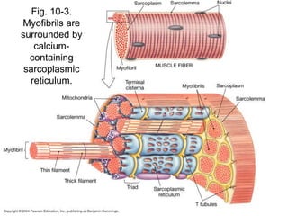
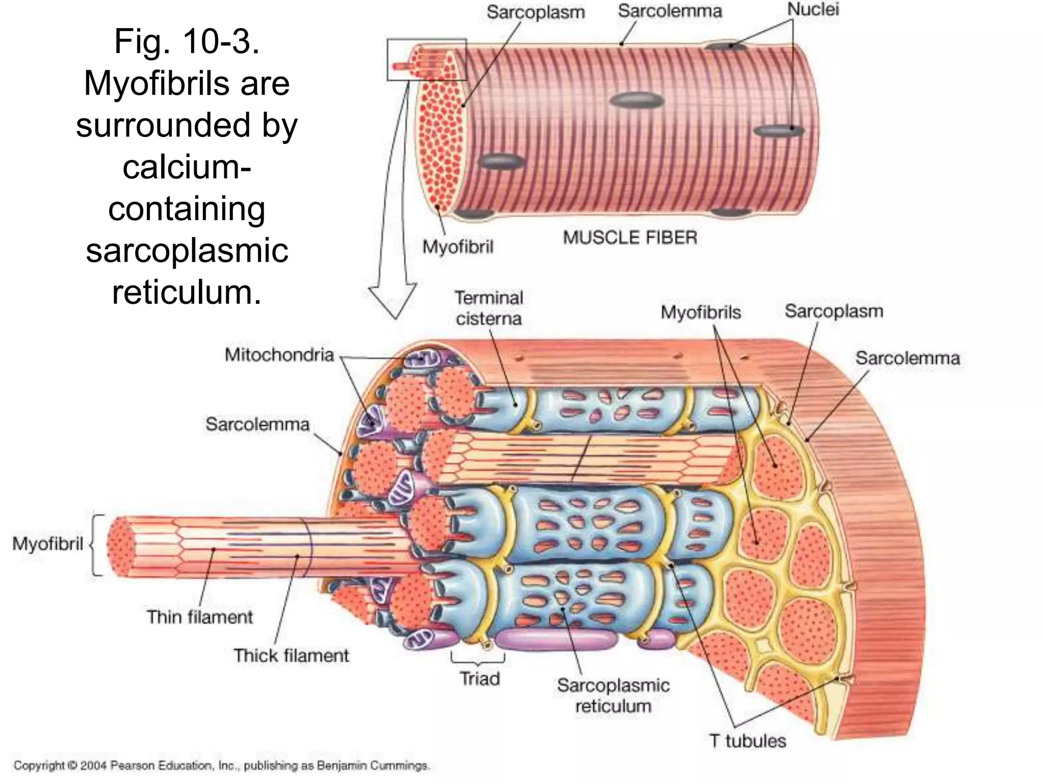
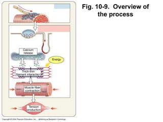
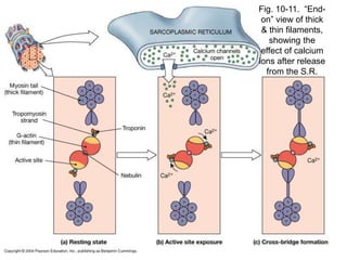
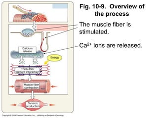
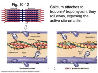
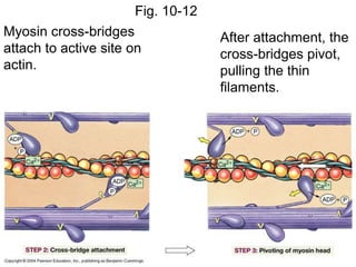
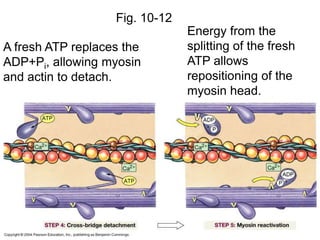
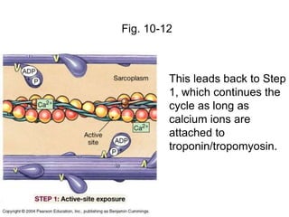
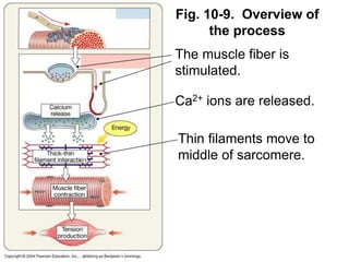
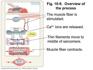
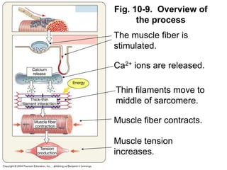
Fig. 10-3.
Myofibrils are
surrounded by
calcium-
containing
sarcoplasmic
reticulum.
Fig. 10-7. Review of thin and
thick filament structure
Fig. 10-9. Overview of
the process
Fig. 10-9. Overview of
the process
The muscle fiber is
stimulated.
Fig. 10-9. Overview of
the process
The muscle fiber is
stimulated.
Ca2+ ions are released.
Fig. 10-11. “End-
on” view of thick
& thin filaments,
showing the
effect of calcium
ions after release
from the S.R.
Fig. 10-9. Overview of
the process
The muscle fiber is
stimulated.
Ca2+ ions are released.
Fig. 10-9. Overview of
the process
The muscle fiber is
stimulated.
Ca2+ ions are released.
Thin filaments move to
middle of sarcomere.
Fig. 10-12
Calcium attaches to
troponin/ tropomyosin; they
roll away, exposing the
active site on actin.
Fig. 10-12
Myosin cross-bridges
attach to active site on
actin.
After attachment, the
cross-bridges pivot,
pulling the thin
filaments.
Fig. 10-12
A fresh ATP replaces the
ADP+Pi, allowing myosin
and actin to detach.
Energy from the
splitting of the fresh
ATP allows
repositioning of the
myosin head.
Fig. 10-12
This leads back to Step
1, which continues the
cycle as long as
calcium ions are
attached to
troponin/tropomyosin.
Fig. 10-9. Overview of
the process
The muscle fiber is
stimulated.
Ca2+ ions are released.
Thin filaments move to
middle of sarcomere.
Fig. 10-9. Overview of
the process
The muscle fiber is
stimulated.
Ca2+ ions are released.
Thin filaments move to
middle of sarcomere.
Muscle fiber contracts.
Fig. 10-9. Overview of
the process
The muscle fiber is
stimulated.
Ca2+ ions are released.
Thin filaments move to
middle of sarcomere.
Muscle fiber contracts.
Muscle tension
increases.

sliding filament.ppt

  • 1.
    Fig. 10-3. Myofibrils are surroundedby calcium- containing sarcoplasmic reticulum.
  • 2.
    Fig. 10-7. Reviewof thin and thick filament structure
  • 3.
    Fig. 10-9. Overviewof the process
  • 4.
    Fig. 10-9. Overviewof the process The muscle fiber is stimulated.
  • 5.
    Fig. 10-9. Overviewof the process The muscle fiber is stimulated. Ca2+ ions are released.
  • 6.
    Fig. 10-11. “End- on”view of thick & thin filaments, showing the effect of calcium ions after release from the S.R.
  • 7.
    Fig. 10-9. Overviewof the process The muscle fiber is stimulated. Ca2+ ions are released.
  • 8.
    Fig. 10-9. Overviewof the process The muscle fiber is stimulated. Ca2+ ions are released. Thin filaments move to middle of sarcomere.
  • 9.
    Fig. 10-12 Calcium attachesto troponin/ tropomyosin; they roll away, exposing the active site on actin.
  • 10.
    Fig. 10-12 Myosin cross-bridges attachto active site on actin. After attachment, the cross-bridges pivot, pulling the thin filaments.
  • 11.
    Fig. 10-12 A freshATP replaces the ADP+Pi, allowing myosin and actin to detach. Energy from the splitting of the fresh ATP allows repositioning of the myosin head.
  • 12.
    Fig. 10-12 This leadsback to Step 1, which continues the cycle as long as calcium ions are attached to troponin/tropomyosin.
  • 13.
    Fig. 10-9. Overviewof the process The muscle fiber is stimulated. Ca2+ ions are released. Thin filaments move to middle of sarcomere.
  • 14.
    Fig. 10-9. Overviewof the process The muscle fiber is stimulated. Ca2+ ions are released. Thin filaments move to middle of sarcomere. Muscle fiber contracts.
  • 15.
    Fig. 10-9. Overviewof the process The muscle fiber is stimulated. Ca2+ ions are released. Thin filaments move to middle of sarcomere. Muscle fiber contracts. Muscle tension increases.