OVERVIEW OF BONES
Bonesare remarkably intricate and dynamic.
They go beyond simply being structural—
constantly adapting, growing, and performing
crucial roles in movement, protection, mineral
homeostasis, and blood formation. Their
structural variety allows for specialized functions
across the body, reflecting a stunning level of
biological design.
3.
FUNCTIONS OF BONES
•Functional Roles of Bones
• Support & Structure: Form the body’s framework and maintain posture.EBSCO
betterhealth.vic.gov.au
• Protection: Shield vital organs—e.g., skull for the brain, ribs for heart and lungs.
EBSCOVerywell Health
• Movement: Work with muscles and joints to enable motion and leverage.
Kenhubbetterhealth.vic.gov.au
• Mineral Storage: Act as reservoirs for essential minerals, notably calcium and
phosphate.EBSCObetterhealth.vic.gov.au
• Hematopoiesis: Red marrow within bones manufactures red and white blood
cells and platelets
STRUCTURES OF BONES
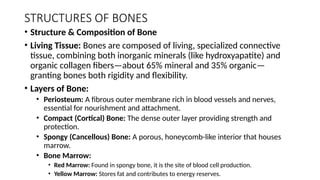
•Structure & Composition of Bone
• Living Tissue: Bones are composed of living, specialized connective
tissue, combining both inorganic minerals (like hydroxyapatite) and
organic collagen fibers—about 65% mineral and 35% organic—
granting bones both rigidity and flexibility.
• Layers of Bone:
• Periosteum: A fibrous outer membrane rich in blood vessels and nerves,
essential for nourishment and attachment.
• Compact (Cortical) Bone: The dense outer layer providing strength and
protection.
• Spongy (Cancellous) Bone: A porous, honeycomb-like interior that houses
marrow.
• Bone Marrow:
• Red Marrow: Found in spongy bone, it is the site of blood cell production.
• Yellow Marrow: Stores fat and contributes to energy reserves.
6.
AXIAL SKELETON
• WhatIs the Axial Skeleton?
-The axial skeleton forms the central core of the human body and comprises
80 bones in adults. It includes the skull, auditory ossicles, hyoid bone,
vertebral column, and the thoracic cage (ribcage).
• Functionally, it:
• Protects vital organs such as the brain, spinal cord, heart, and lungs.
• Serves as the support structure for the body’s axis.
• Acts as an anchoring point for muscles controlling posture, breathing, head
movement, and more.
7.
Breakdown of AxialSkeleton Components
1.Skull (28 Bones)
• Cranial bones (8): Frontal, parietal (2), temporal (2), occipital,
sphenoid, ethmoid—enclose and protect the brain.
• Facial bones (14): Maxillae (2), zygomatic (2), mandible, nasal (2),
palatine (2), inferior nasal concha (2), lacrimal (2), vomer—shape the
face and form entryways for air and food
8.
2.Auditory Ossicles (6Bones)
• Located in the middle ear, these are the smallest bones in the body—
malleus, incus, and stapes (two of each).
• These transmit sound vibrations from the eardrum to the inner ear.
• 3. Hyoid Bone (1 Bone)
• A U-shaped “floating” bone in the neck, supported by muscles and
ligaments.
• Essential for functions like speech, swallowing, and maintaining the
airway.
9.
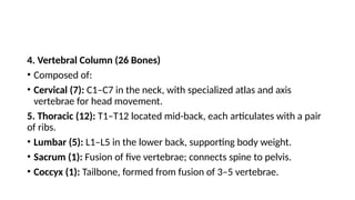
4. Vertebral Column(26 Bones)
• Composed of:
• Cervical (7): C1–C7 in the neck, with specialized atlas and axis
vertebrae for head movement.
5. Thoracic (12): T1–T12 located mid-back, each articulates with a pair
of ribs.
• Lumbar (5): L1–L5 in the lower back, supporting body weight.
• Sacrum (1): Fusion of five vertebrae; connects spine to pelvis.
• Coccyx (1): Tailbone, formed from fusion of 3–5 vertebrae.
10.
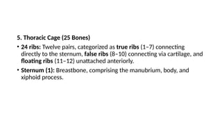
5. Thoracic Cage(25 Bones)
• 24 ribs: Twelve pairs, categorized as true ribs (1–7) connecting
directly to the sternum, false ribs (8–10) connecting via cartilage, and
floating ribs (11–12) unattached anteriorly.
• Sternum (1): Breastbone, comprising the manubrium, body, and
xiphoid process.
11.
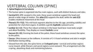
VERTEBRAL COLUMN (SPINE)
1.Spinal Regions & Curvatures
• The human spine is organized into five main regions, each with distinct features and roles:
• Cervical (C1–C7): Located in the neck, these seven vertebrae support the head and
permit a wide range of motion. The atlas (C1) supports the skull, while the axis (C2)
enables rotational movement of the head
• Thoracic (T1–T12): This mid-back segment attaches to the rib cage, providing stability and
protection for vital chest organs. It exhibits a gentle kyphotic outward curve
• Lumbar (L1–L5): These five robust vertebrae in the lower back bear most body weight
and allow flexibility. They display an inward lordotic curve
• Sacrum (S1–S5): Forming the back of the pelvis, these fused vertebrae connect the spine
to the pelvis.
• Coccyx: Often known as the tailbone, it consists of 3–5 fused vertebrae and aids in weight
distribution during sitting.
• Viewed from the side, the spine forms an S-shaped curve—cervical and lumbar regions
curve inward, while thoracic and sacral regions curve outward. This arrangement acts like
a spring, absorbing shock and maintaining balance.
12.
2. Anatomy ofa Vertebra & Supporting
Structures
Each vertebra has a consistent internal structure with variations by
region:
• Vertebral Body: A cylindrical, weight-bearing front section, increasing
in size from cervical to lumbar to better support weight.
• Vertebral Arch: Located at the back, it encloses the vertebral
foramen, collectively forming the spinal canal to protect the spinal
cord
13.
Processes for Attachment:
•Spinous Process: Projects backward; can be felt through the skin and
provides attachment points for muscles and ligaments.
• Transverse Processes: Extend laterally for muscle and ligament
attachment.
• Articular Facets: Paired surfaces (superior and inferior) that form
facet joints, facilitating limited and controlled movement between
neighboring vertebrae
14.
Intervertebral Discs: Locatedbetween vertebral bodies, these include
a tough outer annulus fibrosus and a gel-like inner nucleus pulposus,
acting as shock absorbers and allowing flexibility
Intervertebral Foramina: Small openings between vertebrae through
which spinal nerve roots exit the spinal canal
15.
3. Spinal Cord& Nervous Integration
•The spinal cord, housed inside the vertebral canal, extends from the brainstem to the
lower back and terminates at the conus medullaris, near L1–L2, before continuing as
the cauda equina, a bundle of nerve roots
•About 31 pairs of spinal nerves branch out from the cord through the intervertebral
foramina:
•8 cervical
•12 thoracic
•5 lumbar
•5 sacral
•1 coccygeal
•These nerves transmit sensory input to the brain and motor commands to muscles,
and additional reflexes. The meninges (dura, arachnoid, and pia mater), along with
cerebrospinal fluid, ensure protection and cushioning
16.
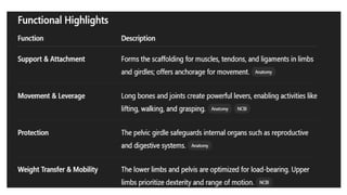
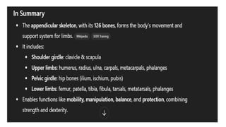
APPENDICULAR SKELETON
• Theappendicular skeleton is one of the two primary divisions of the human skeletal
system (alongside the axial skeleton). It encompasses all bones that support the limbs
and the girdles that connect them to the body's core. There are 126 bones in the
appendicular skeleton, accounting for more than half of the human skeleton’s 206
bones.
Major Regions and Their Key Bones
• 1. Pectoral (Shoulder) Girdle
• Clavicle (2): Acts as a strut linking the upper limb to the axial skeleton at the sternum.
• Scapula (2): The shoulder blade; provides wide attachment for muscles and serves as
part of the shoulder joint with the humerus.
17.
• 2. UpperLimbs (per side) – 30 bones in total per arm:
• Humerus (arm)
• Radius (thumb side of forearm)
• Ulna (pinky side of forearm)
• Carpals (8 wrist bones)
• Metacarpals (5 palm bones)
• Phalanges (14 finger/toe bones per hand; 3 per finger, 2 for thumb)
18.
3. Pelvic Girdle
•Hip bones (2): Each is a fusion of the ilium, ischium, and pubis—forming
a sturdy attachment for lower limbs and protecting pelvic organs.
4. Lower Limbs (per side) – 30 bones in total per leg:
• Femur (thigh; longest, strongest bone)
• Patella (kneecap)
• Tibia (shinbone; weight-bearing)
• Fibula (slim lateral bone)
• Tarsals (7 ankle bones)
• Metatarsals (5 bones forming the foot arch)
• Phalanges (14 bones per foot, arranged similar to fingers)
21.
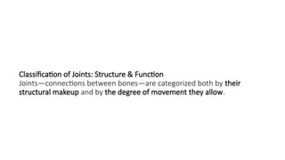
Classification of Joints:Structure & Function
Joints—connections between bones—are categorized both by their
structural makeup and by the degree of movement they allow.
22.
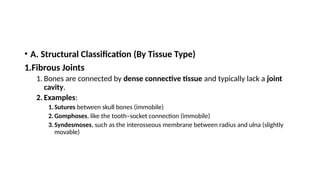
• A. StructuralClassification (By Tissue Type)
1.Fibrous Joints
1. Bones are connected by dense connective tissue and typically lack a joint
cavity.
2. Examples:
1.Sutures between skull bones (immobile)
2.Gomphoses, like the tooth–socket connection (immobile)
3.Syndesmoses, such as the interosseous membrane between radius and ulna (slightly
movable)
23.
• Cartilaginous Joints
•Bones are united by cartilage. There are two subtypes:
• Synchondroses — joined by hyaline cartilage;
generally immobile (e.g., epiphyseal growth plates,
sternum joints)
• Symphyses — joined by fibrocartilage; slightly
movable (e.g., intervertebral discs, pubic symphysis)
24.
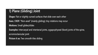
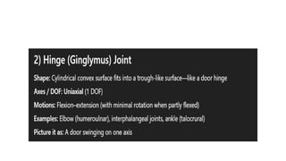
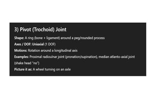
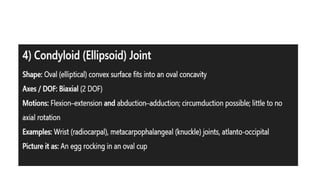
3.Synovial Joints
•Characterized bya joint cavity filled with
synovial fluid, a fibrous capsule, and articular
cartilage covering the bone surfaces
•These are the most common and most mobile
joints in the body
25.
• B. FunctionalClassification (By Mobility)
• Synarthrosis — immovable joint, offering maximum stability (e.g.,
sutures, gomphoses, synchondroses)
• Amphiarthrosis — slightly movable joint, providing limited flexibility
(e.g., intervertebral joints, pubic symphysis)
• Diarthrosis — freely movable joint; all synovial joints fall under this
category