A 32-year-old male patient presented with a single grand mal seizure. Imaging showed extradural masses in the left parietal and frontal regions. Biopsy revealed non-Hodgkin B-cell lymphoma. The CNS manifestations were initially diagnosed as secondary CNS lymphoma. The patient was referred for oncology treatment. The document discusses epidural lymphomatous deposits and provides information on accessing additional case publications and archives on the author's website.
This document discusses a case of subarachnoid hemorrhage and bilateral anterior cerebral artery infarctions caused by a ruptured anterior communicating artery aneurysm in a 40-year-old male patient found unconscious. It provides radiological images showing the location of the aneurysm and resulting hemorrhage. The diagnosis is given as a ruptured anterior communicating artery aneurysm. The discussion section covers complications of cerebral aneurysms including hematoma formation and patterns associated with different aneurysm locations. It describes how aneurysm ruptures can lead to subarachnoid or intracerebral hemorrhage and secondary effects like vasospasm and infarction.
1) Brainmapping provides a quantitative analysis of EEG data by measuring frequency bands and topographic distribution, allowing objective characterization beyond visual analysis.
2) Studies have found focal increases in beta band energy can localize epileptic foci, while decreased alpha/theta with increased beta may indicate surrounding gliosis.
3) Brainmapping uncovered covert epilepsy and subclinical foci, with patterns of diminished and augmented activity suggesting regions of atrophy surrounded by epileptogenic cortex.
Research section...The role of MR imaging in the management of parkinson diseaseProfessor Yasser Metwally
The document discusses a study of 20 patients with idiopathic Parkinson's disease who underwent MRI brain scans and clinical evaluations. Patients were divided into two groups: Group A had a short duration of symptoms and good response to levodopa treatment, while Group B had a longer duration and fluctuating levodopa response. MRI showed changes in the substantia nigra and red nucleus in both groups. Additionally, Group B showed signal changes in the putamen, suggesting more advanced disease involving both nigral and striatal structures.
This document discusses neurological complications that can occur during hematopoietic cell transplantation (HCT). It describes complications that occur during different stages of HCT: 1) During conditioning regimens, complications include encephalopathy, seizures, and cerebral infarction caused by chemotherapy drugs or medical procedures. 2) During bone marrow depletion, complications include encephalopathy, seizures, cerebral infarctions, and hemorrhages due to metabolic issues, drugs, or infections. 3) During chronic immunosuppression in allogeneic HCT, complications include infections by viruses and opportunistic organisms. 4) Late complications include relapse of the original disease, neurological graft-versus-host disease, and second neoplasms.
A 66-year-old male patient presented with non-specific lower back pain. MRI images showed signs of lumbar spondylosis including disc degeneration and bulging in the lower lumbar spine. The document provides figures showing disc herniation and details on accessing additional materials on the author's website, including updates versions of the case publication.
Issues in brainmapping...EEG and brainmap spectral profiles in cortical lesio...Professor Yasser Metwally
Issues in brainmapping...EEG and brainmap spectral profiles in cortical lesions, subcortical white matter lesions and subcortical gray matter (diencephalic) lesions
A 40-year-old patient presented with relapsing-remitting spinal multiple sclerosis. MRI revealed asymptomatic brain involvement and recent progression of myelopathy. The document discusses the diagnosis of spinal MS and differentiating it from transverse myelitis using MRI characteristics such as lesion location and contrast enhancement. Figures show examples of MS plaques and diffuse abnormalities on MRI of the spinal cord.
This document summarizes the pathology of pituitary tumors, including:
1) Pituitary adenomas are benign neoplasms originating from pituitary cells and are the most common tumors of the sella region.
2) Pituitary tumors are currently classified based on their hormonal content and ultrastructural morphology, with 14 recognized subtypes.
3) Growth hormone-secreting adenomas are associated with acromegaly or gigantism and can have various histologic appearances, most commonly densely or sparsely granulated somatotroph adenomas.
This document is a case publication from January 2008 edited by Professor Yasser Metwally about acute disseminated encephalomyelitis (ADEM). It describes an 18-year-old female patient who presented with disturbed consciousness, seizures, optic neuritis and meningism. MRI images show lesions in the brain consistent with ADEM. The publication discusses differentiating ADEM from other conditions like multiple sclerosis, infections and metabolic disorders based on clinical features and test results. It provides guidance on the diagnosis and management of ADEM cases.
This document is a short case publication about a 60-year-old male patient presenting with symptoms of cerebellopontine angle syndrome affecting the 7th, 5th, and bulbar cranial nerves, as well as cerebellar deficits and right ear tinnitus. Imaging revealed a fusiform aneurysm of the vertebrobasilar system with mural thrombosis. Fusiform aneurysms commonly involve the vertebrobasilar system, causing arteries to be diffusely dilated, tortuous, and prolonged with frequent mural thrombosis. They rarely rupture but can cause ischemic manifestations or pressure effects from mass effect. The publication is periodically updated on the author's website.
A 40-year-old female presented with headache, seizures, and altered mental status. MRI showed widespread cerebral venous thrombosis resulting in venous hypertension and congestion. This led to dilated veins and edema in the brain, seen as hyperintensities on T2-weighted MRI. The document discusses the pathophysiology of cerebral venous thrombosis, noting that increased venous pressure can cause edema, hemorrhage, and potentially infarction if pressure exceeds arterial pressure long enough. It describes three stages: initial sinus changes only, early encephalopathy with reversible edema on MRI, and later irreversible infarction if not resolved.
This document summarizes different types of generalized epilepsies based on their EEG patterns and clinical presentations. It discusses primary generalized epilepsies, which have no identifiable cause and normal brain imaging. It also discusses more serious secondary generalized epilepsies, which are caused by diffuse brain injury and often associated with developmental delays. Specific syndromes like Lennox-Gastaut syndrome and West syndrome are examined in detail based on their characteristic EEG patterns including slow spike wave discharges, hypsarrhythmia, and independent multifocal spike discharges.
This document provides an overview of psychiatry disorders in pregnancy. It discusses the prevalence and course of several disorders including anxiety disorders like panic disorder and PTSD, mood disorders like major depression and bipolar disorder, schizophrenia and psychosis, and eating disorders. For many disorders, data on incidence and course during pregnancy is limited. The document highlights that pregnancy does not necessarily improve psychiatric conditions. It also notes potential risks to pregnancy outcomes from conditions like depression, including preterm birth and low birth weight. The importance of understanding psychiatric illnesses and treatments during pregnancy is emphasized to guide clinical decision making.
A 40-year-old female patient presented with a history of migraines. MRI scans revealed two meningioma tumors, one in the parasellar region and one in the cerebellopontine angle. The tumors appeared hyperintense on T2-weighted MRI and showed dense contrast enhancement. Based on the MRI characteristics and location of the tumors, they were diagnosed as multiple syncytial meningiomas. Syncytial meningiomas tend to appear hyperintense on T2-weighted MRI due to high cell count and microcysts.
This document summarizes the classification and treatment of headaches. Headaches are classified as either primary/idiopathic or secondary/symptomatic. Primary headaches include tension headaches, migraines, and cluster headaches and have no underlying cause, while secondary headaches are a symptom of another condition like hypertension or meningitis. The document outlines diagnostic criteria and treatment approaches for different headache types, including acute treatment with medications and preventative treatment.
A 30-year-old male presented with bilateral pyramidal signs and pseudobulbar palsy. MRI showed hyperintense lesions in the crus cerebri and basis pontis, involving the corticospinal tract, more on the left side. This represents demyelination and edema along the corticospinal tract fibers, mainly located in the midbrain/thalamus with extension to the posterior limb of the internal capsule, basis pontis, and medulla. The diagnosis is neuro-Behcet disease.
This case report describes subdural empyema in a 26-year-old male patient presenting with seizures, fever, headache, and nausea. MRI scans showed multiple oval-shaped cystic lesions located between the dura mater and arachnoid mater along the falx cerebri and tentorium cerebelli, consistent with subdural empyema. Subdural empyema is a potentially life-threatening infection most often caused by bacterial spread from sinusitis or mastoiditis. It is important to promptly diagnose and treat subdural empyema surgically in addition to antibiotics due to its rapid spread and high mortality rate if left untreated.
The document describes a case of acute disseminated encephalomyelitis (ADEM) in an 18-year-old female patient. She presented with disturbed consciousness, seizures, optic neuritis, and signs of meningismus. MRI showed multifocal white matter lesions characteristic of ADEM. She was diagnosed with ADEM and recovered fully after two weeks of hospitalization and treatment. ADEM is an immune-mediated inflammatory demyelinating disorder of the CNS that typically follows infections or vaccinations and presents with polysymptomatic neurological dysfunction.
A 16-year-old female patient presented with diminished vision, back pain, seizures, and poor school performance. Examination revealed optic nerve damage, cafe au lait spots on the skin, and skin growths. She was diagnosed with neurofibromatosis type 1 based on these findings. The document describes this genetic disorder and includes figures showing abnormalities in the patient's optic nerves, spine, and brain white matter seen on imaging. It also provides information on typical MRI findings in neurofibromatosis type 1 patients and references for further reading.
- A 15-year-old female developed left-sided optic neuritis followed by transverse myelitis months later. MRI showed periventricular white matter changes atypical for multiple sclerosis.
- The patient was diagnosed with neuromyelitis optica (Devic's disease), a disorder characterized by inflammation of the optic nerve and spinal cord. It is identified by longitudinally extensive spinal cord lesions on MRI and seropositivity for neuromyelitis optica immunoglobulin G.
- Acute transverse myelitis is an inflammatory disorder of the spinal cord that can have various causes, including infections, autoimmune disorders, and neuromyelitis optica. It is characterized clinically by motor,
The document discusses the benefits of exercise for mental health. It states that regular physical activity can help reduce anxiety and depression and improve mood and cognitive function. Exercise causes chemical changes in the brain that may help alleviate symptoms of mental illness.
The document discusses the benefits of exercise for mental health. Regular physical activity can help reduce anxiety and depression and improve mood and cognitive functioning. Exercise causes chemical changes in the brain that may help protect against mental illness and improve symptoms.
The document discusses the benefits of exercise for mental health. Regular physical activity can help reduce anxiety and depression and improve mood and cognitive function. Exercise causes chemical changes in the brain that may help protect against mental illness and improve symptoms.
The document discusses the benefits of exercise for mental health. Regular physical activity can help reduce anxiety and depression and improve mood and cognitive function. Exercise causes chemical changes in the brain that may help protect against mental illness and improve symptoms for those who already suffer from conditions like depression and anxiety.
The document discusses the benefits of exercise for mental health. Regular physical activity can help reduce anxiety and depression and improve mood and cognitive function. Exercise causes chemical changes in the brain that may help protect against mental illness and improve symptoms.
This document discusses the radiological pathology of seizure disorders. It describes various developmental anomalies, neoplasms, infections, immune-mediated disorders, cerebrovascular diseases, and trauma that can cause seizures. Specific conditions mentioned include cortical dysplasia, tuberous sclerosis, Sturge-Weber syndrome, neuronal migration disorders, vascular malformations, infections, and immune-mediated Rasmussen's encephalitis. The document provides detailed descriptions of the histopathological findings and MRI/CT appearances of different lesions that can underlie seizure disorders.
This document discusses cerebral haemorrhage (ICH), which accounts for 10-15% of strokes. ICH can result from several mechanisms, including hypertension (47-66% of cases), cerebral amyloid angiopathy (CAA), and vascular malformations. CAA typically affects the elderly and causes lobar ICH that is often recurrent or involves multiple simultaneous haemorrhages. Vascular malformations like arteriovenous malformations (AVMs) and cavernous angiomas are a common cause of ICH in young, non-hypertensive patients. Imaging techniques like CT and MRI can identify vascular malformations and help determine the underlying cause of ICH.
Cerebral amyloid angiopathy (CAA) refers to the deposition of β-amyloid in the arteries of the cerebral cortex. It is commonly seen in Alzheimer's disease but can also occur in healthy elderly individuals. CAA can cause intracerebral hemorrhage, dementia, or transient neurological symptoms. The deposition damages blood vessels and increases the risk of hemorrhage. Imaging such as CT scans can detect hemorrhages characteristic of CAA, which are often lobar and cortical. Genetic factors like the ApoE genotype can influence the severity and presentation of CAA.
Cerebral microbleeds are small brain hemorrhages detected by MRI that are caused by leakage of blood from damaged small vessel walls. They are increasingly recognized in patients with cerebrovascular disease, Alzheimer's disease, vascular cognitive impairment, and normal elderly populations. Microbleeds in lobar regions may indicate cerebral amyloid angiopathy and link vascular and amyloid neuropathologies, while deep or infratentorial microbleeds often reflect hypertensive vasculopathy. Detection of microbleeds provides insight into cerebral small vessel disease and its relationship to cognitive impairment and dementia.
The document discusses the benefits of exercise for mental health. Regular physical activity can help reduce anxiety and depression and improve mood and cognitive functioning. Exercise causes chemical changes in the brain that may help protect against mental illness and improve symptoms.
A 57-year-old male patient presented with left lower limb weakness that had progressed over three months. MRI images showed bilateral, symmetrical lesions in the posterior parieto-occipital white matter, which had scalloped margins and did not enhance or cause mass effect. Based on the clinical presentation and MRI findings, the patient was diagnosed with progressive multifocal leukoencephalopathy (PML), a demyelinating disease caused by JC virus reactivation that predominantly affects immunocompromised individuals. PML lesions are typically multifocal and located in the white matter of the brain, most often in the parieto-occipital region.
Issues in radiological pathology: Radiological pathology of watershed infarct...Professor Yasser Metwally
The document discusses border zone or watershed infarcts, which occur at the junction between two main arterial territories and constitute approximately 10% of all brain infarcts. There are two types - external (cortical) and internal (subcortical). External infarcts are often embolic in nature while internal infarcts are mainly caused by hemodynamic compromise. Advanced imaging can help identify areas of low perfusion and distinguish the two types. The document then examines the classification, imaging appearance, causal mechanisms, and clinical course of both external and internal border zone infarcts in more detail.
- Video recording of this lecture in English language: https://youtu.be/Pt1nA32sdHQ
- Video recording of this lecture in Arabic language: https://youtu.be/uFdc9F0rlP0
- Link to download the book free: https://nephrotube.blogspot.com/p/nephrotube-nephrology-books.html
- Link to NephroTube website: www.NephroTube.com
- Link to NephroTube social media accounts: https://nephrotube.blogspot.com/p/join-nephrotube-on-social-media.html
share - Lions, tigers, AI and health misinformation, oh my!.pptxTina Purnat
• Pitfalls and pivots needed to use AI effectively in public health
• Evidence-based strategies to address health misinformation effectively
• Building trust with communities online and offline
• Equipping health professionals to address questions, concerns and health misinformation
• Assessing risk and mitigating harm from adverse health narratives in communities, health workforce and health system
Muktapishti is a traditional Ayurvedic preparation made from Shoditha Mukta (Purified Pearl), is believed to help regulate thyroid function and reduce symptoms of hyperthyroidism due to its cooling and balancing properties. Clinical evidence on its efficacy remains limited, necessitating further research to validate its therapeutic benefits.
Local Advanced Lung Cancer: Artificial Intelligence, Synergetics, Complex Sys...Oleg Kshivets
Overall life span (LS) was 1671.7±1721.6 days and cumulative 5YS reached 62.4%, 10 years – 50.4%, 20 years – 44.6%. 94 LCP lived more than 5 years without cancer (LS=2958.6±1723.6 days), 22 – more than 10 years (LS=5571±1841.8 days). 67 LCP died because of LC (LS=471.9±344 days). AT significantly improved 5YS (68% vs. 53.7%) (P=0.028 by log-rank test). Cox modeling displayed that 5YS of LCP significantly depended on: N0-N12, T3-4, blood cell circuit, cell ratio factors (ratio between cancer cells-CC and blood cells subpopulations), LC cell dynamics, recalcification time, heparin tolerance, prothrombin index, protein, AT, procedure type (P=0.000-0.031). Neural networks, genetic algorithm selection and bootstrap simulation revealed relationships between 5YS and N0-12 (rank=1), thrombocytes/CC (rank=2), segmented neutrophils/CC (3), eosinophils/CC (4), erythrocytes/CC (5), healthy cells/CC (6), lymphocytes/CC (7), stick neutrophils/CC (8), leucocytes/CC (9), monocytes/CC (10). Correct prediction of 5YS was 100% by neural networks computing (error=0.000; area under ROC curve=1.0).
These lecture slides, by Dr Sidra Arshad, offer a simplified look into the mechanisms involved in the regulation of respiration:
Learning objectives:
1. Describe the organisation of respiratory center
2. Describe the nervous control of inspiration and respiratory rhythm
3. Describe the functions of the dorsal and respiratory groups of neurons
4. Describe the influences of the Pneumotaxic and Apneustic centers
5. Explain the role of Hering-Breur inflation reflex in regulation of inspiration
6. Explain the role of central chemoreceptors in regulation of respiration
7. Explain the role of peripheral chemoreceptors in regulation of respiration
8. Explain the regulation of respiration during exercise
9. Integrate the respiratory regulatory mechanisms
10. Describe the Cheyne-Stokes breathing
Study Resources:
1. Chapter 42, Guyton and Hall Textbook of Medical Physiology, 14th edition
2. Chapter 36, Ganong’s Review of Medical Physiology, 26th edition
3. Chapter 13, Human Physiology by Lauralee Sherwood, 9th edition
- Video recording of this lecture in English language: https://youtu.be/kqbnxVAZs-0
- Video recording of this lecture in Arabic language: https://youtu.be/SINlygW1Mpc
- Link to download the book free: https://nephrotube.blogspot.com/p/nephrotube-nephrology-books.html
- Link to NephroTube website: www.NephroTube.com
- Link to NephroTube social media accounts: https://nephrotube.blogspot.com/p/join-nephrotube-on-social-media.html
Adhd Medication Shortage Uk - trinexpharmacy.comreignlana06
The UK is currently facing a Adhd Medication Shortage Uk, which has left many patients and their families grappling with uncertainty and frustration. ADHD, or Attention Deficit Hyperactivity Disorder, is a chronic condition that requires consistent medication to manage effectively. This shortage has highlighted the critical role these medications play in the daily lives of those affected by ADHD. Contact : +1 (747) 209 – 3649 E-mail : sales@trinexpharmacy.com
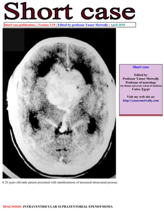
1. Short case publication... Version 3.19 | Edited by professor Yasser Metwally | April 2010
Short case
Edited by
Professor Yasser Metwally
Professor of neurology
Ain Shams university school of medicine
Cairo, Egypt
Visit my web site at:
http://yassermetwally.com
A 26 years old male patient presented with manifestations of increased intracranial pressue.
DIAGNOSIS: INTRAVENTRICULAR SUPRATENTORIAL EPENDYMOMA
2. Figure 1. Precontrast CT scan: Intraventricular ependymoma
filling the lateral and third ventricle, the lesion shows extensive
perivascular calcification. The tumor is a huge one, involving both
lateral ventricles and causing extensivehydrocephalus.
Figure 2. Postcontrast CT scan: Intraventricular ependymoma filling the lateral and third ventricle, the lesion
shows extensive perivascular calcification (Actually perivenous) and is drained by the deep venous system
through the vein of galen. Notice the dense homogeneous enhancement. Hydrocephalic changes are seen with
possible transependymal edema (A). (C is a bone window of B)
3. Figure 3. Angiography shows the extensive tumour hypervascularity that is mainly drained by the deep
venous system through the vein of galen.
Figure 4. The tumor has extended to the third ventricle, ? the interhemispheric fissure, and Probably the
suprasellar and the quadrigeminal cistern. CSF seedling is probably the cause of dissemination through the basal
cisterns and the bihemispheric fissures.
4. Figure 5. Histopathological picture of ependymomas with the
characteristic rosettes (The patient's histopathology)
The patient's tumor was debulked surgically and histopathological examination revealed an ependymoma of the
cellular type with Perivascular pseudorosettes. The patient was given a course of postoperative radiotherapy (Fig.
5). The patient died 7 month following the operation.
References
1. Metwally, MYM: Textbook of neurimaging, A CD-ROM publication, (Metwally, MYM editor) WEB-CD agency for
electronic publishing, version 11.2a April 2010
Addendum
A new version of short case is uploaded in my web site every week (every Saturday and remains available till Friday.)
To download the current version follow the link "http://pdf.yassermetwally.com/short.pdf".
You can download the long case version of this short case during the same week from: http://pdf.yassermetwally.com/case.pdf or visit web
site: http://pdf.yassermetwally.com
To download the software version of the publication (crow.exe) follow the link: http://neurology.yassermetwally.com/crow.zip
At the end of each year, all the publications are compiled on a single CD-ROM, please contact the author to know more
details.
Also to view a list of the previously published case records follow the following link:
(http://wordpress.com/tag/case-record/) or click on it if it appears as a link in your PDF reader
To inspect the patient's full radiological study, click on the attachment icon (the paper clip icon in the left pane) of the acrobat
reader then double click on the attached file
Click here to download the long case version of this short case in PDF format