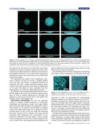
The document summarizes a computational modeling approach for simulating synthetic microbial biofilms at a multiscale level. The approach combines 3D biophysical models of individual cells with models of genetic regulation and intercellular signaling. It was implemented in a software tool called CellModeller that uses parallel GPU computing to simulate over 30,000 cells in a typical biofilm colony within 30 minutes. Simulation results reproduced key features of experimentally observed E. coli biofilm colony morphologies. The modeling framework provides a way to predict the behavior of synthetic biofilms prior to experimental construction.
![ACS Synthetic Biology Research Article
du ⃗
= T[u ⃗] + f (u ⃗)
dt (6)
where u⃗ is now composed of some species that are within a cell
(and are not transported) and those outside the cell, which are
subject to the operator T. For example, in the case of diffusion
T ≡ K∇2, where K is a diagonal matrix of diffusion coefficients
for each species, and for cell autonomous species the
corresponding element of K is zero.
We discretize this system on a regular 3-dimensional grid for
species in the medium and separate variables representing cell-
autonomous species. Cell positions are interpolated linearly in
the spatial grid, and each cell can see its local signal
concentration (see Table 1). The user writes the function
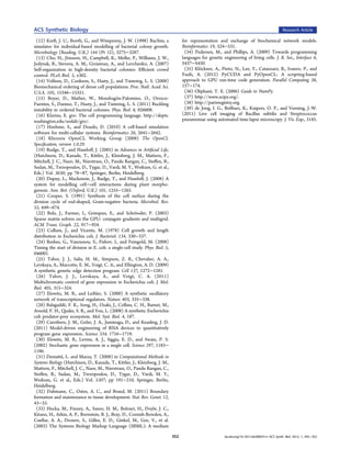
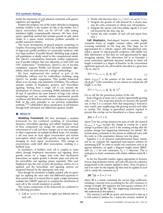
Figure 4. Three snapshots of a simulation of controlled growth, where f(u) for each cell including, for example, importing or exporting
only cells within 10 μm of the rightmost edge of the colony are able to signal and downstream transcriptional regulation. Table 2
grow (yellow). This could be achieved by optical induction of a shows example function definitions for a simple model of the
phytochrome system linked to metabolic control genes. Total
Lux quorum sensing system.
computation time was 28 min. (See Supporting Information for a
video of this simulation.).
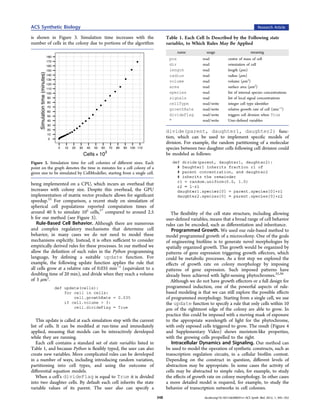
Table 2. Specification of Differential Equations for OpenCL
Solvera
Intracellular dynamics are commonly approximated by
ordinary differential equations, which have been shown to
accurately reproduce experimental observations in a broad
range of synthetic systems. Examples include transcriptional
oscillators,27 quorum-sensing systems with predator−prey
interactions,28 and RNA-regulated genetic devices.29 Despite
this success, the limitations of ordinary differential equations
are well-documented, particularly with regards to variability in
gene expression.30 While variability between cells can be
simulated in CellModeller by introducing randomness at
specific events, such as the partitioning of a molecular species
between daughter cells during cell division, variability at the
level of gene expression would be more accurately modeled
using stochastic simulation methods. Although such methods
are highly computationally intensive, various parallel algorithms
for their implementation have been proposed (see ref 31 for
example). Here we suggest that the stochastic reactions
occurring in each cell could be simulated in parallel, rather
than realizing a single stochastic trajectory. We leave the
implementation of this proposal for future work.
For a given synthetic construct, we represent the processes
that require detailed modeling as a system of differential
equations, with each equation describing the rate of change of a
species. In general, and most commonly for genetic circuits, this
system is nonlinear, where the rate of change of the vector of
species u⃗ is of the form a
Internal species levels and local signal concentrations, as well as cell
surface area and volume, are available to use in the expressions. Here,
du ⃗ in sigRateCL internal AHL is exported into the medium via the
= f (u ⃗ )
dt (5) membrane (hence surface area term), and concentrations must be
scaled for the change in volume. In specRateCL, LuxI production is
Our approach is to solve this general case, with the function f
induced by AHL, and internal AHL is synthesized from LuxI. Function
specified by the user. This is computationally intensive, and we definitions are returned as strings, which are then compiled at run-time
use the OpenCL parallel programming language to compute into OpenCL.
f(ui) for each cell i in parallel. The user must specify simple
OpenCL code to define the rate of change of each species.
Cell signaling is a key part of multicellular organization. In We solve the resulting system of nonlinear partial differential
the biofilm mode of growth, cells communicate via quorum equations using a modified Crank−Nicholson method (see
sensing ligands that diffuse through the biofilm and medium, Supporting Information). Our method solves the update step
after being secreted from the cell. Depending on the numerically, meaning we can define any linear transport
environment in which the biofilm is growing, there may also operator in a modular fashion, such as adding an advection
be other transport processes, such as bulk flow or advection. or bulk flow term in direction n ⃗̂:
We include such signaling through the medium in our
algorithm with a general linear transport operator T: T ≡ K∇2 + Cn ⃗̂·∇ (7)
349 dx.doi.org/10.1021/sb300031n | ACS Synth. Biol. 2012, 1, 345−352](https://image.slidesharecdn.com/rudge2012-13526499777294-phpapp01-121111101157-phpapp01/85/Rudge2012-5-320.jpg)