Reverse shoulder arthroplasty using an implant with a lateral center of rotation
•
2 likes•429 views

Reverse shoulder arthroplasty (RSA) using an implant with a lateral center of rotation was studied to evaluate outcomes, complications, and the influence of surgeon experience. At minimum 2-year follow-up of the first 60 RSAs performed by a single surgeon: - Mean improvements in range of motion and shoulder function scores were significant and comparable to results reported by the implant's design team. - Experience influenced results, with better outcomes in the later cases compared to the initial 15 cases. - Complications including revision surgeries were more common in the early cases, indicating a potential learning curve for the procedure.

Recommended
More Related Content
What's hot
What's hot (19)
Viewers also liked
Viewers also liked (19)
Similar to Reverse shoulder arthroplasty using an implant with a lateral center of rotation
Similar to Reverse shoulder arthroplasty using an implant with a lateral center of rotation (20)
More from Satoshi Kajiyama
More from Satoshi Kajiyama (20)
Reverse shoulder arthroplasty using an implant with a lateral center of rotation
- 1. An Original Study E194 The American Journal of Orthopedics® September 2014 www.amjorthopedics.com Reverse Shoulder Arthroplasty Using an Implant With a Lateral Center of Rotation: Outcomes, Complications, and the Influence of Experience Samer S. Hasan, MD, PhD, Matthew P. Gordon, MD, Jason A. Ramsey, MD, and Martin S. Levy, PhD R everse shoulder arthroplasty (RSA) has revolutionized the treatment of patients with an irreparable rotator cuff tear with glenohumeral arthritis or pseudoparaly- sis; patients with failed rotator cuff surgery with anterosupe- rior escape or glenohumeral instability; and patients with failed shoulder arthroplasty with gross rotator cuff insufficiency.1-10 Multiple RSA systems, each with its own features, including location of center of rotation (COR), are available. The original Grammont design used a glenosphere with medial COR to con- fer protection against baseplate failure,11-13 but this design has been associated with increased risk of scapular notching.10,14-16 The RSP (DjO Global, Inc., Austin, Texas), a reverse shoul- der prosthesis, is characterized by lateral COR5 that improves the deltoid moment arm and tensions the remaining rotator cuff. Multiple studies by its design team have found excellent short- and intermediate-term outcomes and low revision rates in various patient populations.5,7-9,17,18 However, only limited published data on the clinical performance of the RSP have been obtained independent of its design team. We conducted a study of the clinical and self-assessed out- comes, complications, and influence of surgeon learning after the first 60 consecutive RSAs implanted by a single surgeon us- ing the RSP. Our principal hypothesis was that this RSA would significantly improve clinical and self-assessed outcomes and that results would be comparable to those previously reported by its design team. Our secondary hypothesis was that there would be a learning curve for this RSA, with outcomes im- proving with experience. Materials and Methods As this was a retrospective study, we did not submit our study protocol to an outside institutional review board for approval. The protocol was reviewed and approved by the review com- mittee of the Cincinnati Sports Medicine Research and Educa- tion Foundation. We retrospectively reviewed the initial 60 RSAs (57 patients) Abstract Authors’ Disclosure Statement: Dr. Hasan wishes to report that he is a consultant to and receives fellowship funding from DjO Global, Inc., and Cincinnati Sports Medicine Research and Education Foundation receives fellowship funding from DjO Global, Inc. The other authors report no actual or potential conflict of interest in relation to this article. Reverse shoulder arthroplasty (RSA) has revolutionized treatment of arthritis and rotator cuff insufficiency and is performed using implants with either a medial or a lateral center of rotation. We conducted a study of the outcomes and the effect of surgeon learning after the first 60 consecutive lateral- center-of-rotation RSAs implanted by a single surgeon unaffiliated with the design team for this particular reverse shoulder prosthesis. At minimum 2-year follow- up, mean improvements in active forward elevation, abduction, and external rotation were 69°, 55°, and 23°, respectively; mean active internal rotation improved significantly as well (P .001 for all). Mean Simple Shoul- der Test (SST) scores improved from 1.8 (range, 0-6) to 6.9 (range, 0-12) (P .0001), and mean final American Shoulder and Elbow Surgeons score was 72 (range, 27-100). Final radiographs showed scapular notching in 5 shoulders (11%). Gains in SST scores, active forward elevation, and active abduction were lower for the first 15 cases than for the next 45 cases, and 5 of the 8 reop- erations were performed after the first 15 cases. Overall improvements in active motion and self-as- sessed shoulder function in this series are comparable to those previously reported by the design team. Experience with RSA appears to influence efficacy, but the learning curve may not be as steep as previously reported. AJO DO NOT COPY
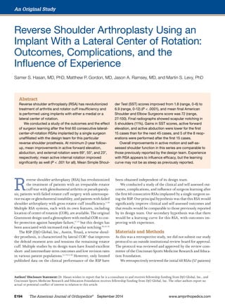
- 2. S. S. Hasan et al www.amjorthopedics.com September 2014 The American Journal of Orthopedics® E195 performed by Dr. Hasan between June 2004 and May 2010. The RSP was used for all RSAs. Mean age at RSA was 75 years (range, 54-92 years), and 44 patients (47 shoulders) were women. Forty-two primary and 18 revision RSAs were per- formed. Revisions were typically performed because of insta- bility or pseudoparalysis with or without glenoid or humeral bone deficiency. Indications for primary and revision RSAs are summarized in Table I. Before surgery, all patients underwent clinical examina- tion, which included measurement of active range of motion (ROM): forward elevation (aFE), abduction (aAB), external ro- tation at the side (aER), and internal rotation to the back (aIR). Standardized Grashey and axillary-lateral radiographs were also obtained. In addition, the 12-item Simple Shoulder Test (SST)19 was used to evaluate self-assessed shoulder function. All RSAs were performed using a deltopectoral approach, incorporating previous incisions whenever practical. The proximal humerus and glenoid were prepared sequentially to accommodate the RSP components, with attention given to inferior glenoid baseplate position and tilt.20-22 All glenoid base- plates were inserted with 3 or 4 locking screws for adjunctive baseplate fixation, and all humeral stems were inserted with antibiotics-impregnated cement. The subscapularis remnant was repaired to the lesser tuberosity using braided sutures through transosseous drill holes, and all shoulders were drained for 24 to 48 hours. Ten- don transfers were not performed in any shoul- der. Revision surgeries were performed using the same principles, but incisions were extended as needed. At time of revision surgery, 1 shoulder with an uncontained glenoid defect required su- perior glenoid augmentation using bulk allograft, and 3 shoulders with proximal humeral bone loss required reconstruction using bulk proximal hu- meral allograft prepared with a step cut23 and af- fixed to the host bone using suture cerclage. After surgery, all patients were admitted for intravenous antibiotics, pain control, and medical management. Then they were discharged home or to an inpatient facility with instructions to immo- bilize the arm in a padded soft brace for 3 weeks and to refrain from weight-bearing for 8 weeks. Supervised physical therapy was offered to select patients at Dr. Hasan’s discretion, but most patients performed patient-directed ex- ercises and then gradually returned to normal activities 8 to 12 weeks after surgery. At most recent follow-up, 10 patients (11 shoulders) were deceased, and 2 patients (2 shoulders) with follow-up of less than 2 years could not be located and were excluded. However, 4 of the 11 shoulders in deceased patients had the minimum 2-year follow-up, and therefore their data were included in the analysis, for a total of 51 shoulders. Self-assessed outcome scores—SST scores and American Shoulder and Elbow Surgeons (ASES) scores—were available for the 51 shoulders at minimum 2-year follow-up (mean, 36 months; range, 23-74 months). Radiographs and shoul- der ROM data were available for 45 shoulders at minimum 2-year follow-up (mean, 33 months; range, 23-69 months). Complications and reoperations for the 60 RSAs included those of patients who subsequently died or who were lost to follow-up. Outcomes and complications for the first 15 shoul- ders (initial group) were compared with those for the next 45 shoulders (second group). Radiographs were evaluated by Dr. Hasan for dislocation, component dissociation or failure, scapular and humeral stress fractures, and scapular notching, which was evaluated us- ing the method of Nerot,16,24 as illustrated in the Figure. Dependent variables included preopera- tive and final aFE, aAB, aER, and aIR; pre- operative and final SST scores; final ASES score; and change in aFE, aAB, aER, aIR, and SST scores. Initial and final outcome measures were compared using the paired t test. Analysis of variance (ANOVA) was per- formed using the main effects of sex, experi- ence (initial vs second group), and surgery (primary vs revision), followed by paired t tests as appropriate. Significance levels for Table I. Reverse Shoulder Arthroplasty (RSA) Indications RSA Indication n Primary Cuff tear arthropathy 36 Failed rotator cuff repair with pseudoparalysis 3 Osteoarthritis with large rotator cuff tear 1 Rheumatoid arthritis with irreparable rotator cuff tear 1 Resection arthroplasty 1 Revision Failed hemiarthroplasty for cuff tear arthropathy 7 Failed hemiarthroplasty for fracture 6 Failed total shoulder arthroplasty 3 Failed humeral head resurfacing with chronic rotator cuff tear 2 Figure. Grashey (true anteroposterior) radiographs show RSP (DjO Global, Inc., Austin, Texas) implants (A) with and (B) without scapular notching. A B AJO DO NOT COPY
- 3. Reverse Shoulder Arthroplasty Using an Implant With a Lateral Center of Rotation E196 The American Journal of Orthopedics® September 2014 www.amjorthopedics.com the comparisons were computed from the ANOVA. Complication and reoperation rates for the groups (initial, second) were compared using the Fisher exact test. All statistical analysis was done at the P .05 significance level using SAS software (SAS Institute, Cary, North Carolina). Results Mean (SD) preoperative aFE was 43° (34°), aAB was 42° (28°), aER was 9° (14°), and aIR was to the buttock. By most recent follow-up, mean (SD) aFE had improved to 112° (31°) (P .0001), aAB to 98° (26°) (P .0001), aER to 29° (21°) (P .0001), and aIR to the L3 spinous process (P .001). Mean number of SST yes re- sponses improved from 1.8 (range, 0-6) to 6.9 (range, 0-12) (P .0001). Mean final ASES score was 72 (range, 27-100). Compared with the women, the men demonstrated significantly more final mean (SD) aFE, 128° (21°) versus 106° (33°) (P .05), and aAB, 116° (20°) ver- sus 92° (25°) (P .01). Mean (SD) SST scores were higher for the men as well, but the differences only approached statisti- cal significance with the numbers avail- able: 8.7 (2.8) versus 6.4 (3.5) (P = .08). Patients who had revision RSA had com- parable outcomes but larger gains in ac- tive ROM than patients who had primary RSA: change in mean (SD) aFE, 95° (27°) versus 56° (45°) (P .01); change in mean (SD) aAB, 73° (31°) versus 47° (34°) (P .05). Mean (SD) ASES scores for pa- tients who had revision RSA and primary RSA were 64 (24) and 76 (20), respectively (P = .08). Table II summarizes the ROM and outcome scores by sex and by status (primary, revision). Mean preoperative aFE and aAB were much lower in the second group (35.5°, 33.5°) than in the initial group (66.2°, 66.7°), and, though the groups’ mean postoperative aFE and aAB were similar, the second group showed sub- stantially more improvement (change in aFE, 77° vs 34°, P = .08; change in aAB, 62° vs 21°, P .05) (Table III). Larger gains in SST scores were also found for the second group, but these were not statistically significant because of the effects of the other confounders. Sixteen complications (14 patients, 24.6%) were identified (Table IV). Six patients (11%) underwent 8 reoperations, in- cluding 4 closed reductions for dislocation (3 patients), 2 open revisions for instability and a dissociated liner (1 patient), 1 evacuation of a hematoma, and 1 fixation of a symptomatic scapular spine nonunion. Five reoperations were performed in the initial group of 15 RSAs, and 3 reoperations (closed reductions) were performed in the second group of 45 RSAs (P .05). Radiographs at minimum 2-year follow-up showed mild scapular notching in 5 (11%) of 45 shoulders, and only 1 additional shoulder demonstrated any scapular notching Table II. Mean Range of Motion, Outcome Scores, and Improvement for All Patients and by Sex and Status (Primary, Revision) Preoperativea Postoperative Improvement Mean SD Mean SD Mean SD Active Forward Elevation, ° Overall 43 34 112 31 69 45 Women 44 32 106 33 63 44 Men 43 41 128 21 85 44 Primary 50 37 108 31 56 45 Revision 29 19 119 33 95 27 Active Abduction, ° Overall 42 28 98 26 55 35 Women 43 29 92 25 49 35 Men 38 29 116 20 75 27 Primary 46 31 96 26 47 34 Revision 33 21 100 27 73 31 Active External Rotation at Side, ° Overall 9 14 29 21 23 22 Women 10 12 28 21 21 21 Men 6 21 32 22 29 25 Primary 10 16 28 20 23 22 Revision 8 11 31 24 24 25 Active Internal Rotation to Back Overall Buttock L3 spinous process — — Women Buttock L3 spinous process — — Men Buttock L3 spinous process — — Primary Buttock L3 spinous process — — Revision Buttock L3 spinous process — — Simple Shoulder Test Score Overall 1.8 1.6 6.9 3.5 5.1 3.4 Women 1.5 1.4 6.4 3.5 4.8 3.6 Men 2.5 2.1 8.7 2.8 6.4 2.4 Primary 2.0 1.7 7.2 3.4 5.0 3.4 Revision 1.2 1.5 6.1 3.6 5.6 3.7 ASES Score Overall — — 72 22 — — Women — — 71 22 — — Men — — 77 22 — — Primary — — 76 20 — — Revision — — 64 24 — — Abbreviation: ASES, American Shoulder and Elbow Surgeons. a N = 51 patients. AJO DO NOT COPY
- 4. S. S. Hasan et al www.amjorthopedics.com September 2014 The American Journal of Orthopedics® E197 on most recent radiographs. To date, no deep infections have been identified, and none of the baseplates or humeral stems have been revised. Discussion Our results confirmed those reported by the design team in other studies,3,5,8,9,17,18,25 which demonstrated a low incidence of scapular notching and improved active ER with the RSP compared with systems having a more medial COR. Frankle and colleagues5 initially reported a mean final ASES score of 68.2, mean FE improvement of 50.1°, and mean ER improve- ment of 29.1°. In a minimum 24-month follow-up study, Cuff and colleagues3 reported an overall complication rate of 6% without baseplate failure or scapular notching. They also re- ported a mean final ASES score of 78, an increase in SST scores from 1.8 to 6.8, and mean aFE and aER gains of 55° and 15°, respectively. Their findings are nearly identical to ours with respect to mean SST scores (improved from 1.8 to 6.9) and mean aFE and aER gains (69°, 23°). In contrast, most studies of Grammont-type implants have not found substantial gains in ER.10,11,14 Boileau and colleagues14 reported a mean aFE gain of 66° but only a mean 4° gain in aER. Simovitch and colleagues26 found a mean 9° improvement in ER among patients with minimal external rotator muscle atrophy but a mean 7° loss among patients with substantial atrophy. More recent reports have documented ER gains with Grammont-type implants and a surgical technique using ei- ther adjunctive modified L’Episcopo transfer of the latissimus dorsi27,28 or a bony increased-offset technique that interposes cancellous autograft between baseplate and glenoid to lateral- ize the prosthetic COR.29 RSP clinical studies independent of the design team are scarce. Levy and Blum30 reported on a single-surgeon experi- ence immediately after fellowship training. Although they identified complications in 10 (25%) of the first 40 consecutive patients who received the RSP, revision surgery was needed in only 2 cases (5%). However, initial and final clinical and self- assessed outcomes were not provided. Clark and colleagues31 Table IV. Complications Complication n Orthopedic Dislocation 4 Dissociation of humeral socket 1 Scapular spine fracture 3 Acromion fracture 1 Humeral stress fracture 1 Postoperative hematoma 1 Deltoid strain 1 Cervical radiculopathy 1 Medical Altered mental status 2 Bowel obstruction 1 Table III. Mean Range of Motion, Outcome Scores, and Improvement for Patients by Group (Initial, Second) Preoperativea Postoperative Improvement Mean SD Mean SD Mean SD Active Forward Elevation, ° Initial group 66 32 106 38 34 45 Second group 36 31 113 30 77 41 Active Abduction, ° Initial group 67 27 99 33 21 29 Second group 34 24 97 24 62 32 Active External Rotation at Side, ° Initial group 10 14 24 25 15 24 Second group 9 15 30 21 22 22 Active Internal Rotation to Back Initial group Buttock L3 spinous process — — Second group Buttock L3 spinous process — — Simple Shoulder Test Score Initial group 1.8 1.8 6.3 3.8 3.4 4.0 Second group 1.8 1.6 7.1 3.4 5.6 3.2 ASES Score Initial group — — 70 21 — — Second group — — 73 22 — — Abbreviation: ASES, American Shoulder and Elbow Surgeons. a N = 51 patients. AJO DO NOT COPY
- 5. Reverse Shoulder Arthroplasty Using an Implant With a Lateral Center of Rotation E198 The American Journal of Orthopedics® September 2014 www.amjorthopedics.com found that subscapularis repair did not influence ROM, dis- location rate, or overall complication rate after RSA using the RSP. At a mean follow-up of about 12 months, active forward flexion was increased 56° in the nonrepair group and 54° in the repair group—improvements comparable to those found in the present study. Enthusiasm for RSA has been dampened by reports of high complication and reoperation rates and a steep learning curve, though the definition of complication has varied widely. Our 24.6% complication rate and 11% reoperation rate are compara- ble to the 20% and 14% rates reported by Clark and colleagues,31 the 17% complication rate reported initially by Frankle and colleagues,5 and the 28% to 32% complication rate reported by Levy and colleagues8,9 for RSA for failed hemiarthroplasty. Our study identified 1 case of component dissociation, which occurred after the first RSA, performed as a revision, and no cases of component loosening or baseplate failure. In their early reports, the design team did not identify any cases of scapular notching.3,5,8,9 In addition, Bries and col- leagues22 did not identify scapular notching in their retrospec- tive review of 138 RSAs. Absence of notching may be related to a larger impingement-free arc of motion afforded by the lateral COR and enhanced by inferior baseplate positioning and tilt.32,33 Length of follow-up may influence the incidence of scapular notching, though most cases occur within the first year after implantation.15 More recently, the design team reported a 13.5% incidence of scapular notching,18 which is comparable to the 11% in the present study and considerably lower than the 44% to 96% re- ported in multiple studies of Grammont-type implants.14-16,34,35 Although its long-term clinical consequences remain incom- pletely understood, scapular notching has been shown to pre- dict an inferior clinical outcome.34,35 Our study findings support the hypothesis that there is an RSA learning curve. With experience, the reoperation rate declined, and the clinical outcome improved (with the num- bers available, however, some of these improvements did not reach statistical significance). Specifically, the initial group in our study demonstrated smaller increases in aAB, a trend toward smaller increases in SST scores and aFE, and a higher incidence of reoperation. Several other studies have aimed to define the RSA learning curve30,36-39 ; their conclusions have varied. Wierks and col- leagues38 reported an overall complication rate of 75% during the first 3 months after their initial 20 RSAs, but this rate was overstated because intraoperative pitfalls were included that did not affect outcome. According to the investigators, intra- operative complications were 10% as likely in their second group (10 RSAs). In a series of 192 RSAs, Kempton and colleagues36 analyzed complications to compute a threshold of 40 cases and reported surgical complication rates of 23.1% for the first 40 cases and 6.5% for the next 152 cases. Riedel and colleagues37 studied the influence of learning in a series of 62 RSAs, comparable in size to the series in the present study. They plotted operative time across their experience to determine that 18 cases were needed for a flat slope or proficiency point. Last, Levy and Blum30 found no difference in complication rates between the first and second 20 patients. However, during fellowship training the surgeon had experience with 131 RSAs using the same implant system, so the learning curve may have already leveled off. Our results suggest a learning curve similar to the curves reported by Kempton and colleagues36 and Riedel and col- leagues.37 With experience, our complication rate decreased, and clinical outcomes appeared to improve, though these re- sults were confounded by patient sex, frequency of revision surgery, and other factors. Our minimum 2-year follow-up was longer than the 3-month follow-up in the study by Wierks and colleagues38 and the 6-month follow-up in the study by Kempton and colleagues.36 Furthermore, our cohort represents the first 60 RSAs of any type performed by Dr. Hasan, so the data truly represent the influence of learning a new surgery. Last, our study of the initial RSA learning curve is different because it evaluated outcomes, including shoulder mobility and self-assessed shoulder function. Nevertheless, we could not identify any specific factors that might explain the im- provements with experience given the heterogeneous patient population and various indications for surgery. RSA learning curve studies, including this study, suggest that the learning curve spans 15 to 20 cases. As Dr. Hasan performed 36 RSAs in 2004 and 105 in 2010, for a mean of 63 per year during the study period, the learning curve may not generalize to low-volume surgeons. Furthermore, we agree with Rockwood40 that RSA should be reserved for surgeons who perform 20 or more shoulder arthroplasties a year (given that typically only a portion of these are RSAs) to ensure that the learning curve does not extend past a few years. This study’s limitations include its retrospective nature and lack of preoperative ASES scores. In addition, radiographs and ROM measurements were not available for 15 of the 60 patients at the minimum 24-month follow-up. Therefore, this study may underestimate the incidence of scapular notching and other late sequelae. Conclusion The RSP improves active shoulder motion and function in care- fully selected older patients with pseudoparalysis or a failed shoulder replacement. Our study replicates the clinical and radiographic outcomes of using the RSP for RSA reported by the design team. Scapular notching is infrequent, and, though reoperations and complications occur, the learning curve may not be as steep as previously reported. Dr. Hasan is Associate Director, Cincinnati Sports Medicine and Orthopaedic Center, Cincinnati, Ohio. Dr. Gordon is Staff Physician, OrthoCare Specialists, Bridgeport, Connecticut. Dr. Ramsey is Staff Physician, Center for Orthopedic Surgery, Lubbock, Texas. Dr. Levy is Professor Emeritus, University of Cincinnati, Cincinnati, Ohio. Address correspondence to: Samer S. Hasan, MD, PhD, Cincinnati Sports Medicine and Orthopaedic Center, 10663 Montgomery Rd, AJO DO NOT COPY
- 6. S. S. Hasan et al www.amjorthopedics.com September 2014 The American Journal of Orthopedics® E199 Cincinnati, OH 45242 (tel, 513-794-8466; fax, 513-792-3230; e-mail, cfleckenstein@csmref.org). Am J Orthop. 2014;43(9):E194-E199. Copyright Frontline Medical Communications Inc. 2014. All rights reserved. References 1. Austin L, Zmistowski B, Chang ES, Williams GR Jr. Is reverse shoulder ar- throplasty a reasonable alternative for revision arthroplasty? Clin Orthop. 2011;469(9):2531-2537. 2. Boileau P, Gonzalez JF, Chuinard C, Bicknell R, Walch G. Reverse total shoulder arthroplasty after failed rotator cuff surgery. J Shoulder Elbow Surg. 2009;18(4):600-606. 3. Cuff D, Pupello D, Virani N, Levy J, Frankle M. Reverse shoulder arthro- plasty for the treatment of rotator cuff deficiency. J Bone Joint Surg Am. 2008;90(6):1244-1251. 4. Cuff DJ, Virani NA, Levy J, et al. The treatment of deep shoulder infec- tion and glenohumeral instability with debridement, reverse shoulder arthroplasty and postoperative antibiotics. J Bone Joint Surg Br. 2008;90(3):336-342. 5. Frankle M, Siegal S, Pupello D, Saleem A, Mighell M, Vasey M. The reverse shoulder prosthesis for glenohumeral arthritis associated with severe rotator cuff deficiency. A minimum two-year follow-up study of sixty patients. J Bone Joint Surg Am. 2005;87(8):1697-1705. 6. Guery J, Favard L, Sirveaux F, Oudet D, Mole D, Walch G. Reverse total shoulder arthroplasty. Survivorship analysis of eighty replacements fol- lowed for five to ten years. J Bone Joint Surg Am. 2006;88(8):1742-1747. 7. Holcomb JO, Hebert DJ, Mighell MA, et al. Reverse shoulder arthro- plasty in patients with rheumatoid arthritis. J Shoulder Elbow Surg. 2010;19(7):1076-1084. 8. Levy J, Frankle M, Mighell M, Pupello D. The use of the reverse shoulder prosthesis for the treatment of failed hemiarthroplasty for proximal hu- meral fracture. J Bone Joint Surg Am. 2007;89(2):292-300. 9. Levy JC, Virani N, Pupello D, Frankle M. Use of the reverse shoulder prosthesis for the treatment of failed hemiarthroplasty in patients with glenohumeral arthritis and rotator cuff deficiency. J Bone Joint Surg Br. 2007;89(2):189-195. 10. Werner CM, Steinmann PA, Gilbart M, Gerber C. Treatment of painful pseudoparesis due to irreparable rotator cuff dysfunction with the Delta III reverse-ball-and-socket total shoulder prosthesis. J Bone Joint Surg Am. 2005;87(7):1476-1486. 11. Boileau P, Watkinson DJ, Hatzidakis AM, Balg F. Grammont reverse prosthesis: design, rationale, and biomechanics. J Shoulder Elbow Surg. 2005;14(1 suppl S):147S-161S. 12. Nyffeler RW, Werner CM, Gerber C. Biomechanical relevance of glenoid component positioning in the reverse Delta III total shoulder prosthesis. J Shoulder Elbow Surg. 2005;14(5):524-528. 13. Vanhove B, Beugnies A. Grammont’s reverse shoulder prosthesis for rotator cuff arthropathy. A retrospective study of 32 cases. Acta Orthop Belg. 2004;70(3):219-225. 14. Boileau P, Watkinson D, Hatzidakis AM, Hovorka I. Neer Award 2005: the Grammont reverse shoulder prosthesis: results in cuff tear arthritis, fracture sequelae, and revision arthroplasty. J Shoulder Elbow Surg. 2006;15(5):527-540. 15. Simovitch RW, Zumstein MA, Lohri E, Helmy N, Gerber C. Predictors of scapular notching in patients managed with the Delta III reverse total shoulder replacement. J Bone Joint Surg Am. 2007;89(3):588-600. 16. Sirveaux F, Favard L, Oudet D, Huquet D, Walch G, Molé D. Grammont inverted total shoulder arthroplasty in the treatment of glenohumeral osteoarthritis with massive rupture of the cuff. Results of a multicentre study of 80 shoulders. J Bone Joint Surg Br. 2004;86(3):388-395. 17. Frankle M, Levy JC, Pupello D, et al. The reverse shoulder prosthesis for glenohumeral arthritis associated with severe rotator cuff deficiency. A minimum two-year follow-up study of sixty patients surgical technique. J Bone Joint Surg Am. 2006;88(suppl 1, pt 2):178-190. 18. Mulieri P, Dunning P, Klein S, Pupello D, Frankle M. Reverse shoulder arthroplasty for the treatment of irreparable rotator cuff tear without glenohumeral arthritis. J Bone Joint Surg Am. 2010;92(15):2544-2556. 19. Matsen FA 3rd, Ziegler DW, DeBartolo SE. Patient self-assessment of health status and function in glenohumeral degenerative joint disease. J Shoulder Elbow Surg. 1995;4(5):345-351. 20. Gutiérrez S, Greiwe RM, Frankle MA, Siegal S, Lee WE 3rd. Biomechani- cal comparison of component position and hardware failure in the reverse shoulder prosthesis. J Shoulder Elbow Surg. 2007;16(3 suppl):S9-S12. 21. Kelly JD 2nd, Humphrey CS, Norris TR. Optimizing glenosphere position and fixation in reverse shoulder arthroplasty, part one: the twelve-mm rule. J Shoulder Elbow Surg. 2008;17(4):589-594. 22. Bries AD, Pill SG, Wade Krause FR, Kissenberth MJ, Hawkins RJ. Ac- curacy of obtaining optimal base plate declination in reverse shoulder arthroplasty. J Shoulder Elbow Surg. 2012;21(12):1770-1775. 23. Chacon A, Virani N, Shannon R, Levy JC, Pupello D, Frankle M. Revision arthroplasty with use of a reverse shoulder prosthesis–allograft composite. J Bone Joint Surg Am. 2009;91(1):119-127. 24. Nicholson GP, Strauss EJ, Sherman SL. Scapular notching: recognition and strategies to minimize clinical impact. Clin Orthop. 2011;469(9):2521- 2530. 25. Walker M, Willis MP, Brooks JP, Pupello D, Mulieri PJ, Frankle MA. The use of the reverse shoulder arthroplasty for treatment of failed total shoul- der arthroplasty. J Shoulder Elbow Surg. 2012;21(4):514-522. 26. Simovitch RW, Helmy N, Zumstein MA, Gerber C. Impact of fatty infiltra- tion of the teres minor muscle on the outcome of reverse total shoulder arthroplasty. J Bone Joint Surg Am. 2007;89(5):934-939. 27. Boileau P, Rumian AP, Zumstein MA. Reversed shoulder arthroplasty with modified L’Episcopo for combined loss of active elevation and external rotation. J Shoulder Elbow Surg. 2010;19(2 suppl):20-30. 28. Boileau P, Chuinard C, Roussanne Y, Neyton L, Trojani C. Modified latissimus dorsi and teres major transfer through a single delto-pectoral approach for external rotation deficit of the shoulder: as an isolated procedure or with a reverse arthroplasty. J Shoulder Elbow Surg. 2007;16(6):671-682. 29. Boileau P, Moineau G, Roussanne Y, O’Shea K. Bony increased-offset reversed shoulder arthroplasty: minimizing scapular impingement while maximizing glenoid fixation. Clin Orthop. 2011;469(9):2558-2567. 30. Levy JC, Blum SM. Reverse shoulder replacement: initial complication rate after fellowship experience. Curr Orthop Pract. 2011;22(3):257-261. 31. Clark JC, Ritchie J, Song FS, et al. Complication rates, dislocation, pain, and postoperative range of motion after reverse shoulder arthroplasty in patients with and without repair of the subscapularis. J Shoulder Elbow Surg. 2012;21(1):36-41. 32. Gutiérrez S, Comiskey CA 4th, Luo ZP, Pupello DR, Frankle MA. Range of impingement-free abduction and adduction deficit after reverse shoulder arthroplasty. Hierarchy of surgical and implant-design–related factors. J Bone Joint Surg Am. 2008;90(12):2606-2615. 33. Gutiérrez S, Levy JC, Frankle MA, et al. Evaluation of abduction range of motion and avoidance of inferior scapular impingement in a reverse shoulder model. J Shoulder Elbow Surg. 2008;17(4):608-615. 34. Lévigne C, Boileau P, Favard L, et al. Scapular notching in reverse shoul- der arthroplasty. J Shoulder Elbow Surg. 2008;17(6):925-935. 35. Lévigne C, Garret J, Boileau P, Alami G, Favard L, Walch G. Scapular notching in reverse shoulder arthroplasty: is it important to avoid it and how? Clin Orthop. 2011;469(9):2512-2520. 36. Kempton LB, Ankerson E, Wiater JM. A complication-based learn- ing curve from 200 reverse shoulder arthroplasties. Clin Orthop. 2011;469(9):2496-2504. 37. Riedel BB, Mildren ME, Jobe CM, Wongworawat MD, Phipatanakul WP. Evaluation of the learning curve for reverse shoulder arthroplasty. Ortho- pedics. 2010;33(4):237-241. 38. Wierks C, Skolasky RL, Ji JH, McFarland EG. Reverse total shoulder replacement: intraoperative and early postoperative complications. Clin Orthop. 2009;467(1):225-234. 39. Walch G, Bacle G, Lädermann A, Nové-Josserand L, Smithers CJ. Do the indications, results, and complications of reverse shoulder arthroplasty change with surgeon’s experience? J Shoulder Elbow Surg. 2012;21(11):1470-1477. 40. Rockwood CA Jr. The reverse total shoulder prosthesis. The new kid on the block. J Bone Joint Surg Am. 2007;89(2):233-235. AJO DO NOT COPY