
Recommended
More Related Content
What's hot
What's hot (20)
Similar to Peptic ulcer
Similar to Peptic ulcer (20)
More from utsav parmar
Recently uploaded
Recently uploaded (20)
Peptic ulcer
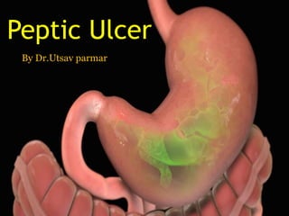
- 1. Peptic Ulcer By Dr.Utsav parmar
- 2. Normal anatomy of stomach
- 3. PEPTIC ULCERS • Ulcer : An open sore that forms on an epithelial surface, e.g., the skin, the mucous membranes, or the lining of the gastrointestinal tract, marked by inflammation, necrosis, and sloughing of damaged tissues • Peptic ulcers are the areas of degeneration and necrosis of gastrointestinal mucosa exposed to acid-peptic secretions • Occur most commonly (98-99%) in either the duodenum or the stomach in the ratio of 4:1.
- 4. Acute Peptic (Stress) Ulcers • multiple, small mucosal erosions, seen most commonly in the stomach • occasionally involving the duodenum.
- 5. Etiology • i) Psychological stress • ii) Physiological stress • Shock • Severe trauma • Septicaemia • Extensive burns • Drugs eg.NSAIDS • Local irritants
- 6. Pathogenesis • Actual hypersecretion of gastric acid is demonstrable in only Cushing’s ulcers occurring from intracranial conditions such as due to brain trauma, intracranial surgery and brain tumours. • In all other etiologic factors, gastric acid secretion is normal or below normal. • In these conditions, the possible hypotheses for genesis of stress ulcers are as under • 1. Ischaemic hypoxic injury to the mucosal cells. • 2. Depletion of the gastric mucus ‘barrier’ rendering the mucosa susceptible to attack by acid-peptic secretions.
- 7. Morphological features • Grossly, acute stress ulcers are multiple (more than three ulcers in 75% of cases). • more common anywhere in the stomach, • followed by first part of duodenum. • They may be oval or circular in shape, usually less than 1 cm in diameter.
- 8. Microscopically • the stress ulcers are shallow and do not invade the muscular layer. • The margins and base may show some inflammatory reaction depending upon the duration . • commonly heal by complete re-epithelialisation without leaving any scars.
- 9. Chronic Peptic Ulcers (Gastric and Duodenal Ulcers) INCIDENCE • Peptic ulcers are more frequent in middle-aged adults. • The peak incidence for duodenal ulcer is 5th decade, • while for gastric ulcer it is a decade later (6th decade). • Duodenal as well as gastric ulcers are more common in males than in females. • Duodenal ulcer is almost four times more common than gastric ulcer
- 10. ETIOLOGY 1. Helicobacter pylori gastritis. 2. NSAIDs-induced mucosal injury. Non-steroidal antiinflammatory 3. Acid-pepsin secretions. 4. Gastritis 5. Dietary factors 6. Psychological factors 7. Genetic factors 8. Hormonal factors.
- 11. PATHOGENESIS. • Although the role of various etiologic factors just described is well known in ulcerogenesis, two most important factors in peptic ulcer are as under: 1. Exposure of mucosa to gastric acid and pepsin secretion. 2. Strong etiologic association with H. pylori infection.
- 12. Duodenal ulcer • There is conclusive evidence to support the role of high acid-pepsin secretions In the causation of duodenal ulcers • 1. There is generally hypersecretion of gastric acid into the fasting stomach at night which takes place under the influence of vagal stimulation. • There is high basal as well as maximal acid output (BAO and MAO) in response to various stimuli.
- 13. • 2. Patients of duodenal ulcer have rapid emptying of the stomach so that the food which normally buffersand neutralises the gastric acid,passes down into the small intestine leaving the duodenal mucosa exposed to the aggressive action of gastric acid.
- 14. 3. Helicobacter gastritis caused by H. pylori is seen in 95-100% cases of duodenal ulcers. The underlying mechanisms are i) Gastric mucosal defense is broken by bacterial elaboration of urease, protease, catalase and phospholipase. ii) Host factors: H. pylori-infected mucosal epithelium releases proinflammatory cytokines such as IL-1, IL-6, IL-8 and tumour necrosis factor-α, all of which incite intense inflammatory reaction. iii) Bacterial factors: Epithelial injury is also induced by cytotoxin-associated gene protein (CagA), while vacuolating cytotoxin (VacA) induces elaboration of cytokines.
- 15. Gastric ulcer The pathological bases are impaired gastric mucosal defenses against acid-pepsin secretions 1. Hyperacidity due to increased serum gastrin levels in response to ingested food in an atonic stomach. 2. However, many patients of gastric ulcer have low- to normal gastric acid levels. 3. Ulcerogenesis in such patients is explained on the basis of damaging influence of other factors such as gastritis, bile reflux, cigarette smoke etc.
- 16. • The normally protective gastric mucus ‘barrier’ against acid-pepsin is deranged in gastric ulcer. • There is depletion in the quantity as well as quality of gastric mucus. • One of the mechanisms for its depletion is colonisation of the gastric mucosa by H. pylori seen in 75-80% patients of gastric ulcer.
- 17. MORPHOLOGIC FEATURES Gross and microscopic changes in gastric and duodenal ulcers are similar and quite characteristic. Gastric ulcers are found predominantly along the lesser curvature in the region of pyloric antrum, more commonly on the posterior than the anterior wall. Most duodenal ulcers are found in the first part of the duodenum, usually immediate post-pyloric, more commonly on the anterior than the posterior wall. Uncommon locations include ulcer in the cardia, marginal ulcer and in the Meckel’s diverticulum
- 18. Distribution of peptic ulcers.
- 19. Grossly, typical peptic ulcers are commonly solitary (80%),small (1-2.5 cm in diameter), round to oval and characteristically ‘punched out’. Benign ulcers have flat margins in level with the surrounding mucosa. The mucosal folds converge towards the ulcer. The ulcers may vary in depth from being superficial (confined to mucosa) to deep ulcers (penetrating into the muscular layer)
- 20. • In about 10-20% of cases, gastric and duodenal ulcers are coexistent. • Vast majority of the peptic ulcers are benign. • Chronic duodenal ulcer never turns malignant, while chronic gastric ulcer may develop carcinoma in less than 1% of cases. • Malignant gastric ulcers are larger, bowl- shaped with elevated and indurated mucosa at the margin
- 21. Benign chronic peptic ulcer. Partial gastrectomy specimen showing a punched out round to oval ulcer on the mucosa, about 1 cm in diameter (arrow) and penetrating into muscularis layer
- 22. Chronic gastric ulcer (A) contrasted with malignant gastric ulcer (B).
- 23. Microscopically, chronic peptic ulcers have 4 histological zones. From within outside 1. Necrotic zone—lies in the floor of the ulcer and is composed of fibrinous exudate containing necrotic debris and a few leucocytes. 2. Superficial exudative zone—The tissue elements show coagulative necrosis giving eosinophilic, smudgy appearance with nuclear debris. 3. Granulation tissue zone— composed of nonspecific inflammatory infiltrate and proliferating capillaries. 4. Zone of cicatrisation—is seen merging into thick layer of granulation tissue. It is composed of dense fibrocollagenic scar tissue over which granulation tissue rests.Thrombosed or sclerotic arteries may cross the ulcer which on erosion may result in haemorrhage.
- 24. • Chronic peptic ulcer. Histologic zones
- 25. CLINICAL FEATURES • Peptic ulcers are remitting and relapsing lesions. Their chronic and recurrent behaviour is summed up the saying: ‘once a peptic ulcer patient, always a peptic ulcer patient.’
- 26. Gastric ulcer Duodenal ulcer Age 6 th decade 5 th decade People at risk laborer stress Periodicity 2-6 weeks attack /1-6 month interval Attacks by work worry weather Pain Immediately after food never at night Late at night relieved by food Vomiting occurs heartburn Hemetemesis and melena 60:40 40:60 Apetite Afraid to eat good Diet Bland diet All kinds weight loss gain Deep tenderness epigastrium hypochondrium
- 27. COMPLICATIONS • Obstruction • Hemorrhage • Perforation • Malignant transformation