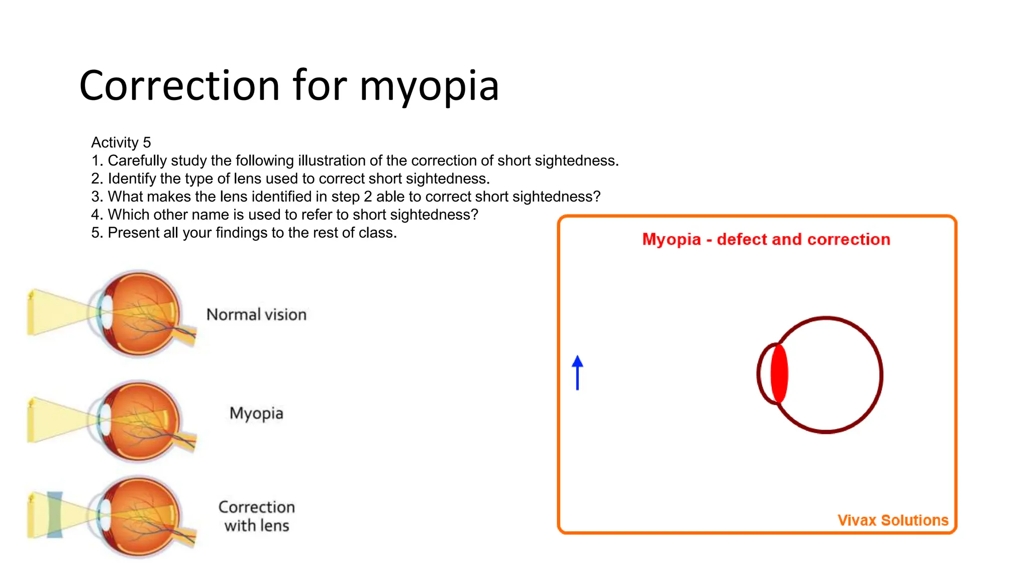
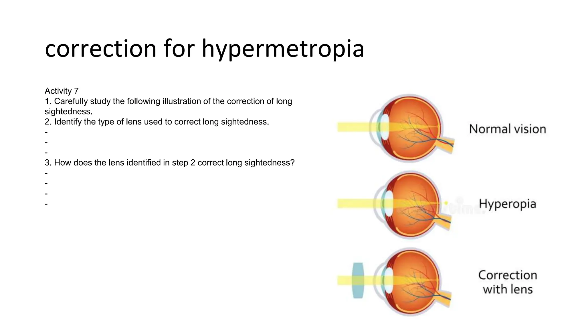
RECEPTOR ORGANS IN MAN
Lesson 1: The Human Eye
What you will learn;
• Competency: The learner appreciates that the sensory organs (eye
and ear) detect stimuli, and enable the body to respond and function
properly.
• LEARNING OUTCOMES: The learner should be able to:
• a) understand the roles and functions of the human eye and ear as sense
organs
• b) understand what short and long sightedness is and how they can be
corrected
The eye; An overview
• Structure of the human eye
• External parts and their functions
• Internal parts and their functions
• Functioning of the eye
• The process of image formation
• Adaptation
• Accommodation
• Eye defects
• Short-sightedness (myopia)
• Long-sightedness (hypermetropia)
• Correction of eye defects
External structure of the eye
Activity 1 Identifying the external features of the human eye
What you need: Model of the human eye, freshly extracted eye of sheep or goat, chart of
human eye, gloves, notebook and pen
Key Question: What are the features of the external parts of the human eye?
What to do:
In groups:
1. Sit while facing each other and look directly into the eye of your partner.
2. Identify and write down the parts of the human eye you are able to see externally include
any hairs associated with the eye.
3. Describe the appearance of the parts you identified in Step 2 and suggest their roles.
Name of part Description Role/function
External structure of the eye
4. Carefully study the features of a fresh mammalian eye or its model provided. Observe and
describe:
a) attachment of the eye,
b) overall shape.
Answers
-
-
-
External structure of the eye
5. Gently press the eyeball and:
a) describe how it feels.
b) suggest the importance of the description of the eye in (a) above.
Answers:
-
-
-
External structure of the eye
6. Look at the chalkboard or any object that is directly in front of you and maintain that
position of your head. Now see the roof without turning your head from its position. What
does this tell you about the muscles illustrated in the Figure?
-
-
-
7. Discuss and agree on:
a) What happens to the eyelids when an object is moved close to the eye.
-
-
-
b) What comes out of the eyes when strong wind or dust is blown directly into the eye.
-
-
-
c) The role of the substance suggested in (b) to the eye.
-
-
-
8. Share your findings with the class for further discussion.
Questions
1. Which of the following is NOT an external part of the human eye? a) Iris
b) Cornea c) Lens d) Pupil
2. What protects the front of the eye? a) Cornea b) Iris c) Pupil
d) Retina
3. _______ control the amount of light entering the eye. a) Eyelids b) Iris c) Pupils d) Lens
4. What is the transparent front part of the eye called? a) Sclera b) Cornea
c) Iris d) Pupil
5. Fill in the blanks: The __________ secretes tears to lubricate the eye.
6. What is the colored part of the eye? _______________
7. What is the role of the eyelashes? ________________________________
8. Name the two muscles that control eyeball movement: ______________ and
_______________
9. How does the corneal reflex protect the eyes?
_____________________________________
10.Which layer of the eye contains blood vessels? ___________________
Internal structure of the eye
Activity 2 Identifying the internal parts of the human eye and their functions
What you need: Chart of human eye, access to the Internet, Biology
textbook, freshly extracted eye of a sheep or goat, notebook and pen
Key Question: What are the internal parts of the human eye and their
functions?
What to do:
1. Carefully study the following illustration of he cross section fo the human
eye.
2. Identify from the cross section of a mammalian eye
provided, the parts labelled in the illustration in step 1
and state their functions in the following table.
Part function
Questions
1. Play this game
2. Which part of the eye focuses light onto the retina? a) Iris
b) Cornea c) Lens d) Pupil
3. Where are the photoreceptor cells located?
a) Iris b) Cornea c) Retina d) Lens
4. The _______________ lining absorbs excess light to prevent reflection.
5. Rods and cones convert light energy into ______________ signals.
6. Name the two fluids that maintain pressure and shape of the eye: _________ and
_________
7. Light passes through the _________________ before striking the retina.
8. The optic nerve carries ______________ from the eye to the brain.
9. What is the name of the point where the optic nerve meets the retina?
___________________
10._____________ are responsible for color vision.
11.______________ detect dim light and provide night vision
Image formation/vision
Activity 3: Formation of images on the retina
1. Trace and list parts of they eye through which light passes
from time of incidence to formation of an image.
2. Use illustration to discuss and describe how human eye
carries out sight.
3. What is relationship between object and its image on retina?
-
-
-
Color vision and color blindness
• Try this simulation
Questions
1. Where is the real inverted image formed in the eye?
a) Cornea
b) Iris c) Retina
d) Lens
2. The image becomes inverted due to ______________ through the lens. a) Accommodation b)
Refraction
c) Convergence d) Divergence
3. At which point do rays of light converge in the eye? _______________
4. Why is the retinal image inverted compared to the object? __________________________
5. How does the brain interpret the inverted image? ____________________________________
6. Circle the option that best completes this statement: "As light passes from the air into the eye, it is
____________." refracted / reflected / absorbed
7. Light is focused on the fovea which is the area of ____________ vision.
8. Name two structures that refract light in the eye: ______________ and _______________
9. The size of the pupil affects the amount of ______________ entering the eye.
10.What causes an image to become upside down as it passes through the eye? _______________
Adaptation of the eye in light and dark
Questions
1. How does the pupil adapt when light levels are low?
a) It contracts b) It expands c) It becomes more opaque d) The lens changes shape
2. Rod cells are more sensitive to ______________ light. a) Bright b) Dim c) Blue
d) Red
3. Why does the pupil dilate in dim light? _______________________________________
4. How does the iris adapt to changes in brightness?
_______________________________
5. Rods contain a pigment called ___________________ that is very sensitive.
6. Cones work best in _____________ light conditions.
7. What causes the pupil size to change? __________________________________
8. Fill in the blanks: Cones detect ____________, while rods detect _______________.
9. Night blindness is due to a deficiency in ________________ cells.
10.Why is peripheral vision better in dim light? ________________________________
Accommodation of the eye
Eye defects-Simulation
• Simulation
Questions
1. What causes the lens to change shape for accommodation? a) Pupil dilation b)
Ciliary muscles contracting/relaxing c) Eyelids squeezing d) External pressure
2. Accommodation allows focusing on near/far objects by
_______________________.
3. How does the ciliary muscle control accommodation?
__________________________
4. When viewing far objects, the ciliary muscle is ________________.
5. The process of altering the lens shape to focus light is called
_______________.
6. What happens to the lens during accommodation?
___________________________
7. Accommodation is achieved by the ______________ and ____________ of the
ciliary muscles.
8. What is the closest point at which the eye can focus clearly?
________________
9. Loss of accommodation with age leads to a condition called
Short sightedness (myopia)
Activity 4:
1. Carefully study the following illustrations of a short sighted eye seeing near
and far objects.
2. Identify the type of object a short sighted eye is able to see clearly.
-
-
Try this simulation also
Correction for myopia
Activity 5
1. Carefully study the following illustration of the correction of short sightedness.
2. Identify the type of lens used to correct short sightedness.
3. What makes the lens identified in step 2 able to correct short sightedness?
4. Which other name is used to refer to short sightedness?
5. Present all your findings to the rest of class.
Questions
1. In myopia, images focus ____________ the retina.
a) Directly on b) In front of c) Behind d) To the side of
2. What type of lens corrects myopia?
a) Converging/convex b) Diverging/concave c) Cylindrical d) Bifocal
3. Myopia is also referred to as ________________.
4. Myopic eyes can see ____________ objects more clearly than far away objects.
5. What defect does myopia cause? _____________________________________
6. Why does image focus in front of the retina in myopia?
____________________________
7. Concave lenses cause light rays to _____________.
8. Myopia results from ______________ or ______________ length.
9. In myopia, near or far vision is clearer? _______________
10. How do concave lenses correct myopia?
_____________________________________
Hypermetropia
• Try this Simulation
Long sightedness (hypermetropia)
Activity 6
1. Carefully study the following illustration of the correction of long sightedness.
2. Identify the type of lens used to correct long sightedness.
-
-
3. What makes the lens identified in step 2 able to correct long sightedness?
-
-
-
4. Which other name is used to refer to long sightedness?
-
-
-
5. Present all your findings to the rest of class.
Try simulation
correction for hypermetropia
Activity 7
1. Carefully study the following illustration of the correction of long
sightedness.
2. Identify the type of lens used to correct long sightedness.
-
-
-
3. How does the lens identified in step 2 correct long sightedness?
-
-
-
-
Questions
1. In hypermetropia, images focus ___________ the retina.
a) Directly on b) Behind c) In front of d) To the side of
2. What type of lens corrects hypermetropia? a) Converging/convex b) Diverging/concave
c) Cylindrical d) Bifocal
3. Hypermetropia is also called ____________________.
4. Hypermetropic eyes can see ____________ objects more clearly than near objects.
5. What defect does hypermetropia cause? ____________________________________
6. Why does image focus behind the retina in hypermetropia?
_________________________
7. Convex lenses cause light rays to __________________.
8. Hypermetropia results from a(n) ____________ or ____________ eyeball.
9. In hypermetropia, near or far vision is clearer? _________________
10 How do convex lenses correct hypermetropia?
___________________________________
Assignment
• Video assignment
ALTERNATIVELY
If you are not signed up on edpuzzle, then watch the video YouTube here and answer these questions

RECEPTOR ORGANS IN MAN.pptx

  • 1.
    RECEPTOR ORGANS INMAN Lesson 1: The Human Eye
  • 2.
    What you willlearn; • Competency: The learner appreciates that the sensory organs (eye and ear) detect stimuli, and enable the body to respond and function properly. • LEARNING OUTCOMES: The learner should be able to: • a) understand the roles and functions of the human eye and ear as sense organs • b) understand what short and long sightedness is and how they can be corrected
  • 3.
    The eye; Anoverview • Structure of the human eye • External parts and their functions • Internal parts and their functions • Functioning of the eye • The process of image formation • Adaptation • Accommodation • Eye defects • Short-sightedness (myopia) • Long-sightedness (hypermetropia) • Correction of eye defects
  • 4.
    External structure ofthe eye Activity 1 Identifying the external features of the human eye What you need: Model of the human eye, freshly extracted eye of sheep or goat, chart of human eye, gloves, notebook and pen Key Question: What are the features of the external parts of the human eye? What to do: In groups: 1. Sit while facing each other and look directly into the eye of your partner. 2. Identify and write down the parts of the human eye you are able to see externally include any hairs associated with the eye. 3. Describe the appearance of the parts you identified in Step 2 and suggest their roles. Name of part Description Role/function
  • 5.
    External structure ofthe eye 4. Carefully study the features of a fresh mammalian eye or its model provided. Observe and describe: a) attachment of the eye, b) overall shape. Answers - - -
  • 6.
    External structure ofthe eye 5. Gently press the eyeball and: a) describe how it feels. b) suggest the importance of the description of the eye in (a) above. Answers: - - -
  • 7.
    External structure ofthe eye 6. Look at the chalkboard or any object that is directly in front of you and maintain that position of your head. Now see the roof without turning your head from its position. What does this tell you about the muscles illustrated in the Figure? - - - 7. Discuss and agree on: a) What happens to the eyelids when an object is moved close to the eye. - - - b) What comes out of the eyes when strong wind or dust is blown directly into the eye. - - - c) The role of the substance suggested in (b) to the eye. - - - 8. Share your findings with the class for further discussion.
  • 8.
    Questions 1. Which ofthe following is NOT an external part of the human eye? a) Iris b) Cornea c) Lens d) Pupil 2. What protects the front of the eye? a) Cornea b) Iris c) Pupil d) Retina 3. _______ control the amount of light entering the eye. a) Eyelids b) Iris c) Pupils d) Lens 4. What is the transparent front part of the eye called? a) Sclera b) Cornea c) Iris d) Pupil 5. Fill in the blanks: The __________ secretes tears to lubricate the eye. 6. What is the colored part of the eye? _______________ 7. What is the role of the eyelashes? ________________________________ 8. Name the two muscles that control eyeball movement: ______________ and _______________ 9. How does the corneal reflex protect the eyes? _____________________________________ 10.Which layer of the eye contains blood vessels? ___________________
  • 9.
    Internal structure ofthe eye Activity 2 Identifying the internal parts of the human eye and their functions What you need: Chart of human eye, access to the Internet, Biology textbook, freshly extracted eye of a sheep or goat, notebook and pen Key Question: What are the internal parts of the human eye and their functions? What to do: 1. Carefully study the following illustration of he cross section fo the human eye. 2. Identify from the cross section of a mammalian eye provided, the parts labelled in the illustration in step 1 and state their functions in the following table. Part function
  • 10.
    Questions 1. Play thisgame 2. Which part of the eye focuses light onto the retina? a) Iris b) Cornea c) Lens d) Pupil 3. Where are the photoreceptor cells located? a) Iris b) Cornea c) Retina d) Lens 4. The _______________ lining absorbs excess light to prevent reflection. 5. Rods and cones convert light energy into ______________ signals. 6. Name the two fluids that maintain pressure and shape of the eye: _________ and _________ 7. Light passes through the _________________ before striking the retina. 8. The optic nerve carries ______________ from the eye to the brain. 9. What is the name of the point where the optic nerve meets the retina? ___________________ 10._____________ are responsible for color vision. 11.______________ detect dim light and provide night vision
  • 11.
    Image formation/vision Activity 3:Formation of images on the retina 1. Trace and list parts of they eye through which light passes from time of incidence to formation of an image. 2. Use illustration to discuss and describe how human eye carries out sight. 3. What is relationship between object and its image on retina? - - -
  • 12.
    Color vision andcolor blindness • Try this simulation
  • 13.
    Questions 1. Where isthe real inverted image formed in the eye? a) Cornea b) Iris c) Retina d) Lens 2. The image becomes inverted due to ______________ through the lens. a) Accommodation b) Refraction c) Convergence d) Divergence 3. At which point do rays of light converge in the eye? _______________ 4. Why is the retinal image inverted compared to the object? __________________________ 5. How does the brain interpret the inverted image? ____________________________________ 6. Circle the option that best completes this statement: "As light passes from the air into the eye, it is ____________." refracted / reflected / absorbed 7. Light is focused on the fovea which is the area of ____________ vision. 8. Name two structures that refract light in the eye: ______________ and _______________ 9. The size of the pupil affects the amount of ______________ entering the eye. 10.What causes an image to become upside down as it passes through the eye? _______________
  • 14.
    Adaptation of theeye in light and dark
  • 15.
    Questions 1. How doesthe pupil adapt when light levels are low? a) It contracts b) It expands c) It becomes more opaque d) The lens changes shape 2. Rod cells are more sensitive to ______________ light. a) Bright b) Dim c) Blue d) Red 3. Why does the pupil dilate in dim light? _______________________________________ 4. How does the iris adapt to changes in brightness? _______________________________ 5. Rods contain a pigment called ___________________ that is very sensitive. 6. Cones work best in _____________ light conditions. 7. What causes the pupil size to change? __________________________________ 8. Fill in the blanks: Cones detect ____________, while rods detect _______________. 9. Night blindness is due to a deficiency in ________________ cells. 10.Why is peripheral vision better in dim light? ________________________________
  • 16.
  • 17.
  • 18.
    Questions 1. What causesthe lens to change shape for accommodation? a) Pupil dilation b) Ciliary muscles contracting/relaxing c) Eyelids squeezing d) External pressure 2. Accommodation allows focusing on near/far objects by _______________________. 3. How does the ciliary muscle control accommodation? __________________________ 4. When viewing far objects, the ciliary muscle is ________________. 5. The process of altering the lens shape to focus light is called _______________. 6. What happens to the lens during accommodation? ___________________________ 7. Accommodation is achieved by the ______________ and ____________ of the ciliary muscles. 8. What is the closest point at which the eye can focus clearly? ________________ 9. Loss of accommodation with age leads to a condition called
  • 19.
    Short sightedness (myopia) Activity4: 1. Carefully study the following illustrations of a short sighted eye seeing near and far objects. 2. Identify the type of object a short sighted eye is able to see clearly. - - Try this simulation also
  • 20.
    Correction for myopia Activity5 1. Carefully study the following illustration of the correction of short sightedness. 2. Identify the type of lens used to correct short sightedness. 3. What makes the lens identified in step 2 able to correct short sightedness? 4. Which other name is used to refer to short sightedness? 5. Present all your findings to the rest of class.
  • 21.
    Questions 1. In myopia,images focus ____________ the retina. a) Directly on b) In front of c) Behind d) To the side of 2. What type of lens corrects myopia? a) Converging/convex b) Diverging/concave c) Cylindrical d) Bifocal 3. Myopia is also referred to as ________________. 4. Myopic eyes can see ____________ objects more clearly than far away objects. 5. What defect does myopia cause? _____________________________________ 6. Why does image focus in front of the retina in myopia? ____________________________ 7. Concave lenses cause light rays to _____________. 8. Myopia results from ______________ or ______________ length. 9. In myopia, near or far vision is clearer? _______________ 10. How do concave lenses correct myopia? _____________________________________
  • 22.
  • 23.
    Long sightedness (hypermetropia) Activity6 1. Carefully study the following illustration of the correction of long sightedness. 2. Identify the type of lens used to correct long sightedness. - - 3. What makes the lens identified in step 2 able to correct long sightedness? - - - 4. Which other name is used to refer to long sightedness? - - - 5. Present all your findings to the rest of class. Try simulation
  • 24.
    correction for hypermetropia Activity7 1. Carefully study the following illustration of the correction of long sightedness. 2. Identify the type of lens used to correct long sightedness. - - - 3. How does the lens identified in step 2 correct long sightedness? - - - -
  • 25.
    Questions 1. In hypermetropia,images focus ___________ the retina. a) Directly on b) Behind c) In front of d) To the side of 2. What type of lens corrects hypermetropia? a) Converging/convex b) Diverging/concave c) Cylindrical d) Bifocal 3. Hypermetropia is also called ____________________. 4. Hypermetropic eyes can see ____________ objects more clearly than near objects. 5. What defect does hypermetropia cause? ____________________________________ 6. Why does image focus behind the retina in hypermetropia? _________________________ 7. Convex lenses cause light rays to __________________. 8. Hypermetropia results from a(n) ____________ or ____________ eyeball. 9. In hypermetropia, near or far vision is clearer? _________________ 10 How do convex lenses correct hypermetropia? ___________________________________
  • 26.
    Assignment • Video assignment ALTERNATIVELY Ifyou are not signed up on edpuzzle, then watch the video YouTube here and answer these questions